📋 Key Information Summary
- Valvular heart disease (VHD) encompasses stenotic and regurgitant lesions of the aortic, mitral, tricuspid, and pulmonary valves — echocardiography is the cornerstone of diagnosis and severity grading.
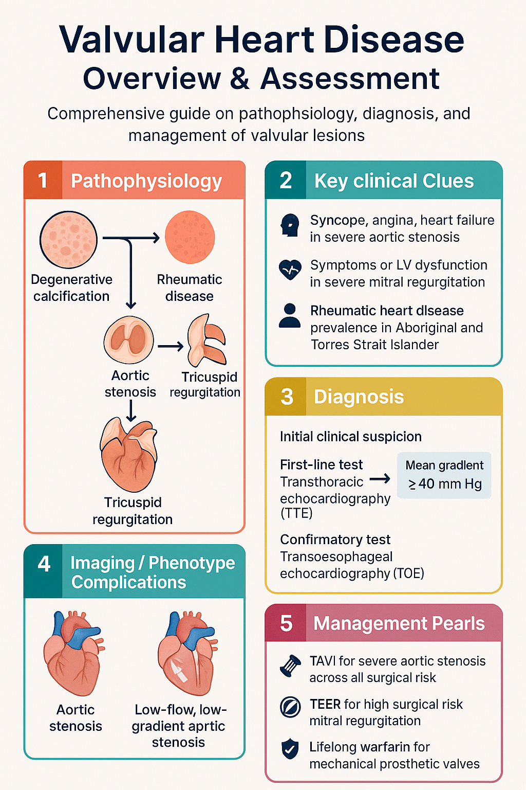
- Degenerative calcific aortic stenosis is the most common valvular lesion in older Australians; rheumatic heart disease remains disproportionately prevalent in Aboriginal and Torres Strait Islander communities.
- Doppler echocardiography quantifies VHD severity using mean gradient, valve area (continuity equation or planimetry), and regurgitant volume/fraction — severity grading drives timing of intervention.
- Class I indications for aortic valve intervention include symptomatic severe AS (syncope, angina, heart failure) and asymptomatic severe AS with LVEF <50% or abnormal exercise test.
- Transcatheter aortic valve implantation (TAVI) is now indicated across all surgical risk categories for severe AS; surgical AVR remains preferred in younger patients (<65 years) or those with complex concomitant pathology.
- Severe primary mitral regurgitation with symptoms or LV dysfunction (LVEF ≤60%, LVESD ≥40 mm) warrants intervention — transcatheter edge-to-edge repair (TEER/MitraClip) is an option for high surgical risk patients.
- Mechanical prosthetic valves require lifelong warfarin anticoagulation (target INR varies by valve position and type); DOACs are contraindicated with mechanical valves.
- Bioprosthetic valve thrombosis (subclinical leaflet thrombosis) may be detected on CT; anticoagulation decisions should be individualised and guided by multidisciplinary heart team discussion.
- Infective endocarditis prophylaxis (amoxicillin 2 g PO or ampicillin 2 g IV pre-procedure) is recommended for patients with prosthetic valves, prior endocarditis, or certain congenital heart diseases per ESC/AHA guidelines.
- All patients with significant VHD should be referred for multidisciplinary Heart Team assessment before intervention — this includes cardiologist, cardiac surgeon, imaging specialist, and anaesthetist.
- Rheumatic heart disease (RHD) control programmes (ARF/RHD register) are essential in ATSI communities; secondary prophylaxis with benzathine penicillin G every 21–28 days is critical.
- Pregnancy in women with VHD requires pre-conception counselling — severe AS and mechanical valves pose the highest maternal and foetal risk.
Introduction & Australian Epidemiology
Valvular heart disease (VHD) encompasses a broad spectrum of structural abnormalities affecting the four cardiac valves, resulting in stenosis, regurgitation, or a combination of both. These lesions impose abnormal haemodynamic loads on the heart, leading to chamber remodelling, heart failure, arrhythmias, and ultimately increased morbidity and mortality if left untreated.
In Australia, VHD is a significant and growing public health burden. The Australian Institute of Health and Welfare (AIHW) reports that diseases of the circulatory system remain a leading cause of death, with valvular heart disease contributing substantially, particularly among older Australians. Degenerative calcific aortic stenosis is the most prevalent valvular lesion in developed nations, driven by an ageing population and the cumulative effects of atherosclerotic risk factors.
Rheumatic heart disease (RHD) continues to represent a major health disparity in Australia. Aboriginal and Torres Strait Islander peoples experience RHD rates that are among the highest in the world — approximately 50–80 times greater than non-Indigenous Australians, with peak incidence in children and young adults in the Northern Territory, Queensland, and Western Australia. The National Rheumatic Fever Strategy and jurisdictional RHD control programmes aim to reduce this burden through register-based care, secondary prophylaxis delivery, and community engagement.
The landscape of valvular intervention has been transformed by transcatheter technologies. Transcatheter aortic valve implantation (TAVI) has expanded from a niche therapy for inoperable patients to a first-line option across all surgical risk categories. Similarly, transcatheter edge-to-edge repair (TEER) for mitral regurgitation and transcatheter tricuspid interventions are increasingly available in Australian tertiary centres. These advances underscore the importance of accurate echocardiographic assessment and timely referral to specialised Heart Teams.
Classification & Aetiology
Classification by Valve Lesion
| Lesion Type | Valve Most Commonly Affected | Haemodynamic Consequence |
|---|---|---|
| Stenosis | Aortic valve (most common); mitral valve | Pressure overload → LV hypertrophy (AS) or LA enlargement (MS) |
| Regurgitation | Mitral valve (most common); aortic valve | Volume overload → LV dilation (AR, MR) or RV dilation (TR, PR) |
| Mixed (stenosis + regurgitation) | Any valve; common in rheumatic and degenerative disease | Combined pressure and volume overload |
Aetiologies by Valve
Aortic Valve
- Calcific (degenerative) aortic stenosis: Most common cause in Australia and other developed nations. Progressive calcification of a trileaflet (or bicuspid) aortic valve. Risk factors include age (>65 years), hypertension, hyperlipidaemia, diabetes, chronic kidney disease, and bicuspid aortic valve morphology.
- Bicuspid aortic valve (BAV): Present in 1–2% of the population; the most common congenital cardiac abnormality. Associated with earlier onset AS or AR, aortopathy, and aortic dissection risk.
- Rheumatic aortic valve disease: Causes commissural fusion leading to stenosis and/or regurgitation. Common in ATSI communities and migrants from endemic regions.
- Aortic regurgitation: May result from leaflet pathology (bicuspid valve, endocarditis, rheumatic disease) or aortic root dilation (Marfan syndrome, bicuspid aortopathy, aortitis, hypertension).
Mitral Valve
- Primary (organic) mitral regurgitation: Degenerative (Barlow disease, fibroelastic deficiency), rheumatic, infective endocarditis, congenital (cleft leaflet), drug-induced (ergotamine, ergot-derived dopamine agonists).
- Secondary (functional) mitral regurgitation: Due to LV dilation/remodelling causing leaflet tethering and annular dilatation — commonly seen in ischaemic or dilated cardiomyopathy.
- Mitral stenosis: Almost always rheumatic in origin in Australia. Other rare causes include severe annular calcification, congenital, and carcinoid.
Tricuspid and Pulmonary Valves
- Tricuspid regurgitation: Most commonly functional (secondary to RV dilation from pulmonary hypertension or left heart disease). Primary causes include endocarditis (particularly in IV drug users), carcinoid, Ebstein anomaly, and pacemaker/ICD lead-induced.
- Tricuspid stenosis: Rare; usually rheumatic.
- Pulmonary valve disease: Most commonly congenital (pulmonary stenosis, repaired tetralogy of Fallot with pulmonary regurgitation). Acquired pulmonary stenosis is rare (carcinoid, external compression).
Echocardiographic Assessment (Doppler, Area, Gradient)
Transthoracic echocardiography (TTE) is the first-line and most important imaging modality for diagnosis, severity grading, and surveillance of VHD. Transoesophageal echocardiography (TOE) is reserved for cases where TTE is inconclusive, for assessment of prosthetic valves, endocarditis, and pre-procedural planning for transcatheter interventions.
Key Echocardiographic Parameters
| Parameter | Method | Clinical Application |
|---|---|---|
| Peak velocity (Vmax) | Continuous-wave Doppler across the valve | Primary measure for AS severity; also used for MS and PR |
| Mean gradient | Modified Bernoulli equation from CW Doppler envelope | Grades stenosis severity (AS, MS); flow-dependent |
| Valve area (AVA/MVA) | Continuity equation (AS); pressure half-time or planimetry (MS) | Flow-independent measure of stenosis severity |
| Vena contracta | Colour Doppler — narrowest jet width at regurgitant orifice | Semi-quantitative assessment of regurgitation severity |
| Regurgitant volume & fraction | PISA method or volumetric (LVOT − aortic flow) | Quantitative grading of MR and AR severity |
| Effective regurgitant orifice area (EROA) | Proximal isovelocity surface area (PISA) method | Quantifies MR severity; EROA ≥40 mm² = severe primary MR |
| LV dimensions & LVEF | M-mode, 2D, or 3D; Simpson biplane method | Assesses ventricular compensation and timing of intervention |
Severity Grading — Aortic Stenosis
Severity Grading — Mitral Regurgitation
Severity Grading — Mitral Stenosis
Severity Grading — Aortic Regurgitation
Additional Imaging Modalities
- Transoesophageal echocardiography (TOE): Superior for mitral valve morphology assessment, prosthetic valve dysfunction, endocarditis vegetations, and pre-TAVI/TEER planning. TOE is available at all Australian public tertiary centres and many private facilities.
- Cardiac CT: Essential for TAVI planning (aortic annulus sizing, vascular access assessment). Also detects subclinical bioprosthetic valve thrombosis (hypo-attenuated leaflet thickening — HALT) and quantifies aortic valve calcification score (useful in discordant AS grading).
- Cardiac MRI: Quantitative assessment of regurgitant volumes (particularly AR and PR), myocardial fibrosis assessment (prognostic in AS), and RV function assessment.
- Exercise stress testing: Unmasked symptomatic AS in apparently asymptomatic patients. An abnormal response (symptom development, fall in BP, ST changes) is a Class IIa indication for intervention.
Surgical vs Transcatheter Approach
The decision between surgical and transcatheter intervention for VHD is guided by a multidisciplinary Heart Team approach, incorporating patient age, surgical risk (STS score, EuroSCORE II), valve pathology, comorbidities, frailty, life expectancy, anatomical suitability, and patient preference. This model is standard across Australian tertiary centres performing structural heart interventions.
Aortic Valve: Surgical AVR vs TAVI
| Factor | Surgical AVR (SAVR) | Transcatheter AVR (TAVI) |
|---|---|---|
| Preferred age | <65 years (or longer life expectancy) | ≥65 years (or higher surgical risk) |
| Surgical risk | Low to intermediate risk (STS <8%) | Any risk category (low to extreme) |
| Valve type | Mechanical or bioprosthetic options | Bioprosthetic only |
| Concomitant surgery | Can address CABG, aortic surgery, other valve pathology | Not applicable |
| Access | Median sternotomy (or minimally invasive) | Transfemoral (preferred), transapical, transaortic |
| Anaesthesia | General anaesthesia | Local + sedation or GA (centre-dependent) |
| Hospital stay | 5–10 days | 2–5 days (often next-day discharge possible) |
| Prosthesis-patient mismatch | Risk in small annuli | Lower risk ( supra-annular design) |
| Long-term data | Decades of durability data | 10+ years for first-generation valves; ongoing registries |
| Key complications | Bleeding, stroke, sternal wound infection | Conduction disturbance (pacemaker 10–25%), vascular complications, paravalvular leak |
Mitral Valve: Surgical Repair vs TEER
| Factor | Surgical Mitral Repair | Transcatheter Edge-to-Edge Repair (TEER / MitraClip) |
|---|---|---|
| Preferred in | Primary MR, low surgical risk, favourable anatomy | Primary MR at prohibitive surgical risk; secondary MR (with HF guideline-directed therapy) |
| Anatomical requirements | Broad — complex repairs possible | Central P2/A2 flail/gap <10 mm; adequate leaflet length; no calcification at grasping zone |
| MR reduction | Near-complete if successful | Reduction to ≤2+ in ~80% of cases |
| Reoperation rate | Low (1–2% per year for repair) | Higher for primary MR; lower for secondary MR |
Tricuspid Valve Interventions
Isolated tricuspid valve surgery is high-risk and historically underperformed. Emerging transcatheter tricuspid interventions (e.g., TriClip TEER, annuloplasty devices) are now available in select Australian centres for severe TR in patients at prohibitive surgical risk. Current evidence supports intervention in symptomatic patients with severe primary TR or severe secondary TR despite optimal medical therapy for left-sided disease and HF.
Indications for Intervention — Summary
Anticoagulation in Prosthetic Valves
Anticoagulation management following valve replacement is determined by prosthesis type (mechanical vs bioprosthetic), valve position, thrombotic risk factors, and bleeding risk. Errors in anticoagulation are a leading cause of preventable prosthetic valve thrombosis and death.
Mechanical Prosthetic Valves
All mechanical valves require lifelong warfarin anticoagulation. Direct oral anticoagulants (DOACs — apixaban, rivaroxaban, dabigatran, edoxaban) are absolutely contraindicated with mechanical prosthetic valves, as demonstrated by the RE-ALIGN trial (dabigatran), which was terminated early due to excess thromboembolic and bleeding events.
Target INR by Valve Type and Position
| Valve Position | Prosthesis Type | Target INR | Notes |
|---|---|---|---|
| Aortic — bileaflet/medtronic hall | Mechanical | 2.0–3.0 | Standard target; lower thrombotic risk than tilting-disc/caged-ball |
| Aortic — tilting-disc or caged-ball | Mechanical | 2.5–3.5 | Higher thrombogenicity |
| Mitral — any mechanical | Mechanical | 2.5–3.5 | Higher thrombosis risk than aortic position; all types |
| Tricuspid — any mechanical | Mechanical | 2.5–3.5 | High thrombosis risk in low-flow right heart position |
Bioprosthetic Valves
| Timeframe | Recommendation | Agent |
|---|---|---|
| First 3–6 months post-op (SAVR) | Warfarin (INR 2.0–3.0) or aspirin 100–150 mg daily | Warfarin preferred if additional risk factors (AF, prior VTE, LV dysfunction, hypercoagulable state) |
| After 3–6 months (SAVR) | Aspirin 75–100 mg daily indefinitely | Consider adding clopidogrel 75 mg daily for first 3–6 months if no warfarin |
| Post-TAVI | Dual antiplatelet therapy (DAPT) — aspirin + clopidogrel for 3–6 months, then aspirin alone | Aspirin 75–100 mg + clopidogrel 75 mg; single antiplatelet after 3–6 months per POPular TAVI trial |
| Post-TAVI with AF | Single antiplatelet + oral anticoagulant (warfarin or DOAC) | Warfarin or DOAC based on AF indication; avoid triple therapy if possible |
Subclinical Bioprosthetic Valve Thrombosis (HALT)
Hypo-attenuated leaflet thickening (HALT) detected on cardiac CT in bioprosthetic valves (both surgical and TAVI) represents subclinical leaflet thrombosis. It is found in 10–15% of TAVI patients and may cause increased gradients. Management is evolving:
- Therapeutic warfarin (INR 2.0–3.0) for 3–6 months has been shown to resolve HALT in most cases.
- Routine CT screening for HALT is not currently recommended; consider CT if unexplained gradient rise on echocardiography.
- Heart Team discussion is essential before initiating anticoagulation for HALT, balancing thrombotic resolution against bleeding risk.
Warfarin — Practical Australian Considerations
Bridging Anticoagulation
Perioperative bridging with unfractionated heparin (UFH) or low-molecular-weight heparin (LMWH — enoxaparin) is required for mechanical valve patients undergoing procedures. Key principles:
- High thrombotic risk (mitral mechanical valve, older-generation aortic valve, recent thrombosis <6 months, AF, LV dysfunction): Bridging mandatory — therapeutic dose UFH or enoxaparin 1 mg/kg BD.
- Low thrombotic risk (bileaflet aortic mechanical valve, no AF, no other risk factors): Bridging may be omitted for low-risk procedures — individualise decision.
- Stop warfarin 5 days pre-procedure; restart 12–24 hours post-procedure when haemostasis is secure.
- LMWH (enoxaparin) is PBS-listed for bridging and is the preferred agent in outpatient bridging. ✔ PBS General Benefit
Special Populations
Aboriginal and Torres Strait Islander Health Considerations
📚 References
- 1. Vahanian A, Beyersdorf F, Praz F, et al. 2021 ESC/EACTS Guidelines for the management of valvular heart disease. Eur Heart J. 2022;43(7):561–632.
- 2. Otto CM, Nishimura RA, Bonow RO, et al. 2020 ACC/AHA Guideline for the Management of Patients With Valvular Heart Disease. Circulation. 2021;143(5):e72–e227.
- 3. Australian Institute of Health and Welfare (AIHW). Heart, stroke and vascular disease — Australian facts. Canberra: AIHW; 2023.
- 4. RHDAustralia (ARF/RHD writing group). The 2020 Australian guideline for prevention, diagnosis and management of acute rheumatic fever and rheumatic heart disease (3rd edition). Darwin: Menzies School of Health Research; 2020.
- 5. Mack MJ, Leon MB, Thourani VH, et al. Transcatheter aortic-valve replacement with a balloon-expandable valve in low-risk patients. N Engl J Med. 2019;380(18):1695–1705 (PARTNER 3 trial).
- 6. Popma JJ, Deeb GM, Yakubov SJ, et al. Transcatheter aortic-valve replacement with a self-expanding valve in low-risk patients. N Engl J Med. 2019;380(18):1706–1715 (EVOLUT Low Risk trial).
- 7. Stone GW, Lindenfeld J, Abraham WT, et al. Transcatheter mitral-valve repair in patients with heart failure. N Engl J Med. 2018;379(24):2307–2318 (COAPT trial).
- 8. De Backer O, Dangas GD, Jilaihawi H, et al. Reduced leaflet motion after transcatheter aortic-valve replacement. N Engl J Med. 2020;382(2):130–139 (PORTICO-NG sub-study on HALT).
- 9. Eikelboom JW, Connolly SJ, Brueckmann M, et al. Dabigatran versus warfarin in patients with mechanical heart valves. N Engl J Med. 2013;369(13):1206–1214 (RE-ALIGN trial).
- 10. Buber J, Cheng A, Leon MB, et al. Aspirin alone versus dual antiplatelet therapy after transcatheter aortic valve implantation. Lancet. 2020;396(10258):1187–1196 (POPular TAVI trial).
- 11. Australian Commission on Safety and Quality in Health Care (ACSQHC). National Safety and Quality Health Service Standards (NSQHS). 2nd edition. Sydney: ACSQHC; 2021.
- 12. Commonwealth of Australia. Pharmaceutical Benefits Scheme (PBS). Available at: pbs.gov.au. Accessed 2024.
- 13. Pibarot P, Dumesnil JG. Prosthetic heart valves: selection of the optimal prosthesis and long-term management. Circulation. 2009;119(7):1034–1048.
- 14. Australian and New Zealand Society of Cardiac and Thoracic Surgeons (ANZSCTS). National Cardiac Surgery Database Program — Annual Report. Melbourne: ANZSCTS; 2023.
- 15. Carapetis JR, Steer AC, Mulholland EK, Weber M. The global burden of group A streptococcal diseases. Lancet Infect Dis. 2005;5(11):685–694.