📋 Key Information Summary
- Mitral regurgitation (MR) is the most common valvular heart disease in Australia, affecting approximately 2–3% of the population, with prevalence increasing with age.
- Primary (degenerative) MR results from intrinsic valve pathology (e.g., myxomatous degeneration, flail leaflet, rheumatic disease); secondary (functional) MR arises from left ventricular remodelling with structurally normal leaflets.
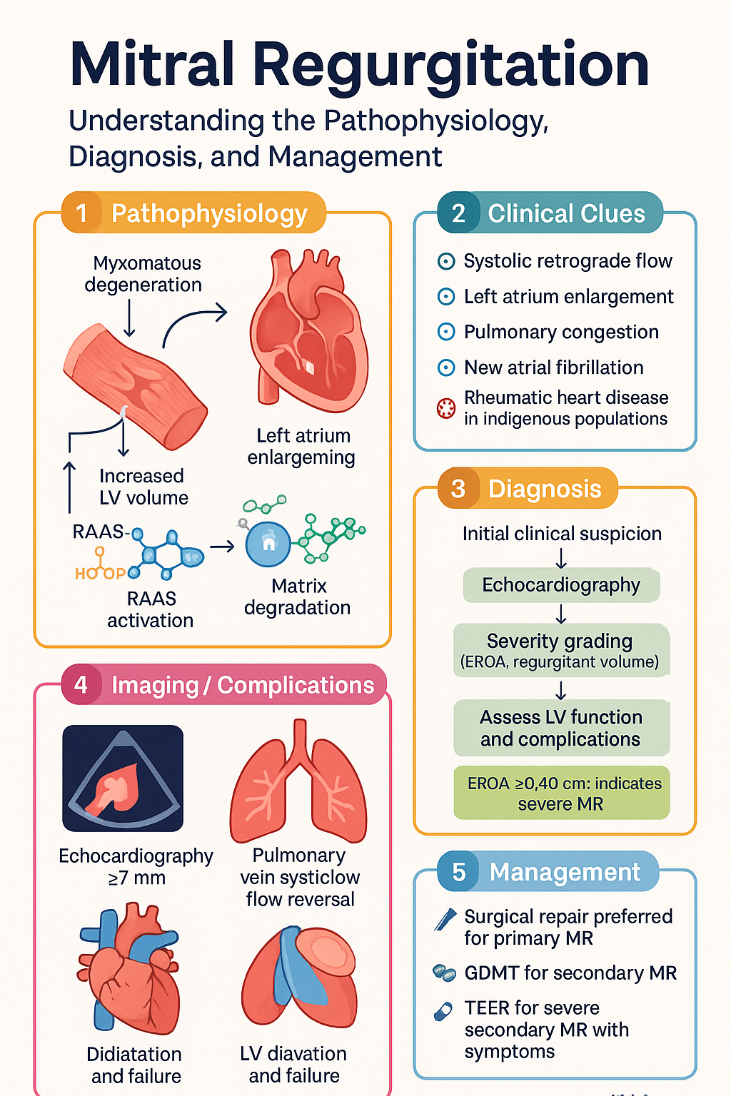
- Echocardiography is the cornerstone of diagnosis — severity grading uses EROA (≥0.40 cm² severe), regurgitant volume (≥60 mL), vena contracta (≥7 mm), and pulmonary vein systolic flow reversal.
- Surgical mitral valve repair is preferred over replacement for primary MR where feasible, offering superior long-term survival, preserved LV function, and freedom from anticoagulation.
- For primary MR, surgery (repair or replacement) is indicated for severe symptomatic disease (Class I) and should be considered for severe asymptomatic disease with LV dysfunction (LVEF ≤60%, LVESD ≥40 mm) or new atrial fibrillation (Class IIa).
- For secondary MR, guideline-directed medical therapy (GDMT) for heart failure — including ACE inhibitors/ARBs/ARNI, beta-blockers, mineralocorticoid receptor antagonists, SGLT2 inhibitors, and device therapy (CRT/ICD) — is first-line treatment.
- Transcatheter edge-to-edge repair (TEER / MitraClip) is recommended for patients with severe secondary MR who remain symptomatic (NYHA II–IV) despite optimal GDMT and are unsuitable for surgery (COAPT trial criteria).
- The COAPT trial demonstrated significant mortality and heart failure hospitalisation benefit with MitraClip in secondary MR with EROA ≥0.30 cm², while MITRA-FR showed no benefit — differences attributed to GDMT optimisation and MR severity thresholds.
- Diuretics are essential for symptom relief in decompensated MR (furosemide 20–80 mg IV/PO), but they do not modify disease progression.
- Aboriginal and Torres Strait Islander Australians have higher rates of rheumatic heart disease–related MR, delayed diagnosis, reduced access to specialist and surgical services, and worse outcomes — culturally appropriate screening and care pathways are critical.
- Timing of intervention is critical — delay in primary MR risks irreversible LV dysfunction; premature intervention in secondary MR without GDMT optimisation may not improve outcomes.
Introduction & Australian Epidemiology
Mitral regurgitation (MR) is the most prevalent valvular heart lesion worldwide and in Australia. The burden of MR is increasing with an ageing population, improved survival after myocardial infarction, and the rising prevalence of heart failure. MR is characterised by systolic retrograde flow from the left ventricle (LV) into the left atrium (LA) through an incompetent mitral valve.
In Australia, moderate-to-severe MR affects approximately 2–3% of the general population, with prevalence rising to >10% in those aged over 75 years. The Australian Institute of Health and Welfare (AIHW) reports that valvular heart disease contributed to over 4,500 hospitalisations and significant mortality in the most recent reporting period. Rheumatic heart disease (RHD) remains an important aetiology, particularly among Aboriginal and Torres Strait Islander communities in the Northern Territory, Queensland, and Western Australia, where RHD-related MR occurs at 20–60 times the rate seen in non-Indigenous Australians.
Mitral valve prolapse (MVP), the leading cause of primary degenerative MR in high-income countries, affects 2–3% of the Australian population. Functional (secondary) MR is increasingly common as a consequence of ischaemic and non-ischaemic cardiomyopathy, reflecting the growing burden of heart failure nationally.
Primary vs Secondary MR
Classification and Pathophysiology
The distinction between primary and secondary MR is fundamental to management. Primary MR arises from structural abnormality of the mitral valve apparatus itself, whereas secondary (functional) MR results from adverse left ventricular remodelling in the setting of a structurally normal valve.
| Feature | Primary (Degenerative) MR | Secondary (Functional) MR |
|---|---|---|
| Aetiology | Myxomatous degeneration (MVP), flail leaflet, rheumatic disease, endocarditis, calcification, congenital cleft | Ischaemic cardiomyopathy (post-MI remodelling), dilated cardiomyopathy (DCM), annular dilatation |
| Valve anatomy | Leaflets/ chordae/ papillary muscles abnormal | Leaflets structurally normal; tethering and/or annular dilatation |
| Jet direction | Variable — posterior prolapse → anterior jet; anterior prolapse → posterior jet | Typically central or posteromedial (ischaemic tethering pattern) |
| LV function | Often preserved initially; LV dilates and fails late | Already impaired (LVEF typically <40%); MR worsens LV failure |
| Hemodynamic | Volume overload on a normal ventricle → eccentric hypertrophy → eventual systolic failure | Volume overload superimposed on a failing ventricle → vicious cycle of adverse remodelling |
| Surgical approach | Valve repair strongly preferred (durability, survival advantage) | Surgery less clearly beneficial; GDMT first-line; TEER for refractory cases |
| Prognosis if untreated | Progressive LV dilatation and failure; sudden death risk with flail leaflet | Driven primarily by underlying cardiomyopathy; MR independently worsens prognosis |
Degenerative MR — Key Subtypes
- Fibroelastic deficiency (FED): Thin, translucent leaflets with elongated or ruptured chordae; typically affects older patients (>60 years), often presents acutely with chordal rupture and flail segment. Repair is usually straightforward with high success rates.
- Myxomatous degeneration (Barlow's disease): Thick, redundant leaflets with excess myxomatous tissue; affects younger patients (<55 years), often bileaflet prolapse. Repair is more complex but highly durable in experienced centres.
- Rheumatic MR: Commissural fusion, chordal thickening and shortening, leaflet restriction. Less amenable to repair; frequently requires replacement. Increasingly rare in non-Indigenous Australians but remains prevalent in Indigenous communities.
Functional MR in Heart Failure
Functional MR is not a valve disease per se but a manifestation of ventricular disease. The two principal mechanisms are:
- Annular dilatation: LV remodelling stretches the mitral annulus, reducing leaflet coaptation. The annular area may increase by >50% in severe DCM.
- Leaflet tethering (displacement of papillary muscles): Apical and lateral displacement of the papillary muscles tenters the leaflets into the LV, creating a coaptation defect. In ischaemic MR, localised posterior wall infarction causes asymmetric posteromedial tethering.
Hemodynamic Differences
In primary MR, the LV ejects into a low-pressure LA, resulting in increased forward stroke volume that initially compensates for the regurgitant volume. LV end-diastolic pressure may remain normal for years. In contrast, in secondary MR, the LV is already volume- and pressure-overloaded, and the added regurgitant volume dramatically increases LA pressure, exacerbating pulmonary congestion and limiting cardiac output.
Severity Assessment
Accurate severity grading of MR is essential for clinical decision-making. Transthoracic echocardiography (TTE) is the initial investigation of choice, with transoesophageal echocardiography (TEE) reserved for inconclusive cases, preoperative planning, and intraoperative guidance. The American Society of Echocardiography (ASE) and European Association of Cardiovascular Imaging (EACVI) recommend an integrative multi-parametric approach.
| Parameter | Mild | Moderate | Severe |
|---|---|---|---|
| Effective Regurgitant Orifice Area (EROA) | <0.20 cm² | 0.20–0.39 cm² | ≥0.40 cm² |
| Regurgitant Volume (RVol) | <30 mL | 30–59 mL | ≥60 mL |
| Regurgitant Fraction (RF) | <30% | 30–49% | ≥50% |
| Vena Contracta Width | <3 mm | 3–6 mm | ≥7 mm |
| Colour Flow Jet Area | Small central jet <4 cm² | Intermediate | Large central jet >10 cm² or wall-impinging jet of any size |
| Pulmonary Vein Flow | Systolic dominance (S > D) | Systolic blunting (S < D) | Systolic flow reversal |
| Mitral Inflow E-wave Velocity | <1.0 m/s | 1.0–1.2 m/s | >1.2 m/s |
| LA/LV Size | Normal | Mildly dilated | LA volume index >40 mL/m²; LV dilated |
Quantitative Methods
- PISA (Proximal Isovelocity Surface Area) method: The most widely used quantitative technique. Measures the radius of the convergence zone on the LV side of the valve using colour Doppler. EROA = (2π × r² × Va) / Vpeak, where r = PISA radius, Va = aliasing velocity, Vpeak = peak MR jet velocity. Regurgitant volume = EROA × VTI of MR jet.
- Volumetric method: Calculates RVol as (mitral inflow volume) − (LV outflow volume), obtained from pulsed-wave Doppler at the mitral annulus and LVOT respectively. More labour-intensive but useful when PISA is unreliable.
Additional Assessment Modalities
Carpentier Classification of Mitral Valve Dysfunction
| Type | Leaflet Motion | Mechanism | Examples |
|---|---|---|---|
| Type I | Normal | Annular dilatation or leaflet perforation | Functional MR in DCM, post-endocarditis perforation |
| Type II | Excessive (prolapse / flail) | Chordal elongation or rupture, papillary muscle dysfunction | MVP, Barlow's disease, FED, post-MI papillary muscle rupture |
| Type IIIa | Restricted (diastole & systole) | Commissural fusion, leaflet thickening, chordal shortening | Rheumatic heart disease |
| Type IIIb | Restricted (systole only) | Papillary muscle displacement, LV dilatation | Ischaemic MR, dilated cardiomyopathy |
Surgical Repair vs Replacement
Principles
For primary (degenerative) MR, mitral valve repair is the gold standard surgical approach and should be performed whenever anatomically feasible. Repair preserves the native valve, avoids prosthetic valve complications (thromboembolism, endocarditis, haemolysis), maintains LV geometry and function, and offers superior long-term survival compared with replacement.
Repair Success Rates and Durability
| Outcome | Mitral Valve Repair | Mitral Valve Replacement |
|---|---|---|
| Operative mortality | 1–2% (elective, primary MR) | 3–6% (elective, primary MR) |
| 10-year freedom from reoperation | 90–95% (degenerative, experienced centres) | 85–90% (mechanical); 75–85% (bioprosthetic) |
| LVEF preservation | Maintains or improves LV function | LV function may decline post-replacement due to chordal disruption |
| Anticoagulation | Not required (unless AF) | Warfarin lifelong (mechanical); 3 months (bioprosthetic) |
| Endocarditis risk | Low (native valve preserved) | Higher (prosthetic valve) |
| LV remodelling | Favourable reverse remodelling | May have persistent or worsening dilatation |
Surgical Indications (ACC/AHA 2020 / ESC 2021)
Primary MR — Class I (Indicated)
- Severe symptomatic MR (NYHA II–IV) — regardless of LVEF
- Severe asymptomatic MR with LVEF ≤60% or LVESD ≥40 mm
- Severe MR undergoing other cardiac surgery (CABG, AVR)
Primary MR — Class IIa (Reasonable)
- Severe asymptomatic MR with preserved LV function (LVEF >60%, LVESD <40 mm) and:
- New-onset atrial fibrillation
- Pulmonary hypertension (PASP >50 mmHg at rest)
- Repair likelihood ≥95% with <1% mortality at an experienced centre
Valve Morphology Suitability for Repair
Choice of Prosthetic Valve (When Replacement Is Required)
Chordal-Sparing Replacement
When mitral replacement is performed for primary MR, preservation of the posterior (and ideally both) subvalvular chordae tendineae is strongly recommended. Chordal-sparing techniques maintain LV geometry and systolic function, resulting in improved postoperative LVEF and survival compared with conventional chordal transection.
Transcatheter Interventions
Transcatheter Edge-to-Edge Repair (TEER / MitraClip)
TEER uses a clip (MitraClip, Abbott) delivered transseptally to grasp both mitral leaflets and create a double orifice, reducing regurgitation. It is the most widely studied and deployed transcatheter mitral intervention, with over 150,000 procedures performed worldwide and growing availability across major Australian cardiac centres.
Patient Selection
| Criterion | Primary (Degenerative) MR | Secondary (Functional) MR |
|---|---|---|
| Indication | Severe symptomatic MR in patients deemed prohibitive or high risk for surgery by the Heart Team | Severe symptomatic MR (NYHA II–IV) despite ≥3 months optimised GDMT and CRT (if indicated); prohibitive or high surgical risk |
| EROA threshold | ≥0.40 cm² (standard severe MR) | ≥0.30 cm² (COAPT criterion) — ≥0.20 cm² per ESC 2021 |
| Anatomical requirements | Adequate leaflet length (≥10 mm), no severe calcification at grasping zone, MVA ≥4.0 cm² | Central/coaptation defect ≤10 mm; absence of severe tethering with coaptation depth ≥10 mm is unfavourable |
| ACC/AHA 2020 class | Class IIa (prohibitive surgical risk) | Class IIa (persistent severe symptomatic MR despite GDMT, prohibitive surgical risk) |
Landmark Trials: COAPT vs MITRA-FR
| Parameter | COAPT Trial (2018) | MITRA-FR Trial (2018) |
|---|---|---|
| n | 614 | 304 |
| Population | Severe secondary MR (EROA ≥0.30 cm²); LVEF 20–50%; optimised GDMT ≥1 month | Moderate-to-severe or severe secondary MR (EROA ≥0.20 cm²); LVEF 15–40% |
| Primary endpoint | All-cause mortality + HF hospitalisation at 2 years | All-cause mortality + unplanned HF hospitalisation at 1 year |
| Result | Positive: 46% relative reduction in HF hospitalisation; mortality reduced (29.1% vs 46.1%) | Negative: No significant difference in composite endpoint (54.6% vs 51.3%) |
| Key differences | More severe MR (EROA ≥0.30); better GDMT optimisation; MR was "disproportionate" to LV severity | Less severe MR (EROA ≥0.20); less GDMT optimisation; MR was "proportionate" to LV severity |
| Implication | TEER benefits patients with truly severe MR that is disproportionate to the degree of LV dysfunction, after thorough GDMT optimisation. MR that is proportionate to LV disease may not benefit. | |
Other Transcatheter Technologies (Emerging / Under Investigation)
- Tendyne (Abbott): Transcatheter mitral valve replacement (TMVR) — tethered prosthesis, anchored to the LV apex. Tendyne has CE Mark (Europe) and is under investigation in Australia for patients unsuitable for TEER or surgical repair.
- Cardioband (Edwards): Transcatheter annuloplasty — reduces annular diameter via a percutaneous band placed along the posterior annulus. CE Mark approved; may complement TEER in selected cases.
- PASCAL (Edwards): TEER device alternative to MitraClip with a central spacer and independent clasping. CLASP IID/IIF trial data support non-inferiority for primary and secondary MR.
- Highlife / AltaValve (TMVR): Next-generation transcatheter valve replacement devices under early clinical investigation.
Australian Access
MitraClip (TEER) is available at approximately 15–20 centres across Australia, predominantly in capital cities (Sydney, Melbourne, Brisbane, Perth, Adelaide, Hobart). The procedure is funded through the MBS (item 38553, inserted 2020) and some state-based heart fund programs. Access remains limited in regional and remote areas. The Australian Cardiac Outcomes Registry (ACOR) monitors TEER outcomes nationally.

Medical Management
Guideline-Directed Medical Therapy (GDMT) for Secondary MR
In secondary (functional) MR, GDMT is the first-line and foundation therapy. All patients should be on optimised heart failure therapy before considering any intervention. Medical therapy reduces afterload, improves LV geometry, and may reduce MR severity through reverse remodelling.
Device Therapy for Secondary MR
- Cardiac Resynchronisation Therapy (CRT): Indicated for HFrEF with LVEF ≤35%, LBBB with QRS ≥150 ms, NYHA II–IV despite GDMT. CRT reduces functional MR through improved ventricular synchrony and reverse remodelling. Should be optimised before considering TEER or surgery.
- Implantable Cardioverter-Defibrillator (ICD): Recommended for primary prevention of sudden cardiac death in HFrEF with LVEF ≤35% after ≥3 months optimised GDMT.
Timing of Intervention
Monitoring
Echocardiographic Surveillance
| MR Severity | Primary MR | Secondary MR |
|---|---|---|
| Mild | Every 2–3 years | Every 2–3 years |
| Moderate | Annually | Annually |
| Severe | Every 6–12 months; earlier if symptoms change | Every 3–6 months (more frequent due to LV dynamics) |
| Post-intervention | At 1 month, 6 months, then annually after repair/replacement | At 1 month, 6 months, then annually after TEER |
Post-Surgical Monitoring
- Mitral valve repair: TTE at 1 month post-op, then annually. Assess residual MR, LVEF, LV dimensions, mitral valve gradient. Endocarditis prophylaxis for the first 6 months (prosthetic ring).
- Mechanical valve replacement: INR monitoring (target 2.5–3.5 for mitral position). TTE at 1 month, then annually. Lifelong anticoagulation.
- Bioprosthetic valve replacement: TTE at 1 month, 1 year, then annually. No long-term anticoagulation (3 months post-op warfarin only). Monitor for structural valve deterioration.
- Post-TEER (MitraClip): TTE at 1 month, 6 months, then annually. Monitor residual MR severity, transmitral gradient (threshold for concern: >5 mmHg), and NYHA class.
Biomarkers
- NT-proBNP / BNP: Useful for assessing haemodynamic burden and monitoring response to medical therapy in secondary MR. Rising BNP with stable or worsening MR severity suggests need for intervention.
- Troponin: May be elevated in acute severe MR (papillary muscle dysfunction/rupture); serial monitoring useful in acute presentations.
Special Populations
Pregnancy
- Severe MR in pregnancy increases risk of pulmonary oedema, arrhythmias, and haemodynamic decompensation, particularly in the second and third trimesters when blood volume peaks.
- ACE inhibitors/ARBs/ARNI are contraindicated (teratogenic — fetopathy, anuria, death). Switch to hydralazine + methyldopa for afterload reduction.
- Spironolactone is contraindicated (anti-androgen effects). Use furosemide cautiously for fluid management.
- Mechanical valve patients require warfarin management (teratogenic in first trimester; but superior thromboprophylaxis). Multidisciplinary planning with obstetrics, cardiology, and haematology is essential.
- Pre-pregnancy surgical repair for severe primary MR is preferred. Vaginal delivery is generally safe for mild-moderate MR; caesarean section for severe haemodynamic instability.
- Post-partum endocarditis prophylaxis is not routinely recommended for MR alone.
Paediatrics
- Paediatric MR most commonly results from congenital anomalies (cleft mitral valve, parachute valve, AVSD), rheumatic heart disease (particularly in Indigenous communities), or post-endocarditis.
- ACE inhibitors (e.g., enalapril 0.1 mg/kg/day, titrate to 0.5 mg/kg/day) can reduce afterload and regurgitant volume in children with severe MR and LV dilation.
- Surgical repair is preferred over replacement; valve-sparing techniques are prioritised to avoid prosthetic valve–patient mismatch with growth.
- Indications for surgery include: symptoms, LV dilation (LV end-diastolic dimension >3.5 cm/m² BSA), declining LVEF, or failure to thrive.
- TEER (MitraClip) is not yet established in paediatric populations. Transcatheter options are limited to specialised paediatric cardiac centres.
Elderly
- MR prevalence increases significantly with age (>10% in those >75 years). Degenerative (calcific) MR and functional MR from ischaemic cardiomyopathy predominate.
- Surgical risk is higher in the elderly (EuroSCORE II should be calculated). TEER (MitraClip) offers a less invasive alternative for high-risk and prohibitive-risk patients.
- Diuretics should be used cautiously — risk of hypotension, falls, renal impairment, and electrolyte disturbance. Start low, titrate slowly.
- Bioprosthetic valves are preferred in patients >65 years to avoid lifelong anticoagulation and associated bleeding risks.
- Shared decision-making is particularly important in the elderly, balancing procedural risk, life expectancy, quality of life, and patient preferences.
Renal Impairment
- CKD is common in HF populations and increases operative risk. eGFR should be assessed before all interventions.
- ACE inhibitors/ARBs: start at half dose if eGFR 30–60; avoid if eGFR <30 (or use under specialist supervision with monitoring). Hold if K⁺ >5.5 mmol/L.
- SGLT2 inhibitors: can be initiated at eGFR ≥20 mL/min/1.73 m² (dapagliflozin/empagliflozin); no dose adjustment needed.
- MRAs: contraindicated if eGFR <30 or K⁺ >5.0 mmol/L.
- Contrast use for TEER: ensure adequate hydration; consider iso-osmolar contrast; monitor creatinine post-procedure.
Hepatic Impairment
- Congestive hepatopathy is common in severe MR due to chronic right heart failure and elevated CVP. Liver function tests (LFTs) should be monitored.
- Warfarin (for mechanical valve replacement): use with caution in severe hepatic dysfunction — impaired clotting factor synthesis increases bleeding risk. INR monitoring is unreliable.
- Spironolactone: risk of hyperkalaemia and gynaecomastia is increased in hepatic impairment. Eplerenone may be preferred (less anti-androgen effect).
- Surgical risk is elevated in patients with cirrhosis (Child-Pugh B or C). TEER may be preferable as a less invasive option.
Immunocompromised
- Immunocompromised patients (transplant recipients, chemotherapy, HIV, biologics) have increased risk of infective endocarditis, which may cause or exacerbate MR.
- Infective endocarditis prophylaxis should follow current guidelines for high-risk patients with prosthetic valves or prior endocarditis.
- Postoperative infection risk is higher; consider prolonged wound surveillance and prophylactic antibiotics tailored to the patient's immune status.
- Drug interactions: calcineurin inhibitors (cyclosporine, tacrolimus) interact with many cardiac medications. Dose adjustment of beta-blockers and statins may be needed.
Aboriginal and Torres Strait Islander Health Considerations
📚 References
- 1. Otto CM, Nishimura RA, Bonow RO, et al. 2020 ACC/AHA guideline for the management of patients with valvular heart disease. Circulation. 2021;143(5):e72–e86.
- 2. Vahanian A, Beyersdorf F, Praz F, et al. 2021 ESC/EACTS guidelines for the management of valvular heart disease. Eur Heart J. 2022;43(7):561–632.
- 3. Stone GW, Lindenfeld J, Abraham WT, et al. Transcatheter mitral-valve repair in patients with heart failure (COAPT). N Engl J Med. 2018;379(24):2307–2318.
- 4. Obadia JF, Messika-Zeitoun D, Leurent G, et al. Percutaneous repair or medical treatment for secondary mitral regurgitation (MITRA-FR). N Engl J Med. 2018;379(24):2297–2306.
- 5. Mack MJ, Lindenfeld J, Abraham WT, et al. 5-year outcomes of transcatheter mitral valve repair in patients with heart failure. J Am Coll Cardiol. 2023;81(11):1045–1057.
- 6. Zoghbi WA, Adams D, Bonow RO, et al. Recommendations for noninvasive evaluation of native valvular regurgitation: a report from the American Society of Echocardiography. J Am Soc Echocardiogr. 2017;30(4):303–371.
- 7. Australian Institute of Health and Welfare. Heart, stroke and vascular disease — Australian facts. AIHW; 2023.
- 8. RHDAustralia (ARF/RHD writing group). The 2020 Australian guideline for prevention, diagnosis and management of acute rheumatic fever and rheumatic heart disease. 3rd ed. Darwin: Menzies School of Health Research; 2020.
- 9. National Heart Foundation of Australia and Cardiac Society of Australia and New Zealand. Australian clinical guidelines for heart failure 2018. Heart Lung Circ. 2018;27(10):1123–1208.
- 10. Heart Foundation of Australia. Diagnosis and management of heart failure: Quick reference guide. Melbourne: National Heart Foundation of Australia; 2018.
- 11. David TE, David CM, Manlhiot C, et al. Outcomes of mitral valve repair for degenerative disease. J Thorac Cardiovasc Surg. 2019;157(6):2133–2142.
- 12. Gammie JS, Chikwe J, Badhwar V, et al. Isolated mitral valve surgery: the Society of Thoracic Surgeons adult cardiac surgery database analysis. Ann Thorac Surg. 2018;106(3):716–727.
- 13. Australian and New Zealand Society of Cardiac and Thoracic Surgeons (ANZSCTS). National cardiac surgery database annual report. Melbourne: ANZSCTS; 2023.
- 14. Marwick TH, Shah SJ, Thomas JD. Myocardial mechanics in mitral regurgitation: principles and implications for management. J Am Coll Cardiol. 2019;73(18):2291–2303.
- 15. Colquhoun SM, Nolan TM, Carapetis JR, et al. Progress towards eliminating rheumatic heart disease in Indigenous Australians. Med J Aust. 2020;212(4):161–163.