📋 Key Information Summary
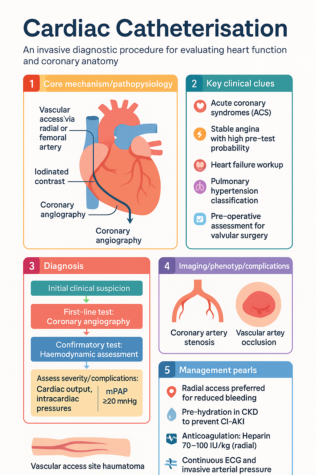
- Cardiac catheterisation is an invasive diagnostic procedure enabling direct measurement of intracardiac pressures, oxygen saturations, cardiac output, and coronary angiography.
- Left heart catheterisation (coronary angiography) is the gold standard for defining coronary anatomy and is indicated for acute coronary syndromes (ACS), stable angina with high pre-test probability, and pre-operative assessment prior to valvular surgery.
- Right heart catheterisation (haemodynamic assessment) measures right-sided pressures, pulmonary artery wedge pressure, and cardiac output via thermodilution or Fick method — essential in heart failure workup, pulmonary hypertension classification, and pre-transplant evaluation.
- Radial artery access is the preferred first-line approach for coronary angiography (reduced bleeding, shorter stay) unless specific contraindications exist; femoral access is standard for right heart catheterisation.
- Major vascular complications occur in <1% of cases with radial access and 2–3% with femoral access; risk is minimised by ultrasound-guided puncture, ACT-guided anticoagulation, and vascular closure devices.
- All patients require pre-procedure assessment including renal function (eGFR), coagulation screen, FBC, and consideration of contrast allergy prophylaxis.
- Chronic kidney disease patients require pre-hydration with isotonic sodium chloride 1 mL/kg/h for ≥6 hours pre- and post-procedure to prevent contrast-induced acute kidney injury (CI-AKI).
- Antiplatelet and anticoagulation management must be planned: heparin 70–100 IU/kg (radial) or 50–70 IU/kg (femoral) for ACT 250–350 seconds; dual antiplatelet therapy (DAPT) is continued peri-procedure for stable patients.
- Standard monitoring includes continuous ECG, pulse oximetry, and invasive arterial pressure during the procedure.
- Haemodynamic data — including mean arterial pressure, mean pulmonary artery pressure, pulmonary capillary wedge pressure, and calculated cardiac index — guide diagnosis and management in heart failure and pulmonary hypertension.
- In Australia, cardiac catheterisation is performed in public and private catheterisation laboratories, typically managed by interventional cardiologists with procedural volumes ≥200 cases/year as per CPCA standards.
- Aboriginal and Torres Strait Islander patients experience higher rates of ischaemic heart disease, present later, and have reduced access to catheterisation services in remote regions — early referral is critical.
Introduction & Australian Epidemiology
Cardiac catheterisation is an invasive diagnostic and potentially therapeutic procedure that allows direct measurement of intracardiac pressures, oxygen saturations, cardiac output, and detailed visualisation of coronary anatomy. It encompasses both left heart catheterisation (coronary angiography ± left ventriculography) and right heart catheterisation (haemodynamic assessment), and remains the reference standard for the definitive evaluation of structural, valvular, and coronary artery disease.
In Australia, over 95,000 coronary angiography procedures are performed annually across public and private catheterisation laboratories, with approximately 40–45% of these proceeding to percutaneous coronary intervention (PCI) in the same sitting. The Cardiac Society of Australia and New Zealand (CSANZ) and the Conjoint Committee for the Recognition of Training in Cardiac Interventional Procedures (CPCA) oversee quality standards and procedural training requirements.
The shift toward radial artery access has been transformative: Australian data from the RIVAL and MATRIX trials have confirmed a significant reduction in major bleeding, vascular complications, and mortality with radial-first approaches. Current Australian practice sees >80% of elective coronary angiography performed via radial access, with femoral access reserved for specific anatomical or procedural indications.
Indications for cardiac catheterisation span the full spectrum of cardiovascular disease: acute coronary syndromes (ACS), stable ischaemic heart disease with high pre-test probability, heart failure with suspected coronary aetiology, valvular heart disease requiring haemodynamic assessment, pulmonary hypertension workup, and pre-operative evaluation prior to cardiac surgery or transcatheter interventions such as TAVI or MitraClip.
This guideline provides a comprehensive overview of left and right heart catheterisation techniques, indications, access strategies, haemodynamic interpretation, and complication management within the Australian clinical context, including consideration of access disparities affecting Aboriginal and Torres Strait Islander communities.
Left Heart Catheterisation (Coronary Angiography)
Left heart catheterisation involves cannulation of the left ventricle via retrograde arterial access, most commonly to perform coronary angiography (visualisation of the coronary arteries with iodinated contrast) and/or left ventriculography (assessment of LV systolic function and mitral regurgitation).
Technique
- Arterial access is obtained percutaneously using the Seldinger technique — radial (right preferred, left if needed) or femoral (common femoral artery, ideally at the femoral head under fluoroscopy or ultrasound guidance).
- A diagnostic catheter (e.g., Judkins Left, Judkins Right, or Amplatz) is advanced over a guidewire under fluoroscopic guidance into the aortic root.
- Selective cannulation of the left and right coronary ostia is performed with hand injection of non-ionic iodinated contrast (iopromide or iohexol), with cineangiographic recording at 15–25 frames/second.
- Left ventriculography (if indicated) uses a pigtail catheter advanced retrogradely across the aortic valve into the LV; contrast is injected at 12–15 mL/s to assess wall motion and ejection fraction.
- Haemodynamic pullback across the aortic valve can identify transvalvular gradients (aortic stenosis).
Views and Biplane Angiography
| Coronary Segment | Essential Views | Purpose |
|---|---|---|
| Left main (LM) | AP cranial, LAO cranial | Bifurcation assessment, ostial disease |
| LAD (proximal) | RAO caudal, LAO cranial | Diagonal/mid-LAD lesions |
| LAD (distal) | LAO caudal (spider view) | Distal LAD and D1/D2 bifurcation |
| Circumflex / OM | LAO caudal (cranberry view) | OM origin, proximal Cx |
| RCA (proximal–mid) | LAO, AP | RCA body and PDA |
| RCA (distal) | RAO | Crux, PDA, PLV |
Contrast Agents Used in Australia
Anticoagulation During Left Heart Catheterisation
Spasm Prophylaxis (Radial Access)
Right Heart Catheterisation (Haemodynamic Assessment)
Right heart catheterisation (RHC) involves placement of a balloon-tipped, flow-directed catheter (typically a Swan–Ganz catheter) via central venous access into the pulmonary artery to obtain direct intracardiac pressure measurements, mixed venous oxygen saturation, and thermodilution or Fick cardiac output.
Indications
- Heart failure: Differentiation of cardiac vs. non-cardiac causes of dyspnoea; assessment of filling pressures to guide therapy; evaluation for advanced therapies (LVAD, transplantation).
- Pulmonary hypertension: Confirm diagnosis (mPAP ≥20 mmHg at rest), classify aetiology (pre-capillary vs. post-capillary), and assess vasoreactivity (acute vasodilator challenge with inhaled nitric oxide or IV epoprostenol).
- Valvular heart disease: Severity grading of aortic stenosis (Gorlin formula) and mitral stenosis (pressure half-time, Gorlin).
- Cardiac tamponade: Equalisation of diastolic pressures across chambers.
- Constrictive pericarditis: Discordant ventricular filling with respiration, enhanced ventricular interdependence (Kussmaul sign).
- Shock: Differentiation of cardiogenic, distributive, and obstructive shock.
- Pre-operative: Assessment for cardiac surgery or transcatheter interventions.
Access Route
RHC is typically performed via right internal jugular vein (IJV) under ultrasound guidance. Alternative sites include femoral vein and left IJV. The right IJV is preferred because it provides a direct path to the right atrium and is associated with fewer complications.
Normal Haemodynamic Values
| Parameter | Normal Range | Unit |
|---|---|---|
| Right atrial pressure (RAP) | 2–8 | mmHg |
| Right ventricular systolic pressure | 15–30 | mmHg |
| Pulmonary artery systolic (PASP) | 15–30 | mmHg |
| Pulmonary artery diastolic (PADP) | 4–12 | mmHg |
| Mean pulmonary artery pressure (mPAP) | 9–18 | mmHg |
| Pulmonary capillary wedge pressure (PCWP) | 6–12 | mmHg |
| Left ventricular end-diastolic pressure (LVEDP) | 5–12 | mmHg |
| Cardiac index (CI) | 2.5–4.0 | L/min/m² |
| Stroke volume index (SVI) | 33–47 | mL/m² |
| Systemic vascular resistance (SVR) | 800–1200 | dyn·s·cm⁻⁵ |
| Pulmonary vascular resistance (PVR) | 20–130 | dyn·s·cm⁻⁵ |
Derived Haemodynamic Calculations
| Derived Parameter | Formula |
|---|---|
| Cardiac Output (CO) | Thermodilution: CO = (V × (Tb − Ti) × K) / ∫ΔTb dt |
| Fick Cardiac Output | CO = VO₂ / (CaO₂ − CvO₂) × 10 |
| Pulmonary vascular resistance (PVR) | PVR = 80 × (mPAP − PCWP) / CO |
| Systemic vascular resistance (SVR) | SVR = 80 × (MAP − RAP) / CO |
| Transpulmonary gradient (TPG) | TPG = mPAP − PCWP |
| Diastolic pressure gradient (DPG) | DPG = PADP − PCWP |
Indications, Access & Complications
Indications for Cardiac Catheterisation
Access Site Comparison
| Feature | Radial Access | Femoral Access |
|---|---|---|
| Preferred for | Coronary angiography, diagnostic LHC | RHC, complex PCI, haemodynamic support |
| Bleeding risk | Low (~0.5%) | Higher (~2–3%) |
| Access site complication | Radial artery occlusion (1–5%, usually clinically silent) | Haematoma, pseudoaneurysm, AV fistula |
| Patient mobility | Immediate ambulation | 4–6 hours bed rest (closure device) / 6–8 hours (manual) |
| Contraindications | Negative Allen test, Raynaud's, prior radial harvest (CABG) | Severe PAD, aortic aneurysm, recent groin surgery |
| Closure | TR Band® or radial compression device | Manual pressure, Angioseal™, Perclose ProGlide™ |
Complications
| Complication | Incidence | Management |
|---|---|---|
| Access site haematoma | 2–6% | Compression, observation. Surgical evacuation if expanding or haemodynamically significant. |
| Pseudoaneurysm | 0.5–2% | Ultrasound-guided thrombin injection (first-line). Surgical repair if failed. |
| Radial artery occlusion | 1–5% | Usually clinically silent. Patency haemostasis technique reduces risk. Anticoagulation if symptomatic. |
| Contrast-induced AKI | 2–5% (higher if CKD, DM) | Pre-hydration with 0.9% NaCl, minimise contrast volume (<3× eGFR mL). Avoid nephrotoxins 48 h peri-procedure. |
| Allergic reaction | 0.2–0.7% | Mild: antihistamine ± corticosteroid. Anaphylaxis: adrenaline IM, oxygen, IV fluids. |
| Stroke | 0.05–0.1% | Immediate neurological assessment. Stroke team activation. CT brain ± thrombolysis. |
| Coronary dissection / perforation | 0.1–0.3% | Prolonged balloon inflation, covered stent, pericardiocentesis, emergency cardiac surgery. |
| Ventricular fibrillation | 0.5–1% | Immediate defibrillation. Often transient with catheter manipulation. |
| Vasovagal reaction | 3–5% | Atropine 600 µg IV, IV fluid bolus, leg elevation. |
Pre-procedure Checklist
Interpretation of Results
Coronary Angiography Interpretation
Coronary lesions are assessed by visual estimation of diameter stenosis (%DS), location (segment numbering per CAD-RADS/SYNTAX), morphology (eccentric vs concentric, calcified, thrombotic, bifurcation), and flow characteristics.
| Stenosis Severity | % Diameter Stenosis | Clinical Significance |
|---|---|---|
| Normal | 0% | No atherosclerotic disease |
| Mild | 1–49% | Atherosclerosis present; unlikely flow-limiting; medical therapy |
| Moderate | 50–69% | Indeterminate; consider FFR/iFR to assess physiological significance |
| Severe | 70–89% | Likely flow-limiting; revascularisation usually indicated if symptomatic |
| Critical | 90–99% | Haemodynamically significant; high-grade obstruction |
| Occlusion (CTO) | 100% | Chronic total occlusion; antegrade or retrograde CTO-PCI techniques |
Fractional Flow Reserve (FFR) & Instantaneous Wave-Free Ratio (iFR)
Physiological lesion assessment is indicated for angiographically moderate (50–70%) stenoses to guide revascularisation decisions. FFR and iFR are available in Australian tertiary centres.
| Parameter | Normal | Significant | Grey Zone |
|---|---|---|---|
| FFR (hyperaemic) | ≥0.80 | <0.75 (definite ischaemia) | 0.75–0.80 |
| iFR (resting) | ≥0.89 | <0.85 (definite ischaemia) | 0.85–0.89 |
Thrombolysis in Myocardial Infarction (TIMI) Flow Grading
| TIMI Grade | Description |
|---|---|
| TIMI 0 | No perfusion — no antegrade flow beyond the occlusion |
| TIMI 1 | Penetration without perfusion — contrast passes but does not opacify distal bed |
| TIMI 2 | Partial perfusion — contrast opacifies distal bed but fills more slowly than normal |
| TIMI 3 | Complete perfusion — normal filling and washout of contrast |
SYNTAX Score
The SYNTAX score quantifies coronary artery disease complexity based on lesion location, dominance, number of lesions, and morphological characteristics (total occlusion, bifurcation, calcification, thrombus, diffuse disease).
Interpretation of Right Heart Haemodynamic Data
| Haemodynamic Pattern | Key Findings | Diagnosis |
|---|---|---|
| Post-capillary PH | mPAP ≥20, PCWP >15, PVR <3 WU | Left heart disease (Group 2 PH) |
| Pre-capillary PH | mPAP ≥20, PCWP ≤15, PVR ≥3 WU | PAH, CTEPH, lung disease (Groups 1, 4, 5) |
| Combined pre- and post-capillary | mPAP ≥20, PCWP >15, PVR ≥3 WU | Combined PH (Group 2 with reactive component) |
| Constrictive pericarditis | Dip-and-plateau, LVEDP–RVEDP equalisation, discordant ventricular filling | Constrictive pericarditis (differentiate from restrictive cardiomyopathy) |
| Cardiac tamponade | Equal diastolic pressures (RA ≈ RV ≈ PA ≈ PCWP), pulsus paradoxus, RA waveform loss of Y descent | Pericardial effusion with tamponade physiology |
| Cardiogenic shock | CI <2.2, PCWP >18, SVR >1500 | Pump failure — consider MCS (IABP, Impella, ECMO) |
Shunt Assessment
Oxygen saturation step-up across cardiac chambers detects intracardiac shunts. A step-up of ≥7% at the atrial level suggests an atrial septal defect (ASD); ≥5% at the ventricular level suggests a ventricular septal defect (VSD). The Qp:Qs ratio (pulmonary-to-systemic flow ratio) quantifies shunt magnitude:
- Qp:Qs <1.5: haemodynamically insignificant
- Qp:Qs 1.5–2.0: moderate shunt — consider intervention if symptomatic
- Qp:Qs >2.0: large shunt — intervention usually indicated
Special Populations
Aboriginal and Torres Strait Islander Health Considerations
📚 References
- 1. Chew DP, Scott IA, Cullen L, et al. National Heart Foundation of Australia & Cardiac Society of Australia and New Zealand: Australian Clinical Guidelines for the Management of Acute Coronary Syndromes 2016. Heart Lung Circ. 2016;25(9):895–951.
- 2. Jolly SS, Yusuf S, Cairns J, et al. Radial versus femoral access for coronary angiography and intervention in patients with acute coronary syndromes (RIVAL): a randomised, parallel group, multicentre trial. Lancet. 2011;377(9775):1409–1420.
- 3. Valgimigli M, Gagnor A, Calabró P, et al. Radial versus femoral access in patients with acute coronary syndromes undergoing invasive management: a randomised multicentre trial (MATRIX). Lancet. 2015;385(9986):2465–2476.
- 4. Tonino PAL, De Bruyne B, Pijls NHJ, et al. Fractional flow reserve versus angiography for guiding percutaneous coronary intervention (FAME). N Engl J Med. 2009;360(3):213–224.
- 5. Davies JE, Sen S, Dehbi H-M, et al. Use of the instantaneous wave-free ratio or fractional flow reserve in PCI (DEFINE-FLAIR). N Engl J Med. 2017;376(19):1824–1834.
- 6. Serruys PW, Morice M-C, Kappetein AP, et al. Percutaneous coronary intervention versus coronary-artery bypass grafting for severe coronary artery disease (SYNTAX). N Engl J Med. 2009;360(10):961–972.
- 7. Task Force for the Diagnosis and Treatment of Pulmonary Hypertension of the ESC. 2022 ESC/ERS Guidelines for the diagnosis and treatment of pulmonary hypertension. Eur Heart J. 2022;43(38):3618–3731.
- 8. Nijjer SS, Sen S, Petraco R, Davies JE. Advances in coronary physiology. Circ J. 2015;79(6):1172–1184.
- 9. Australian Institute of Health and Welfare (AIHW). Aboriginal and Torres Strait Islander Health Performance Framework 2023 — summary report. Canberra: AIHW; 2023.
- 10. Brown A, O'Shea RL, Mott K, et al. Essential service standards for equitable national cardiovascular care for Aboriginal and Torres Strait Islander people. Heart Lung Circ. 2015;24(2):126–141.
- 11. Hallan SI, Orth SR. The concept of contrast-induced nephropathy — moving forward. Nephrol Dial Transplant. 2020;35(4):571–574.
- 12. Runciman WB, Coiera EW, Day RO, et al. Towards the safe and efficient use of medicines in Australia — the Australian Commission on Safety and Quality in Health Care. Med J Aust. 2013;198(10):511–513.
- 13. Tonelli M, Wiebe N, Culleton B, et al. Chronic kidney disease and mortality risk: a systematic review. J Am Soc Nephrol. 2006;17(7):2034–2047.
- 14. O'Gara PT, Kushner FG, Ascheim DD, et al. 2013 ACCF/AHA guideline for the management of ST-elevation myocardial infarction. Circulation. 2013;127(4):e362–e425.
- 15. Collet J-P, Thiele H, Barbato E, et al. 2020 ESC Guidelines for the management of acute coronary syndromes in patients presenting without persistent ST-segment elevation. Eur Heart J. 2021;42(14):1289–1367.