📋 Key Information Summary
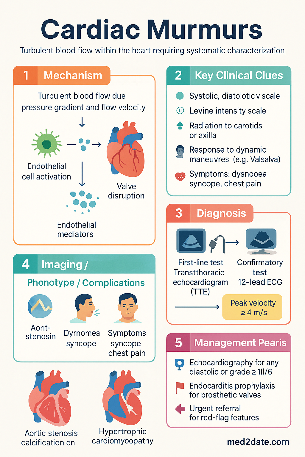
- Cardiac murmurs arise from turbulent blood flow and require systematic characterisation by timing (systolic vs diastolic vs continuous), intensity (Levine I–VI/6), quality, radiation, and response to dynamic manoeuvres.
- Innocent (functional) murmurs are common in children (up to 80%) and young adults; they are always systolic, grade ≤ III/6, non-radiating, and disappear with positional change or Valsalva.
- Any diastolic murmur is pathological until proven otherwise and mandates echocardiography.
- Systolic murmurs are classified as ejection (crescendo–decrescendo) or pansystolic (holosystolic); ejection murmurs may be innocent or pathological, while pansystolic murmurs are always pathological.
- The most common pathological murmurs in Australia are aortic stenosis in the elderly, mitral regurgitation, and hypertrophic obstructive cardiomyopathy in young athletes.
- Bedside dynamic manoeuvres — Valsalva, squat-to-stand, passive leg elevation, and amyl nitrite inhalation — can differentiate murmurs when echocardiography is not immediately available.
- Transthoracic echocardiography (TTE) is the gold-standard investigation (MBS item 55124) and should be performed for any murmur graded ≥ III/6, any diastolic murmur, or any systolic murmur with symptoms.
- A 12-lead ECG (MBS item 11700) is essential for assessing chamber enlargement, conduction abnormalities, and arrhythmias associated with valvular disease.
- Continuous murmurs (e.g., patent ductus arteriosus, arteriovenous fistula) are always pathological and require urgent investigation.
- Rheumatic heart disease remains disproportionately prevalent in Aboriginal and Torres Strait Islander communities — echocardiographic screening programmes are in place across Northern Territory, Queensland, and Western Australia.
- Endocarditis prophylaxis (Australian Dental Association / Cardiac Society consensus) is indicated for prosthetic valves, previous infective endocarditis, and certain congenital heart diseases — NOT for native valvular disease alone.
- Red-flag features warranting urgent referral: new murmur with haemodynamic instability, syncope, chest pain, signs of heart failure, or sepsis.
Introduction & Australian Epidemiology
Cardiac murmurs are audible vibrations produced by turbulent blood flow within the heart or great vessels. While many murmurs are benign, others signify significant structural heart disease that may require medical or surgical intervention. A systematic approach to murmur characterisation — incorporating timing, character, radiation, dynamic manoeuvres, and associated clinical findings — is essential for accurate bedside diagnosis.
Australian epidemiology: Innocent murmurs are detected in up to 80% of healthy children and 10–15% of adults during routine examination. Degenerative calcific aortic stenosis affects approximately 3% of Australians aged >75 years, making it the most common valvular lesion requiring intervention in the elderly. Rheumatic heart disease (RHD) persists as a major health burden in Aboriginal and Torres Strait Islander communities, with notification rates exceeding 100 per 100,000 in some Northern Territory communities compared with <1 per 100,000 in the non-Indigenous population. Mitral valve prolapse affects 2–3% of the general population, while bicuspid aortic valve (present in 1–2% of Australians) is the most common congenital cardiac abnormality.
This guideline provides a structured approach to murmur classification, bedside differentiation of innocent from pathological murmurs, key murmur features, and appropriate investigation pathways within the Australian healthcare context.
Pathophysiology of Cardiac Murmurs
Murmur generation requires two conditions: (1) a pressure gradient across an orifice and (2) sufficient blood flow velocity to produce laminar flow disruption. The physical determinants are described by the simplified Bernoulli equation (ΔP = 4V²), where the pressure gradient is proportional to the square of flow velocity.
Mechanisms of Murmur Generation
- Valvular stenosis: Narrowing of a valve orifice accelerates flow, producing turbulence distal to the obstruction (e.g., aortic stenosis produces a systolic ejection murmur).
- Valvular regurgitation: Incomplete valve closure permits retrograde flow across a pressure gradient (e.g., mitral regurgitation produces a pansystolic murmur).
- Increased flow volume: Conditions such as anaemia, thyrotoxicosis, pregnancy, and arteriovenous fistulae increase cardiac output and flow velocity through normal valves.
- Structural abnormalities: Ventricular septal defects, patent ductus arteriosus, and hypertrophic cardiomyopathy create abnormal flow pathways.
- Tethering or prolapse: Elongated chordae tendineae or myxomatous degeneration alter valve geometry, causing regurgitation.
Classification (Systolic, Diastolic, Continuous)
The first step in murmur assessment is timing within the cardiac cycle. Palpation of the carotid upstroke simultaneously with auscultation helps identify the first heart sound (S1) and hence the onset of systole.
| Timing | Subtype | Character | Causes |
|---|---|---|---|
| Systolic | Ejection (crescendo–decrescendo) | Diamond-shaped; begins after S1, peaks mid-systole, ends before S2 | Aortic stenosis, pulmonary stenosis, bicuspid aortic valve, innocent flow murmur, HOCM, aortic sclerosis |
| Pansystolic (holosystolic) | Flat intensity from S1 to S2 (or beyond) | Mitral regurgitation, tricuspid regurgitation, ventricular septal defect | |
| Diastolic | Early diastolic (decrescendo) | High-pitched, blowing; begins with S2, diminishes | Aortic regurgitation, pulmonary regurgitation |
| Mid-to-late diastolic (low-pitched rumble) | Best heard with bell at apex in left lateral decubitus | Mitral stenosis, Austin Flint murmur, Carey–Coombs murmur (acute rheumatic fever) | |
| Continuous | Throughout systole and diastole | Machinery-like; present in both phases | Patent ductus arteriosus, ruptured sinus of Valsalva, arteriovenous fistula, mammary soufflé (pregnancy) |
Murphy Classification of Systolic Murmurs (Australian teaching standard)
- Group 1 — Early systolic: Begins with S1 and fades before S2. Causes: small muscular VSD, tricuspid regurgitation in the absence of pulmonary hypertension.
- Group 2 — Ejection systolic: Begins after S1, ends before S2. A gap exists at both ends. Causes: aortic stenosis, pulmonary stenosis, innocent murmur, HOCM, aortic sclerosis.
- Group 3 — Pansystolic: Begins with S1 and continues to or beyond S2. No gap. Causes: mitral regurgitation, tricuspid regurgitation, VSD.
- Group 4 — Late systolic: Begins mid-to-late systole, extends to S2. Causes: mitral valve prolapse, HOCM with late peaking.
Innocent vs Pathological Murmurs
Differentiating innocent (functional/benign) murmurs from pathological murmurs is one of the most important clinical skills in cardiology. Misclassification may lead to unnecessary anxiety and investigation, or conversely, missed serious pathology.
| Feature | Innocent Murmur | Pathological Murmur |
|---|---|---|
| Timing | Systolic only | Any timing (systolic, diastolic, continuous) |
| Grade | ≤ III/6 | Any grade; ≥ III/6 often pathological |
| Quality | Soft, musical, vibratory, blowing | Harsh, rough, rumbling |
| Radiation | Localised, no radiation to neck/axilla | Radiates to carotids (AS), axilla (MR), back (coarctation) |
| S1 / S2 | Normal S1 and S2; no added sounds | May have abnormal splitting, S3, S4, ejection click, opening snap |
| Positional change | Disappears or markedly changes with position | Persists across positions |
| Val manoeuvre | Diminishes | HOCM increases; AS and MR decrease |
| Symptoms | Asymptomatic; normal growth/development | Dyspnoea, syncope, chest pain, exercise intolerance, failure to thrive (paediatric) |
| Thrill | Absent (thrill = grade ≥ IV/6) | May be present |
Common Innocent Murmurs
| Murmur | Population | Features |
|---|---|---|
| Still's murmur | Children 3–8 years | Grade I–III/6, vibratory/groaning, lower left sternal border, supine → disappears sitting/standing |
| Pulmonary flow murmur | Children and young adults | Ejection systolic, left upper sternal border, accentuated by expiration |
| Venous hum | Children 3–10 years | Continuous, supraclavicular, abolished by turning head or compressing jugular vein |
| Mammary soufflé | Pregnant/postpartum women | Continuous, overbreast area, abolished by stethoscope pressure |
| Physiological flow murmur | All ages (fever, anaemia, pregnancy) | Soft ejection systolic, base, resolves when underlying condition treated |
Key Murmurs & Their Features
The following table summarises the hallmark auscultatory findings, dynamic manoeuvre responses, and associated signs for the most clinically important murmurs encountered in Australian practice.
| Murmur | Location & Radiation | Pitch / Quality | Dynamic Response | Associated Signs |
|---|---|---|---|---|
| Aortic stenosis | Right upper sternal border → carotids (both); apex (Gallavardin phenomenon) | Harsh, rough, crescendo–decrescendo | ↓ Valsalva, ↓ squatting; ↑ handgrip may paradoxically soften | Slow-rising carotid upstroke (pulsus parvus et tardus), S4, narrow S2 splitting, ejection click if bicuspid valve |
| Mitral regurgitation | Apex → axilla; may radiate to left sternal border if posterior leaflet | Blowing, high-pitched, pansystolic | ↑ Handgrip; ↓ Valsalva; ↑ squatting; ↑ leg elevation | Displaced apex beat, S3, wide-apart S2 (early P2), AF common in chronic severe MR |
| Aortic regurgitation | Left lower sternal border (Erb's point); best heard sitting forward, end-expiration | High-pitched, blowing, decrescendo early diastolic | ↑ Sitting/leaning forward, end-expiration; ↑ handgrip increases murmur | Wide pulse pressure, collapsing (water-hammer) pulse, de Musset sign, Quincke sign, Duroziez sign, Austin Flint murmur at apex |
| Mitral stenosis | Apex (bell), left lateral decubitus; no radiation | Low-pitched rumbling, mid-diastolic ± presystolic crescendo | ↑ Exercise; ↑ left lateral decubitus; best heard with bell | Loud S1, opening snap (shorter OS–S2 interval = more severe stenosis), tapping apex beat, AF, signs of pulmonary hypertension |
| Hypertrophic obstructive cardiomyopathy (HOCM) | Left lower sternal border; no radiation to carotids | Harsh, crescendo–decrescendo, ejection systolic | ↑ Valsalva (classical distinguishing feature); ↓ squatting; ↑ standing from squat | Bifid pulse, double-apex beat, S4, associated with systolic anterior motion (SAM) of mitral valve on echo |
| Ventricular septal defect (VSD) | Left lower sternal border; may be localised or diffuse | Harsh, loud, pansystolic (small defects = louder) | Minimal dynamic change | Palpable thrill at left sternal border; large VSD → S3, displaced apex, signs of heart failure |
| Patent ductus arteriosus (PDA) | Left infraclavicular / upper left sternal border | Machinery-like continuous murmur | Minimal; may accentuate with inspiration | Bounding pulse, widened pulse pressure, displaced apex, S3 if large |
| Mitral valve prolapse (MVP) | Apex; may radiate to axilla if significant MR | Mid-to-late systolic click ± late systolic crescendo murmur | ↑ Valsalva/standing (click and murmur move earlier); ↓ squatting (click and murmur move later) | Click timing varies with preload; associated with Marfan syndrome, Ehlers–Danlos |
Grading Murmur Intensity (Levine Scale)
Clinical Presentation & Diagnostic Approach
Bedside Assessment Protocol
- Confirm presence and timing: Palpate carotid pulse while auscultating; synchronise S1 with pulse upstroke. Determine if murmur is systolic, diastolic, or continuous.
- Characterise intensity: Grade I–VI using the Levine scale. Note presence of a palpable thrill (≥ grade IV).
- Describe quality: Harsh, blowing, musical, rumbling, rough, high-pitched vs low-pitched.
- Map radiation: Carotids (aortic stenosis), axilla (mitral regurgitation), back (coarctation), neck (pulmonary stenosis).
- Listen for associated sounds: S3, S4, ejection click, opening snap, fixed splitting of S2 (ASD), wide paradoxical splitting (aortic stenosis).
- Perform dynamic manoeuvres: Valsalva, squat-to-stand, passive leg elevation, handgrip, amyl nitrite inhalation (where available).
- Assess for signs of haemodynamic consequence: Pulse character and volume, blood pressure (both arms if coarctation suspected), JVP, apex beat location and character, peripheral oedema, hepatomegaly.
- Evaluate symptoms: Exertional dyspnoea, syncope or presyncope, anginal chest pain, palpitations, exercise intolerance, orthopnoea, paroxysmal nocturnal dyspnoea.
Red Flags Warranting Urgent Investigation
- New murmur with haemodynamic instability (hypotension, tachycardia, poor perfusion)
- New murmur in the setting of fever, rigors, or positive blood cultures — suspect infective endocarditis
- Acute severe mitral regurgitation (papillary muscle rupture post-MI, ruptured chordae tendineae) — loud, often short pansystolic murmur with pulmonary oedema
- Aortic dissection with new aortic regurgitation — early diastolic murmur, pulse deficits, unequal arm pressures
- Any diastolic murmur — always pathological
- Murmur with syncope — may indicate critical aortic stenosis or HOCM
Investigations
First-Line Investigations
Second-Line / Specialist Investigations
Laboratory Investigations (Supportive)
| Test | Indication |
|---|---|
| Full blood count (FBC) | Anaemia (flow murmur), infective endocarditis (↑ WCC, ↓ Hb) |
| Inflammatory markers (CRP, ESR) | Infective endocarditis, acute rheumatic fever |
| Blood cultures (3 sets) | Suspected infective endocarditis — before antibiotics |
| BNP / NT-proBNP | Assess heart failure severity; serial monitoring in valvular disease |
| Iron studies, TFT | Exclude high-output causes (iron deficiency anaemia, thyrotoxicosis) |
| ASO titre, anti-DNase B | Confirm acute rheumatic fever (revised Jones criteria) |
Special Populations
Aboriginal and Torres Strait Islander Health Considerations
Management Principles
Management of cardiac murmurs depends entirely on the underlying aetiology. The following provides a framework for initial management and referral.
Innocent Murmurs
- Reassurance — explain the benign nature to the patient/parents.
- Document findings clearly in the medical record (grade, location, character, absence of associated abnormalities).
- Echocardiography is NOT required unless there is clinical doubt or parental/patient anxiety.
- Repeat auscultation at subsequent visits — innocent murmurs may vary with physiological state.
Pathological Murmurs — Referral Criteria
Endocarditis Prophylaxis
- Prosthetic cardiac valves (including transcatheter)
- Previous infective endocarditis
- Certain congenital heart diseases (unrepaired cyanotic CHD, completely repaired CHD with prosthetic material within 6 months, repaired CHD with residual defects at or adjacent to prosthetic material)
- Cardiac transplant recipients with valvulopathy
Monitoring & Follow-Up
The frequency of clinical and echocardiographic follow-up depends on valve lesion severity and symptoms. The following intervals align with Australian Cardiac Society and ESC/AHA recommendations:
| Valve Lesion | Severity | Clinical Review | Echo Interval |
|---|---|---|---|
| Aortic stenosis | Mild (Vmax 2.0–2.9 m/s) | Every 12 months | Every 3–5 years |
| Aortic stenosis | Moderate (Vmax 3.0–3.9 m/s) | Every 6–12 months | Every 1–2 years |
| Aortic stenosis | Severe (Vmax ≥4.0 m/s) | Every 6 months | Every 6–12 months (or sooner if symptomatic) |
| Mitral regurgitation | Severe (primary) | Every 6 months | Every 6–12 months; annual if asymptomatic with preserved LV |
| Mitral stenosis | Moderate–severe | Every 6–12 months | Every 1–2 years |
| Aortic regurgitation | Severe | Every 6 months | Every 6–12 months; annual if asymptomatic with preserved LV |
📚 References
- 1. Nishimura RA, Otto CM, Bonow RO, et al. 2014 AHA/ACC guideline for the management of patients with valvular heart disease. J Am Coll Cardiol. 2014;63(22):e57–185.
- 2. Vahanian A, Beyersdorf F, Praz F, et al. 2021 ESC/EACTS guidelines for the management of valvular heart disease. Eur Heart J. 2022;43(7):561–632.
- 3. Australian Institute of Health and Welfare (AIHW). Rheumatic heart disease in Australia, 2023. AIHW, Canberra. Available at: https://www.aihw.gov.au/reports/heart-stroke-vascular-diseases/rheumatic-heart-disease-in-australia
- 4. RHDAustralia (ARF/RHD writing group). The 2020 Australian guideline for prevention, diagnosis and management of acute rheumatic fever and rheumatic heart disease (3rd edition). Menzies School of Health Research, Darwin.
- 5. Heart Foundation of Australia. Position statement: diagnosis and management of aortic stenosis in adults. Heart Foundation, 2021.
- 6. Royal Australian College of General Practitioners (RACGP). Red Book: cardiovascular risk assessment. In: Guidelines for preventive activities in general practice (10th edition). RACGP, Melbourne, 2024.
- 7. National Heart Foundation of Australia and Cardiac Society of Australia and New Zealand. Australian clinical guidelines for the diagnosis and management of heart failure. Med J Aust. 2018;209(8):363–369.
- 8. Chambers JB, Garbi M, Nieman K, et al. Appropriateness criteria for the use of cardiovascular imaging in heart valve disease in adults: a European Association of Cardiovascular Imaging report. Eur Heart J Cardiovasc Imaging. 2017;18(3):241–254.
- 9. Roberts-Thomson KC, Lau DH, Sanders P. The diagnosis and management of cardiac murmurs in Australian general practice. Aust Fam Physician. 2010;39(9):647–652.
- 10. Bowyer JJ, Butto E, Sholler GF. Echocardiographic screening for rheumatic heart disease in high-risk Australian Aboriginal children. J Paediatr Child Health. 2019;55(3):278–283.
- 11. Medicines Australia / PBAC. Pharmaceutical Benefits Schedule — search. Australian Government Department of Health. Available at: https://www.pbs.gov.au
- 12. MBS Online. Medicare Benefits Schedule — Australian Government Department of Health. Available at: http://www.mbsonline.gov.au
- 13. Maganti K, Rigolin VH, Sarano ME, Bonow RO. Valvular heart disease: diagnosis and management. Mayo Clin Proc. 2010;85(5):483–500.