Home Cardiology Restrictive and Infiltrative Cardiomyopathies

Restrictive and Infiltrative Cardiomyopathies

🎧 Restrictive and Infiltrative Cardiomyopathies — deep-dive podcast

📋 Key Information Summary

📋
  • Restrictive cardiomyopathies (RCM) are characterised by abnormal diastolic function with normal or reduced ventricular volumes; infiltrative cardiomyopathies involve extracellular or intracellular substance deposition within the myocardium.
  • Cardiac amyloidosis — AL (light-chain) and ATTR (transthyretin) — is the most common infiltrative cause; Tc-99m pyrophosphate scintigraphy distinguishes ATTR from AL when serum free light chains and urine immunofixation are negative.
  • ATTR cardiac amyloidosis (both wild-type and hereditary) is increasingly recognised in Australia, particularly in males >65 years with heart failure and prior carpal tunnel syndrome.
  • Tafamidis (Vyndaqel®) is PBS-listed (Authority Required) for ATTR cardiomyopathy and reduces all-cause mortality and cardiovascular hospitalisations.
  • AL amyloidosis requires urgent haematology referral; first-line treatment is bortezomib-based chemotherapy (CyBorD) with consideration of autologous stem cell transplant.
  • Cardiac sarcoidosis presents with heart failure, conduction disease, or ventricular arrhythmias; FDG-PET/CT is the key diagnostic and monitoring tool, and immunosuppression with corticosteroids is first-line.
  • Hereditary haemochromatosis (HFE-related) is common in Australians of Northern European descent; cardiac iron overload causes dilated or restrictive cardiomyopathy and is detected by cardiac MRI T2* mapping.
  • Iron chelation with deferasirox or deferoxamine, combined with serial venesection, reverses cardiac iron loading if initiated before irreversible fibrosis.
  • Fabry disease (α-galactosidase A deficiency) causes LVH mimicking HCM with restrictive physiology; enzyme replacement therapy (agalsidase beta) and chaperone therapy (migalastat) are available.
  • Endomyocardial fibrosis is rare in Australia but seen in some migrant communities from tropical regions; glycogen storage disorders (PRKAG2, Danon disease) cause severe paediatric cardiomyopathy.
  • All patients with infiltrative cardiomyopathy require genetic counselling and cascade screening where an hereditary aetiology is confirmed.
  • Aboriginal and Torres Strait Islander peoples have higher rates of iron overload syndromes and rheumatic heart disease, which may mimic or coexist with infiltrative cardiomyopathy; culturally safe cardiac screening is essential.
🎬 Restrictive and Infiltrative Cardiomyopathies — clinical explainer

Introduction & Australian Epidemiology

Restrictive cardiomyopathies (RCM) represent a heterogeneous group of myocardial diseases characterised by increased ventricular stiffness, impaired diastolic filling with preserved or reduced ventricular volumes, and ultimately elevated filling pressures. Infiltrative cardiomyopathies constitute a major subset in which specific substances — either extracellular (amyloid, fibrosis) or intracellular (iron, glycosphingolipids, glycogen) — accumulate within the myocardium, disrupting normal architecture and function.

In Australia, the epidemiology of infiltrative cardiomyopathies has shifted significantly over the past decade, driven by improved diagnostic imaging and heightened clinical awareness:

  • Cardiac amyloidosis: Estimated prevalence of wild-type ATTR (ATTRwt) is 16–25% in autopsy studies of elderly males with heart failure. The Australian Amyloidosis Network reports increasing referral rates since the availability of Tc-99m PYP imaging and disease-modifying therapies.
  • Hereditary ATTR: The Val122Ile variant is found in ~3–4% of African-descent populations; other TTR variants (Val30Met, Thr60Ala) occur in Australian families of Celtic and other European ancestry. Genetic testing through national services (e.g., Victorian Clinical Genetics Services) is available.
  • Cardiac sarcoidosis: Sarcoidosis prevalence in Australia is estimated at 15–20 per 100,000; cardiac involvement occurs in 5–10% of systemic sarcoidosis cases, though autopsy studies suggest subclinical cardiac involvement in up to 25%.
  • Hereditary haemochromatosis: The C282Y homozygous HFE mutation affects approximately 1 in 200 Australians of Northern European descent, making it one of the most common autosomal recessive conditions in Australia. Cardiac iron overload occurs in 15–20% of untreated homozygotes, typically presenting between ages 30–50.
  • Fabry disease: Estimated incidence 1 in 40,000–117,000 live births; cardiac involvement is the leading cause of death. Newborn screening pilot programmes have been conducted in some Australian states.
  • Endomyocardial fibrosis: Rare in the general Australian population but seen in migrant communities from sub-Saharan Africa, South Asia, and Latin America.

The Australian Institute of Health and Welfare (AIHW) data show that cardiomyopathies contribute to approximately 5,500 hospitalisations per year nationally. Early identification and accurate subtyping of infiltrative cardiomyopathies is critical because specific disease-modifying therapies now exist for many conditions, and treatment outcomes are markedly better when initiated before irreversible myocardial damage occurs.
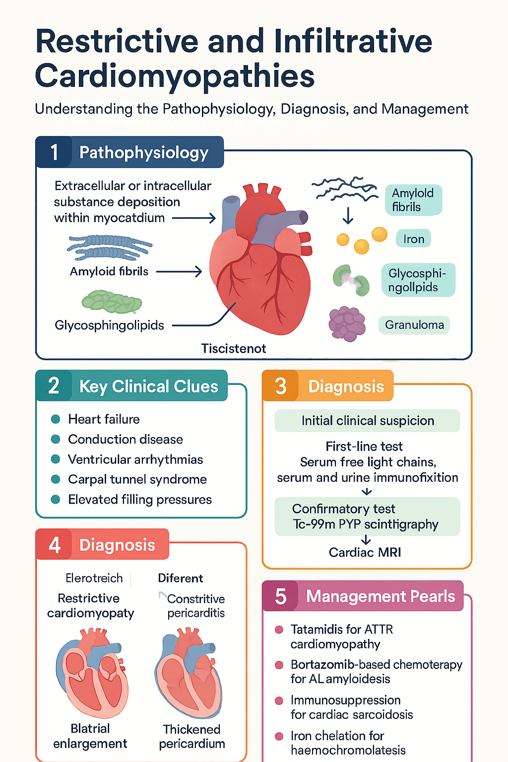
Restrictive and Infiltrative Cardiomyopathies clinical infographic — pathophysiology, clinical clues, diagnosis, imaging, and management
Tap or click image to enlarge — Restrictive and Infiltrative Cardiomyopathies: pathophysiology, clinical clues, diagnosis, imaging, and management.
Restrictive and Infiltrative Cardiomyopathies infographic, full size

Pathophysiology of Restrictive & Infiltrative Cardiomyopathies

The restrictive phenotype results from increased myocardial stiffness that impairs ventricular filling during diastole. This may be caused by:

  • Extracellular infiltration: Amyloid fibrils (AL or ATTR) deposit in the interstitium, displacing cardiomyocytes and causing wall thickening with a stiff, non-compliant ventricle. Endomyocardial fibrosis (EMF) involves progressive fibrotic obliteration of the ventricular apex.
  • Intracellular infiltration: Iron (haemochromatosis) deposits within cardiomyocyte lysosomes and mitochondria, generating reactive oxygen species and causing oxidative damage. Glycosphingolipids (Fabry disease) accumulate in lysosomes due to deficient α-galactosidase A activity. Glycogen (PRKAG2, Danon disease) accumulates within lysosomes and cytoplasm.
  • Granulomatous infiltration: Non-caseating granulomas in cardiac sarcoidosis replace normal myocardium and cause fibrosis, conduction system disruption, and ventricular arrhythmias.

Regardless of aetiology, the haemodynamic consequences converge: elevated ventricular end-diastolic pressures transmit retrogradely to the atria and venous circulation, causing pulmonary congestion and systemic venous congestion. Atrial dilatation predisposes to atrial fibrillation. The "dip-and-plateau" or "square root sign" on cardiac catheterisation is a hallmark of restrictive physiology.

⚠️
Key distinction: Restrictive cardiomyopathy must be differentiated from constrictive pericarditis, as the latter is surgically curable while the former generally is not. Differentiation requires multimodality imaging (echocardiography with tissue Doppler, CT/MRI, and sometimes cardiac catheterisation with simultaneous RV/LV pressure measurement).

Cardiac Amyloidosis

AL vs ATTR Differentiation

Cardiac amyloidosis is caused by the deposition of misfolded protein fibrils in the myocardium. Accurate typing is mandatory because treatments differ fundamentally between subtypes:

Feature AL Amyloidosis ATTR Amyloidosis (Wild-Type) ATTR Amyloidosis (Hereditary)
Precursor protein Immunoglobulin light chains (κ or λ) Transthyretin (normal sequence) Mutant transthyretin
Age at onset 50–70 years (median ~60) >65 years (predominantly male) 30–60 years (variant-dependent)
Sex predominance M:F ≈ 1:1 M:F ≈ 15–50:1 Variable
Associated features Nephrotic syndrome, macroglossia, periorbital purpura, peripheral neuropathy, hepatomegaly Bilateral carpal tunnel syndrome, spinal stenosis, HFpEF in elderly male Peripheral/autonomic neuropathy, cardiac involvement (variant-dependent)
Serum free light chains Abnormal κ/λ ratio Normal Normal
Serum/urine immunofixation Monoclonal protein detected Negative Negative
Tc-99m PYP scintigraphy Negative (or weakly positive with confirmed non-AL) Strongly positive (Perugini grade 2–3) Strongly positive
Tissue biopsy confirmation Positive Congo red + mass spectrometry typing Positive Congo red + TTR typing; genetic testing Positive Congo red + TTR gene sequencing
🚨
Critical diagnostic algorithm: All patients with suspected cardiac amyloidosis MUST have serum free light chains, serum and urine immunofixation electrophoresis performed FIRST. If monoclonal protein is detected → suspect AL amyloidosis → urgent bone marrow biopsy and fat aspirate for Congo red staining → refer to haematology for staging and treatment. Tc-99m PYP scintigraphy should only be interpreted as supporting ATTR diagnosis when AL has been excluded by negative free light chain assays and immunofixation.

Technetium-99m Pyrophosphate (Tc-99m PYP) Imaging

Tc-99m PYP (or DPD/HMDP) scintigraphy has transformed the non-invasive diagnosis of ATTR cardiac amyloidosis. The Perugini visual scoring system grades cardiac uptake:

Grade 0
No Cardiac Uptake
Normal cardiac uptake equal to surrounding bone/soft tissue.
Amyloidosis unlikely
Grade 1–2
Mild to Moderate Uptake
Cardiac uptake greater than bone but less than or equal to bone. Requires AL exclusion and confirmatory testing.
Needs further workup
Grade 2–3
Strong Uptake
Cardiac uptake greater than bone, with visible soft tissue uptake. Highly specific for ATTR cardiac amyloidosis when AL has been excluded.
ATTR cardiac amyloidosis (if AL excluded)

Quantitative analysis using the heart-to-contralateral-lung (H/CL) ratio at 1 hour (≥1.5 on planar imaging or ≥1.3 with SPECT/CT) further improves diagnostic accuracy. Tc-99m PYP imaging is available at most major nuclear medicine centres in Australia, including Royal Melbourne Hospital, Royal Prince Alfred Hospital, Flinders Medical Centre, and Royal Brisbane and Women's Hospital. MBS item 61318 applies for myocardial perfusion studies and related cardiac nuclear medicine.

Cardiac MRI in Amyloidosis

Cardiovascular MRI (CMR) demonstrates characteristic findings:

  • Global subendocardial or transmural late gadolinium enhancement (LGE) in a non-coronary distribution
  • Elevated native T1 mapping values (>1,050 ms at 1.5T) and extracellular volume (ECV >40%)
  • Difficulty nulling the myocardium on inversion-recovery sequences (a "red flag" sign)
  • Biventricular wall thickening with small chamber sizes, biatrial enlargement

Treatment: ATTR Cardiac Amyloidosis

💊
Tafamidis
Vyndaqel® · Pfizer · TTR stabiliser
Adult dose 61 mg PO once daily (tafamidis meglumine 80 mg equivalent to tafamidis free acid 61 mg)
Paediatric dose Not established; specialist use only
Renal adjustment None required
Hepatic adjustment No dose adjustment; use with caution in severe hepatic impairment (Child-Pugh C)
Key evidence ATTR-ACT trial: 30% reduction in all-cause mortality and 32% reduction in cardiovascular hospitalisations at 30 months
PBS status ⚑ Authority Required — For ATTR cardiac amyloidosis (wild-type or hereditary) with NYHA class I–III heart failure
💊
Patisiran
Onpattro® · Alnylam · TTR gene silencer (siRNA)
Adult dose 0.3 mg/kg IV infusion every 3 weeks
Renal adjustment None required
Key evidence APOLLO trial demonstrated improved neuropathy and cardiac outcomes in hATTR; cardiac subgroup data show LV wall thickness reduction and improved global longitudinal strain
PBS status ✘ Not PBS-listed — Available through Special Access Scheme or manufacturer compassionate programme in Australia
💊
Inotersen
Tegsedi® · Ionis · TTR gene silencer (ASO)
Adult dose 284 mg SC once weekly
Key evidence NEURO-TTR trial; effective in hATTR with neuropathy; risk of thrombocytopaenia and glomerulonephritis
PBS status ✘ Not PBS-listed — Limited availability in Australia
💊
Eplontersen
Wainua® · Ionis/AstraZeneca · GalNAc-conjugated ASO
Adult dose 45 mg SC once monthly
Key evidence NEURO-TTR 2 trial: TTR reduction >80%; non-inferior to patisiran with more convenient monthly dosing
PBS status ✘ Not PBS-listed — Anticipated TGA approval; available through clinical trials

Treatment: AL Cardiac Amyloidosis

AL amyloidosis is a haematological emergency with a median survival of <6 months when cardiac involvement is advanced (Mayo Stage IIIb). Treatment is directed at the underlying clonal plasma cell disorder.

🚨
Emergency referral: All patients with confirmed AL amyloidosis and cardiac involvement must be referred urgently to a specialist amyloidosis centre. In Australia, established centres include: Royal Melbourne Hospital (Victorian Amyloidosis Centre), Royal Prince Alfred Hospital (Sydney), Royal Adelaide Hospital, and Princess Alexandra Hospital (Brisbane).
💊
Bortezomib + Cyclophosphamide + Dexamethasone (CyBorD)
Velcade® + Endoxan® + dexamethasone · First-line regimen
Regimen Bortezomib 1.5 mg/m² SC D1, 8, 15, 22; Cyclophosphamide 300 mg/m² PO D1, 8, 15, 22; Dexamethasone 40 mg PO weekly; 28-day cycles × 6–8
Renal adjustment Cyclophosphamide requires dose reduction for eGFR <30 mL/min; bortezomib no adjustment
Key evidence Overall haematological response rate 65–75%; cardiac response in ~30–40% with organ improvement
PBS status ⚑ Authority Required — Bortezomib PBS-listed for multiple myeloma and AL amyloidosis
💊
Daratumumab + CyBorD (Dara-CyBorD)
Darzalex® · Janssen · Anti-CD38 monoclonal antibody
Regimen Daratumumab 180 mg SC weekly × 8, then fortnightly × 16, then monthly, in combination with CyBorD backbone
Key evidence ANDROMEDA trial: superior haematological CR rate (~53% vs ~18%) compared to CyBorD alone; now preferred first-line for Mayo Stage III AL amyloidosis
PBS status ⚑ Authority Required — Listed for AL amyloidosis in combination with bortezomib-based regimen

Heart Failure Management in Amyloidosis

⚠️
Caution with standard HF therapies: Beta-blockers, ACE inhibitors, and ARBs are poorly tolerated in cardiac amyloidosis due to reliance on preload to maintain cardiac output. Digoxin binds to amyloid fibrils and is contraindicated in AL amyloidosis (risk of toxicity even at therapeutic levels). Calcium channel blockers also bind to amyloid and should be avoided.
  • Diuretics: Loop diuretics (furosemide 20–80 mg PO daily, torsemide 10–20 mg PO daily) are the mainstay of volume management. Titrate to maintain euvolaemia.
  • Anticoagulation: Consider for atrial fibrillation (common) — CHA₂DS₂-VASc scoring applies; direct oral anticoagulants (DOACs) are generally preferred over warfarin.
  • Pacemaker/ICD: Conduction disease and ventricular arrhythmias are common in ATTR. Pacemaker implantation for advanced AV block. ICD consideration for secondary prevention of sudden cardiac death; primary prevention role is uncertain.
  • Advanced therapies: Heart transplantation may be considered in selected AL patients who achieve haematological complete response, or in hereditary ATTR with combined heart-liver transplant (liver being the source of TTR production).

Cardiac Sarcoidosis

Diagnosis of Cardiac Involvement

Cardiac sarcoidosis (CS) is notoriously difficult to diagnose because endomyocardial biopsy has low sensitivity (~20–30%) due to the patchy nature of granulomatous infiltration. The 2017 Japanese Circulation Society (JCS) criteria and the Heart Rhythm Society (HRS) expert consensus provide the most widely used diagnostic frameworks:

Diagnosis is established by either:

  1. Histological diagnosis: Non-caseating granulomas on endomyocardial biopsy with no alternative cause identified.
  2. Clinical diagnosis (JCS criteria): Two or more major criteria, OR one major plus two or more minor criteria from the following:
Category Criteria
Major Advanced AV block (including complete heart block) or sustained ventricular tachycardia; basal thinning of the interventricular septum or regional wall motion abnormality on imaging; LGE on CMR in a non-coronary distribution; ⁶⁷Ga or ¹⁸F-FDG uptake in the myocardium
Minor Non-sustained ventricular tachycardia; abnormal ECG (fragmented QRS, ST-T changes, pathological Q waves, or axis deviation); reduced LVEF (<50%); LV diastolic dysfunction; elevated cardiac biomarkers (troponin, BNP/NT-proBNP)

PET Imaging in Cardiac Sarcoidosis

¹⁸F-FDG PET/CT is the cornerstone of non-invasive diagnosis and disease activity monitoring in CS:

  • Preparation: Prolonged fasting (>12 hours) with a high-fat, low-carbohydrate diet for 24–48 hours prior to suppress physiological myocardial glucose uptake. Some centres use unfractionated heparin (50 IU/kg IV) 15 minutes before tracer injection.
  • Positive scan: Focal or focal-on-diffuse FDG uptake in the myocardium (SUVmax >2× blood pool) in a non-coronary pattern, typically with perfusion defects on ⁸²Rb or ¹³N-ammonia rest perfusion (indicating scar).
  • Sensitivity/Specificity: Sensitivity ~85–89% and specificity ~78–95% for CS when combined with perfusion imaging.
  • Serial monitoring: FDG-PET is used to assess treatment response during immunosuppression; reduction in FDG uptake indicates disease control.

Cardiac MRI complements PET: LGE in a mid-myocardial or epicardial distribution (particularly basal inferolateral and inferoseptal walls) is the most sensitive technique for detecting myocardial fibrosis. T2-weighted imaging and T2 mapping identify active oedema/inflammation. CMR is preferred when FDG-PET is unavailable or the scan is equivocal.

Immunosuppression

💊
Prednisolone
Panafcortelone® · Generic · Corticosteroid — First-line
Adult dose 0.5 mg/kg/day PO (typically 30–40 mg daily) for 4–8 weeks, then taper over 6–12 months to 5–10 mg daily maintenance
Duration Minimum 12–18 months; many patients require low-dose long-term maintenance
Monitoring Blood glucose (steroid-induced diabetes risk), bone density (bisphosphonate prophylaxis), repeat FDG-PET at 3–6 months
PBS status ✔ PBS General Benefit
💊
Methotrexate
Methoblastin® · Generic · Steroid-sparing immunosuppressant
Adult dose 7.5–15 mg PO once weekly with folic acid 5 mg weekly (on a non-methotrexate day)
Renal adjustment Avoid or reduce dose if eGFR <30 mL/min
Key use Steroid-sparing agent for CS; used in combination with corticosteroids for steroid-refractory or relapsing disease
PBS status ✔ PBS General Benefit
💊
Mycophenolate mofetil
CellCept® · Roche · Alternative steroid-sparing agent
Adult dose 500–1000 mg PO BD, titrated to target 1000–1500 mg BD
Renal adjustment Caution if eGFR <25 mL/min; no specific dose adjustment
PBS status ⚑ Authority Required — For transplant and specific autoimmune indications
ℹ️
Treatment algorithm for CS: Active inflammation on FDG-PET → start corticosteroids. If persistent FDG uptake at 3–6 months → add methotrexate or mycophenolate. Refractory disease → consider azathioprine, cyclophosphamide, or infliximab in consultation with a sarcoidosis specialist. TNF-α inhibitors (infliximab) are increasingly used for refractory CS but require TB screening and monitoring.

Arrhythmia Management

Arrhythmias are the leading cause of sudden cardiac death in CS and require aggressive management:

  • Conduction disease: High-degree AV block is the most common presentation. Permanent pacemaker implantation is indicated for persistent complete heart block. Conduction may recover with immunosuppression, but permanent pacing is generally recommended given the unpredictable course.
  • Ventricular tachycardia: Antiarrhythmic drugs — sotalol (80–160 mg PO BD) or amiodarone (loading 200 mg TDS × 10 days, then 200 mg daily) — are first-line. Catheter ablation is increasingly used for recurrent VT refractory to medical therapy; however, recurrence rates are higher than in ischaemic VT due to the diffuse and progressive nature of granulomatous infiltration.
  • ICD implantation: Recommended for patients with sustained VT, cardiac arrest, or LVEF <35%. Consider for LVEF 36–50% with positive electrophysiology study or extensive LGE on CMR (a major predictor of arrhythmic events).
  • Atrial fibrillation: Common; rate control with cautious beta-blockade (avoiding excessive negative inotropy), or rhythm control with amiodarone. Anticoagulation per CHA₂DS₂-VASc score.

Haemochromatosis — Cardiac Iron Overload

Diagnosis

Hereditary haemochromatosis (HH) is caused by mutations in the HFE gene (most commonly C282Y homozygosity) leading to inappropriately increased intestinal iron absorption. Cardiac involvement occurs when total body iron exceeds storage capacity:

1
Screening Tests
Transferrin saturation (TSAT) >45% and serum ferritin >300 μg/L (men) or >200 μg/L (women). Elevated ferritin alone is non-specific (inflammation, metabolic syndrome).
2
Genetic Testing
HFE gene analysis: C282Y/C282Y homozygosity confirms HH. C282Y/H63D compound heterozygosity has lower penetrance. Referral to clinical genetics for family cascade screening.
3
Liver Assessment
Hepatic iron concentration by MRI (R2* or proton density fat fraction) or liver biopsy if MRI unavailable. Liver elastography for fibrosis staging. FibroScan® widely available in Australia.
4
Cardiac Assessment
Cardiac MRI with T2* mapping is the gold standard for myocardial iron quantification. T2* <20 ms indicates iron overload; <10 ms indicates severe iron loading with high risk of heart failure and arrhythmia. ECG, echocardiography, and 24-hour Holter monitoring for baseline arrhythmia assessment.
⚠️
Cardiac T2* interpretation: T2* >20 ms = normal myocardial iron; 10–20 ms = mild-to-moderate iron overload requiring intensified chelation; <10 ms = severe iron overload with high risk of heart failure — initiate intensive combination chelation and consider ICU monitoring.

Iron Chelation Therapy

💊
Deferasirox
Exjade® (dispersible tablet) / Jadenu® (film-coated) · Novartis · Oral iron chelator — Preferred first-line
Adult dose Exjade: 20–40 mg/kg/day PO (dispersible tablet on empty stomach). Jadenu: 14–28 mg/kg/day PO (film-coated, can be taken with food). Start at 20 mg/kg and titrate based on ferritin response.
Paediatric dose ≥2 years: 20 mg/kg/day, titrated up to 40 mg/kg if needed
Renal adjustment Contraindicated if eGFR <40 mL/min; dose reduction if eGFR 40–60 mL/min
Hepatic adjustment Avoid in severe hepatic impairment (Child-Pugh C)
Monitoring Serum creatinine and eGFR fortnightly for first month, then monthly. LFTs monthly. Urinalysis for proteinuria. Ferritin monthly to guide dosing.
PBS status ⚑ Authority Required — For transfusional haemosiderosis and non-transfusion-dependent thalassaemia with iron overload
💊
Deferoxamine (Desferrioxamine)
Desferal® · Novartis · Parenteral iron chelator — For severe cardiac iron overload
Adult dose Severe cardiac iron: 40–60 mg/kg/day by continuous SC infusion pump (8–12 hours overnight, 5–7 days/week) or 2–4 g/day IV in divided doses for acute cardiac decompensation
Paediatric dose 20–40 mg/kg/day SC infusion; adjust based on ferritin and growth
Renal adjustment Use with caution if eGFR <30 mL/min; avoid in anuria
Monitoring Annual ophthalmology and audiology (retinal and auditory toxicity). Serum ferritin to guide dose. Growth monitoring in children.
PBS status ⚑ Authority Required — For iron overload states
💊
Deferiprone
Ferriprox® · Chiesi · Oral iron chelator — Used as adjunct for cardiac iron
Adult dose 75–100 mg/kg/day PO in 3 divided doses (max 100 mg/kg/day)
Key evidence Evidence suggests deferiprone has superior cardiac iron penetration compared to deferasirox; combination with deferoxamine is used for severe cardiac iron loading
Monitoring FBC weekly for first 6 months (risk of agranulocytosis — discontinue if neutrophils <1.5 × 10⁹/L), then fortnightly. LFTs monthly.
PBS status ⚑ Authority Required — For thalassaemia major with cardiac iron loading

Cardiac Monitoring Protocols

Investigation Frequency Purpose
Cardiac MRI with T2* Every 6–12 months during chelation; annually once T2* >20 ms Quantify myocardial iron; guide chelation intensity
Echocardiography Baseline; then annually or with symptoms LVEF, diastolic function, wall thickness, RV function
12-lead ECG Baseline; every 6 months during active chelation Low voltage complexes, T-wave changes, arrhythmia detection
24-hour Holter Annually; more frequently if symptomatic Atrial fibrillation, non-sustained VT, bradyarrhythmias
NT-proBNP / BNP Every 3–6 months during active treatment Monitor for heart failure progression
Serum ferritin Monthly during chelation Target <50 μg/L (or 300 μg/L if combined with phlebotomy)
Transferrin saturation Every 3 months Confirm adequate iron reduction

Venesection (Phlebotomy)

Serial venesection remains the primary treatment for HFE-related hereditary haemochromatosis without significant cardiac involvement:

  • Induction phase: Remove 500 mL blood (≈200–250 mg iron) weekly until serum ferritin <50 μg/L.
  • Maintenance phase: Venesection every 2–4 months to keep ferritin <50 μg/L. Frequency guided by haemoglobin and ferritin monitoring.
  • Cardiac benefit: Venesection alone may improve early cardiac iron loading but is insufficient for severe myocardial iron deposition (T2* <10 ms), where chelation is required.
  • Availability: Venesection is available through the Australian Red Cross Lifeblood service and most GP practices. Bulk-billed under Medicare.
ℹ️
Cardiac-specific management: In patients presenting with heart failure secondary to iron overload, intensive IV deferoxamine (2–4 g/day continuous infusion) combined with oral deferiprone (75–100 mg/kg/day) is the most aggressive regimen and can reverse cardiac dysfunction if initiated before irreversible fibrosis. Heart failure medications (ACE inhibitors, beta-blockers) should be used cautiously as for other restrictive cardiomyopathies.

Other Infiltrative Diseases

Fabry Disease

Fabry disease is an X-linked lysosomal storage disorder caused by deficiency of α-galactosidase A (α-Gal A), leading to accumulation of globotriaosylceramide (Gb3) in cardiomyocytes, endothelial cells, and conduction tissue. Cardiac involvement is the leading cause of death in both classical and later-onset phenotypes.

Cardiac manifestations:

  • Concentric left ventricular hypertrophy (LVH) — often misdiagnosed as hypertrophic cardiomyopathy (HCM)
  • Restrictive diastolic dysfunction with preserved EF until late stages
  • Conduction abnormalities (short PR interval, AV block)
  • Atrial fibrillation, ventricular tachycardia, and sudden cardiac death
  • Myocardial fibrosis on CMR (LGE in basal inferolateral wall is characteristic)
  • Valvular disease (mitral and aortic regurgitation)

Diagnosis: Low α-Gal A enzyme activity in males (leucocyte or plasma assay); genetic testing of GLA gene in both sexes (females may have normal enzyme activity due to X-inactivation). Newborn screening pilot studies in Taiwan and some European countries have identified high prevalence of later-onset variants. In Australia, diagnostic testing is available through Victorian Clinical Genetics Services (VCGS) and SA Pathology.

💊
Agalsidase beta
Fabrazyme® · Sanofi Genzyme · Enzyme replacement therapy (ERT)
Adult dose 1 mg/kg IV infusion every 2 weeks
Paediatric dose ≥2 years: 1 mg/kg IV every 2 weeks
Key evidence Reduces Gb3 deposits, stabilises renal function, improves cardiac outcomes when started before significant fibrosis. FOS Registry data support early initiation.
PBS status ⚑ Authority Required — For confirmed Fabry disease with clinical manifestations
💊
Agalsidase alfa
Replagal® · Shire · Enzyme replacement therapy
Adult dose 0.2 mg/kg IV infusion every 2 weeks
PBS status ✘ Not PBS-listed — Available through Special Access Scheme
💊
Migalastat
Galafold® · Amicus Therapeutics · Oral chaperone therapy
Adult dose 123 mg PO every other day (alternate days)
Eligibility Only for patients with amenable GLA gene mutations (~30–50% of Fabry patients). Requires in vitro amenability testing.
Key evidence ATTRACT study: comparable renal and cardiac outcomes to ERT over 24 months with convenience of oral dosing
PBS status ✘ Not PBS-listed — TGA-approved; available through patient assistance programmes

Glycogen Storage Disorders with Cardiac Involvement

Several glycogen storage disorders cause severe cardiomyopathy in childhood:

Disorder Gene Cardiac Features Key Extra-Cardiac Features
Pompe disease (GSD II) GAA Massive LVH, restrictive physiology, conduction abnormalities; infantile form presents with severe hypertrophic cardiomyopathy and heart failure by 2–6 months Hypotonia, macroglossia, hepatomegaly, respiratory failure; elevated CK, AST, LDH
PRKAG2 syndrome PRKAG2 Massive LVH, Wolff-Parkinson-White syndrome, progressive conduction disease requiring pacemaker; glycogen-laden cardiomyocytes Type 2 diabetes in young adults; relatively preserved systolic function early
Danon disease LAMP2 Severe hypertrophic cardiomyopathy progressing to dilated cardiomyopathy; ventricular pre-excitation; ventricular arrhythmias; male patients typically require transplantation by age 19 Skeletal myopathy (elevated CK), intellectual disability (variable), retinal degeneration
Forbes disease (GSD III) AGL Cardiomyopathy in ~15% (usually hypertrophic); generally milder cardiac course Hepatomegaly, hypoglycaemia, elevated transaminases; may improve with age
💊
Alglucosidase alfa
Myozyme® / Lumizyme® · Sanofi Genzyme · ERT for Pompe disease
Infantile dose 20 mg/kg IV every 2 weeks
Late-onset dose 20 mg/kg IV every 2 weeks
Key evidence Significantly improved survival in infantile Pompe disease (from <1 year median survival to >70% survival at 5 years); LVH regression; improved motor outcomes
PBS status ⚑ Authority Required — For confirmed Pompe disease with clinical manifestations
💊
Avalglucosidase alfa
Nexviazyme® · Sanofi Genzyme · Next-generation ERT for Pompe disease
Adult dose 20 mg/kg IV every 2 weeks
Key evidence COMET trial: non-inferior respiratory outcomes and superior motor outcomes compared to alglucosidase alfa in treatment-naïve late-onset Pompe patients; improved lysosomal uptake
PBS status ✘ Not PBS-listed — TGA-approved; available through patient assistance programmes

Endomyocardial Fibrosis (EMF)

Endomyocardial fibrosis is a neglected tropical cardiomyopathy endemic to tropical and subtropical regions (sub-Saharan Africa, South Asia, South America). It is characterised by progressive fibrotic obliteration of the right and/or left ventricular apices with associated AV valve dysfunction.

Epidemiology in Australia: Rare in the general population but should be considered in migrants from endemic regions presenting with heart failure, restrictive physiology, and eosinophilia. No specific Australian prevalence data exist.

Diagnosis:

  • Echocardiography: apical obliteration, AV valve regurgitation, thrombus in obliterated apex
  • Cardiac MRI: endocardial fibrosis with characteristic apical thrombus; LGE in the subendocardial layer
  • Cardiac catheterisation: restrictive physiology with elevated filling pressures
  • Endomyocardial biopsy: fibrotic endocardium with eosinophilic infiltration (early stages)

Management:

  • Medical: diuretics for congestion; anticoagulation if thrombus present; corticosteroids for active eosinophilic myocarditis (early disease)
  • Surgical: endocardectomy with AV valve repair/replacement is the definitive treatment (Davies procedure); requires cardiothoracic referral; outcomes best in experienced centres
  • No disease-modifying medical therapy exists; heart transplantation may be considered for end-stage biventricular failure

Clinical Presentation & Diagnostic Criteria

Common Clinical Features of Restrictive Cardiomyopathy

Symptoms
  • Exertional dyspnoea (most common presenting symptom)
  • Orthopnoea and paroxysmal nocturnal dyspnoea
  • Fatigue and exercise intolerance
  • Peripheral oedema, ascites (right heart failure)
  • Palpitations (atrial fibrillation, ventricular arrhythmias)
  • Syncope or pre-syncope
  • Symptoms related to underlying systemic disease (e.g., peripheral neuropathy in amyloidosis, skin rash in sarcoidosis, arthralgia in haemochromatosis)
Signs
  • Elevated JVP with rapid x and y descents
  • Kussmaul's sign (paradoxical rise in JVP with inspiration)
  • S3 and/or S4 gallop
  • Mitral and tricuspid regurgitation murmurs
  • Hepatomegaly with hepatojugular reflux
  • Peripheral oedema, ascites, pleural effusions
  • Low voltage ECG (despite apparent wall thickening on echo — a red flag for amyloidosis)
⚠️
The "amyloid paradox": Echocardiographic wall thickening with low voltage on ECG is a classic clue to cardiac amyloidosis. In contrast, hypertrophic cardiomyopathy typically shows high voltage ECG with similar wall thickness. Any patient with HFpEF, wall thickening, and low voltage ECG should be investigated for amyloidosis.

Key Diagnostic "Red Flags" for Specific Aetiologies

Clinical Clue Consider First-Line Investigation
Elderly male with HFpEF, bilateral carpal tunnel, low voltage ECG, wall thickening Wild-type ATTR amyloidosis SFLC, serum/urine IFE → Tc-99m PYP
Heart failure + nephrotic syndrome + macroglossia AL amyloidosis SFLC, serum/ urine IFE → bone marrow biopsy
Young patient with heart block + bilateral hilar lymphadenopathy Cardiac sarcoidosis CMR → FDG-PET/CT
Dilated/restrictive cardiomyopathy + bronze skin + diabetes + liver disease Haemochromatosis Ferritin, TSAT, HFE gene → cardiac MRI T2*
Young male with LVH + acroparaesthesia + angiokeratomas + proteinuria Fabry disease α-Gal A enzyme activity, GLA gene sequencing
Infant with massive LVH + hypotonia + macroglossia Pompe disease GAA enzyme activity, GAA gene testing
Migrant from tropical region with heart failure + eosinophilia + apical obliteration Endomyocardial fibrosis Echo with apical views → CMR
🖼️ Restrictive and Infiltrative Cardiomyopathies — visual summary
Restrictive and Infiltrative Cardiomyopathies visual summary infographic

Investigations

Essential 12-Lead ECG Low voltage (amyloid), short PR/WPW (Fabry, PRKAG2, Danon), conduction disease, pathological Q waves, fragmented QRS (sarcoidosis). MBS item 11600.
Essential Transthoracic Echocardiography Assess wall thickness, cavity size, systolic and diastolic function (tissue Doppler e′ velocity), atrial size, valvular function, pericardial effusion. Speckle tracking for global longitudinal strain (GLS) — reduced in all infiltrative cardiomyopathies. MBS item 55118.
Essential Cardiac MRI (CMR) Gold standard for myocardial tissue characterisation. LGE patterns, T1 mapping, T2 mapping (oedema/inflammation), ECV quantification. T2* mapping for iron quantification. Available at major tertiary centres. MBS item 63333.
Available Tc-99m Pyrophosphate / DPD Scintigraphy Non-invasive diagnosis of ATTR cardiac amyloidosis. Available at major nuclear medicine centres (Melbourne, Sydney, Brisbane, Adelaide, Perth). Requires prior exclusion of AL amyloidosis. MBS item 61318.
Available ¹⁸F-FDG PET/CT Cardiac sarcoidosis diagnosis and activity assessment. Requires specialised cardiac preparation protocol (prolonged fasting, high-fat/low-carb diet). Available at major PET centres. MBS item 61402 (PET for malignancy); cardiac indications may require prior authorisation.
Essential Serum Free Light Chains + Immunofixation Mandatory first-line test for all suspected amyloidosis. Serum κ and λ free light chains, κ/λ ratio, serum and urine immunofixation electrophoresis. Available through all major pathology services (Sullivan Nicolaides, Douglass Hanly Moir, etc.).
Available HFE Gene Testing For suspected hereditary haemochromatosis. C282Y and H63D mutation analysis. Available through VCGS, SA Pathology, and commercial genetic testing. MBS item 73298 (genetic test for haemochromatosis).
Available GLA Gene Sequencing / α-Gal A Enzyme Activity For Fabry disease diagnosis. Enzyme assay in males; gene sequencing essential for females. Available through VCGS and specialist biochemical genetics laboratories.
Specialist Endomyocardial Biopsy Sensitivity is low for patchy infiltrative diseases (sarcoidosis ~20–30%). Reserved for cases where non-invasive testing is inconclusive. Immunohistochemistry and mass spectrometry for amyloid typing.
Available NT-proBNP / BNP Elevated in all forms of restrictive cardiomyopathy with raised filling pressures. Prognostic in amyloidosis (Mayo staging uses NT-proBNP ≥1,800 ng/L for Stage III). MBS item 66834.
Available Troponin (high-sensitivity) Elevated in active myocarditis, cardiac sarcoidosis, and amyloidosis. Used in amyloidosis staging (Mayo staging incorporates hs-troponin T).

Risk Stratification & Severity Scoring

AL Amyloidosis — Mayo Clinic Staging (2012 Revised)

Stage I
Low Risk
NT-proBNP <1,800 ng/L AND hs-TnT <0.025 μg/L AND difference between involved and uninvolved free light chains (dFLC) <180 mg/L. Median survival: 94 months.
Setting: Haematology outpatient
Stage II
Intermediate Risk
One or two adverse markers above thresholds. Median survival: 40 months.
Setting: Haematology specialist centre
Stage III–IV
High Risk
Three adverse markers (NT-proBNP ≥1,800, hs-TnT ≥0.025, dFLC ≥180). Stage IV (IIIb): NT-proBNP ≥8,500 ng/L. Median survival: 6–14 months. Consider daratumumab-based regimen.
Setting: Inpatient amyloidosis centre; consider clinical trial

Cardiac Sarcoidosis — Risk of Arrhythmic Events

Risk Factor Implication
LVEF <35% Indication for ICD (as per standard HF guidelines)
LVEF 36–54% Consider ICD if extensive LGE on CMR or inducible VT on EP study
Extensive LGE on CMR (≥2 segments) Independent predictor of arrhythmic events; warrants close monitoring and lower ICD threshold
History of syncope High risk; evaluate for arrhythmogenic cause; lower threshold for ICD
Active inflammation on FDG-PET Higher arrhythmic risk; intensify immunosuppression; closer monitoring

Haemochromatosis — Cardiac Risk

Cardiac T2* Iron Loading Clinical Risk Management Intensity
>20 ms Normal Low Routine monitoring; venesection for systemic iron
10–20 ms Mild-to-moderate Moderate Oral chelation + venesection; repeat T2* in 6 months
<10 ms Severe High (heart failure, arrhythmia, sudden death) Intensive IV deferoxamine + oral deferiprone; cardiology monitoring

Pharmacological Management — General Principles

The management of restrictive and infiltrative cardiomyopathies has two parallel goals: (1) treating the underlying infiltrative disease process, and (2) managing the resultant heart failure and arrhythmias. Disease-specific therapies are detailed in the individual subtopic sections above.

Heart Failure Management — Common Principles

⚠️
Important drug interactions/cautions in infiltrative CM: Standard HFrEF therapies (beta-blockers, ACEi/ARB, sacubitril/valsartan, MRAs) are often poorly tolerated in restrictive cardiomyopathies due to dependence on heart rate and preload to maintain cardiac output. Initiate cautiously at low doses. Digoxin is contraindicated in AL amyloidosis. Calcium channel blockers bind to amyloid fibrils and should be avoided in amyloidosis.
💊
Furosemide
Lasix® / Frusemide · Generic · Loop diuretic — Mainstay of volume management
Adult dose 20–80 mg PO daily (oral); 20–40 mg IV for acute decompensation; titrate to maintain euvolaemia
Renal adjustment May require higher doses with eGFR <30 mL/min; consider continuous IV infusion for diuretic resistance
PBS status ✔ PBS General Benefit
💊
Torsemide
Torem® · Generic · Loop diuretic — Better bioavailability alternative
Adult dose 10–20 mg PO daily; more predictable oral absorption than furosemide
PBS status ✔ PBS General Benefit
💊
Amiodarone
Cordarone-X® · Generic · Antiarrhythmic — For atrial and ventricular arrhythmias
Adult dose Loading: 200 mg PO TDS for 10 days, then 200 mg BD for 7 days, then 200 mg daily maintenance. IV loading for acute VT: 300 mg over 1 hour, then 900 mg over 23 hours.
Monitoring Thyroid function (TSH) every 6 months; LFTs every 6 months; annual ophthalmology; chest X-ray at baseline; ECG monitoring for QT prolongation
PBS status ✔ PBS General Benefit

Quick Reference — Disease-Specific Therapies

ATTR Cardiac Amyloidosis
Tafamidis 61 mg PO daily
Lifelong
PBS Authority Required; does not reverse existing damage
AL Amyloidosis
Dara-CyBorD (daratumumab + bortezomib + cyclophosphamide + dexamethasone)
6–8 cycles; then maintenance
Urgent haematology referral; Mayo staging guides intensity
Cardiac Sarcoidosis (active)
Prednisolone 0.5 mg/kg/day → taper
12–18 months minimum
Monitor FDG-PET response; add steroid-sparing agent
Cardiac Iron Overload (T2* 10–20 ms)
Deferasirox 20–30 mg/kg/day PO
Until T2* >20 ms; then maintenance
Monitor renal function; ferritin monthly
Cardiac Iron Overload (T2* <10 ms)
IV deferoxamine 40–60 mg/kg/day + deferiprone 75 mg/kg/day PO
Intensive; reassess at 6 months
ICU monitoring; highest risk of arrhythmia and death
Fabry Disease (cardiac)
Agalsidase beta 1 mg/kg IV q2w OR migalastat 123 mg q2d
Lifelong
Migalastat only for amenable mutations; ERT for all others
Pompe Disease (infantile)
Alglucosidase alfa 20 mg/kg IV q2w
Lifelong
PBS Authority Required; early initiation critical

Monitoring

Baseline
Comprehensive echocardiography (including GLS), 12-lead ECG, 24-hour Holter, NT-proBNP, troponin, full blood count, renal and liver function, disease-specific biomarkers (SFLC/IFE, ferritin/TSAT, α-Gal A enzyme). CMR with tissue characterisation. Specialist referral as appropriate.
3 months
Clinical review for symptom response. Repeat biomarkers (NT-proBNP, disease-specific markers). Assess treatment tolerance and adverse effects. Repeat FDG-PET for cardiac sarcoidosis if on immunosuppression.
6 months
Repeat echocardiography. Assess for cardiac response (wall thickness, GLS, LVEF in AL amyloidosis). Cardiac MRI T2* for haemochromatosis. Ferritin target review. Holter monitor if prior arrhythmia.
12 months
Full reassessment: echo, ECG, Holter, CMR if indicated, biomarkers. Assess treatment response and plan ongoing management. Genetic counselling if hereditary aetiology confirmed. Review need for device therapy.
Annually (ongoing)
Clinical review every 3–6 months. Echocardiography and biomarkers at minimum annually. Disease-specific monitoring: Tc-99m PYP for ATTR (if not on therapy), FDG-PET for sarcoidosis (if active disease), cardiac MRI T2* for haemochromatosis, enzyme activity and GLA genotyping cascade for Fabry families.
ℹ️
Cardiac amyloidosis treatment targets: In AL amyloidosis, haematological complete response (negative serum/urine IFE and normal SFLC ratio) is associated with improved organ response and survival. Organ cardiac response is defined as ≥30% reduction in NT-proBNP (if baseline ≥650 ng/L). In ATTR, treatment aims to stabilise disease progression — cardiac response (reduced wall thickness, improved GLS) may take 12–24 months.

Special Populations

🤰

Pregnancy

General considerations Restrictive cardiomyopathies carry very high risk in pregnancy (NYHA class III–IV is WHO Category IV — pregnancy contraindicated). Pre-pregnancy counselling is mandatory. Haemodynamic changes of pregnancy (increased blood volume, tachycardia, reduced SVR) poorly tolerated.
AL amyloidosis Chemotherapy is teratogenic. Pregnancy must be avoided during treatment. Contraception essential. If diagnosed in pregnancy, multidisciplinary team (cardiology, haematology, obstetrics, neonatology) must manage jointly.
Haemochromatosis Venesection is safe in pregnancy. Chelators (deferasirox, deferoxamine, deferiprone) are contraindicated in pregnancy (Category D/X). Iron absorption naturally decreases in pregnancy.
Fabry disease ERT may be continued in pregnancy with caution (limited data). Migalastat is not recommended in pregnancy. Genetic counselling regarding X-linked inheritance and female carrier status is essential.
👶

Paediatrics

Pompe disease Infantile-onset Pompe disease is a paediatric emergency. ERT with alglucosidase alfa (20 mg/kg IV q2w) must be initiated as early as possible. Cross-reactive immunological material (CRIM)-negative patients require immune tolerance induction. Avalglucosidase alfa is emerging as preferred option.
Danon disease Male patients typically require cardiac transplantation by late adolescence. Female carriers have later onset. Genetic counselling essential. No disease-modifying therapy exists; gene therapy trials underway.
Fabry disease Paediatric onset typically involves neuropathic pain and proteinuria before cardiac manifestations. ERT initiated when symptoms develop. Regular cardiac screening from age 10 in confirmed cases.
Haemochromatosis Neonatal haemochromatosis (gestational alloimmune liver disease) is distinct from HFE-related HH. Juvenile haemochromatosis (HJV or HAMP mutations) presents in 2nd–3rd decade with cardiac involvement. Paediatric dosing of chelators per weight-based protocols.
🧓

Elderly

Wild-type ATTR Most common infiltrative cardiomyopathy in the elderly. High index of suspicion needed in males >65 years with HFpEF and bilateral carpal tunnel syndrome. Tafamidis is effective regardless of age. Standard HF medications often poorly tolerated — start low, go slow.
Polypharmacy considerations Avoid digoxin and calcium channel blockers in amyloidosis. Be cautious with beta-blockers (may worsen fatigue and exercise tolerance). Rate control for AF may be challenging — amiodarone often preferred.
AL amyloidosis Treatment decisions must balance disease severity against frailty and comorbidities. Reduced-intensity chemotherapy regimens may be appropriate. Autologous SCT typically not offered to patients >65–70 years or with significant cardiac involvement.
🫘

Renal Impairment

AL amyloidosis Nephrotic syndrome is common. Cyclophosphamide dose reduction required for eGFR <30. Daratumumab requires no renal adjustment. Consider dialysis if ESRD develops — does not preclude chemotherapy.
Deferasirox Contraindicated if eGFR <40 mL/min. Dose reduction if eGFR 40–60. Switch to deferoxamine if renal impairment limits use.
Fabry disease Renal involvement (proteinuria, declining eGFR) is common alongside cardiac disease. ACE inhibitors/ARBs are indicated for renal protection even if cardiac function is borderline. ERT dose not adjusted for renal impairment.
🫁

Hepatic Impairment

Haemochromatosis Hepatic iron loading is the primary site; cirrhosis increases risk of hepatocellular carcinoma (HCC). Surveillance ultrasound and AFP every 6 months for established cirrhosis. Deferasirox avoided in Child-Pugh C.
AL amyloidosis Hepatic amyloid infiltration causes hepatomegaly and cholestatic liver dysfunction. Dose adjustment for hepatic impairment is regimen-specific; haematology guidance required.
🛡️

Immunocompromised

Cardiac sarcoidosis + immunosuppression Corticosteroids increase infection risk, diabetes risk, and bone loss. Pneumocystis jirovecii prophylaxis (trimethoprim-sulfamethoxazole 160/800 mg PO three times weekly) should be considered for patients on prolonged high-dose corticosteroids. Bone density monitoring and calcium/vitamin D supplementation mandatory.
AL amyloidosis + chemotherapy Bortezomib and daratumumab increase infection risk. Antiviral prophylaxis (valaciclovir 500 mg daily) for herpes zoster reactivation. IV immunoglobulin replacement if hypogammaglobulinaemia develops (common with daratumumab).

Aboriginal and Torres Strait Islander Health Considerations

Aboriginal and Torres Strait Islander Health
Prevalence considerations
Aboriginal and Torres Strait Islander peoples have higher rates of rheumatic heart disease (RHD) which can mimic or coexist with restrictive cardiomyopathy. Iron overload patterns differ from HFE-related haemochromatosis in non-Indigenous populations. Acquired iron overload from repeated blood transfusions (for haemoglobinopathies) and dietary iron loading may be relevant in some communities.
Diagnostic access barriers
Advanced cardiac imaging (CMR, Tc-99m PYP, FDG-PET) is concentrated in metropolitan tertiary centres. Aboriginal and Torres Strait Islander peoples in remote and very remote communities face significant barriers to accessing these investigations, including long travel distances, cultural dislocation from Country, and limited culturally safe services. Telehealth and fly-in/fly-out cardiology services partially bridge this gap but are suboptimal.
Genetic testing considerations
HFE gene mutations (C282Y, H63D) have lower prevalence in Aboriginal and Torres Strait Islander populations compared to Australians of Northern European descent. However, other forms of iron overload (including non-HFE-related) may occur. Genetic testing should be offered in a culturally safe framework with appropriate consent processes, with consideration of community-level implications of genetic data for First Nations peoples. Genetic counselling should be available.
Treatment adherence and continuity
Many infiltrative cardiomyopathies require long-term, complex treatment regimens (e.g., tafamidis, ERT infusions, chelation, immunosuppression). Ensuring treatment continuity for Aboriginal and Torres Strait Islander patients who may move between remote communities and urban centres requires coordinated care plans, patient-held records, and integration with Aboriginal Community Controlled Health Organisations (ACCHOs). Home medication delivery services and local health worker training can improve adherence.
RHD vs infiltrative cardiomyopathy differentiation
Rheumatic heart disease remains endemic in Aboriginal and Torres Strait Islander communities, particularly in the Northern Territory, Western Australia, and Far North Queensland. RHD causes valvular disease with secondary restrictive physiology and must be differentiated from primary infiltrative cardiomyopathy. Echocardiographic screening through RHDAustralia and RHD Control Programmes should be integrated with broader cardiac assessment.
Culturally safe care
Cardiac investigations (imaging, biopsies, genetic testing) require culturally appropriate communication and informed consent. Male health workers may be needed for male patients (men's business considerations). Family-based approaches to cascade screening should respect kinship structures. Integration with ACCHOs and Aboriginal health practitioners enhances trust and engagement. End-of-life discussions in cardiac amyloidosis require particular cultural sensitivity and engagement with family and community Elders where appropriate.
National strategies
The National Agreement on Closing the Gap (2020) and the Implementation Plan for the National Aboriginal and Torres Strait Islander Health Plan 2021–2031 include cardiovascular disease as a priority area. AIHW data consistently show Aboriginal and Torres Strait Islander peoples have 2–3 times the cardiovascular mortality of non-Indigenous Australians. Targeted screening programmes for infiltrative cardiomyopathies should be embedded within existing chronic disease management programmes in ACCHOs.
📊 Restrictive and Infiltrative Cardiomyopathies — slide deck

Open slides PDF in new tab

📚 References

  1. 1. Maurer MS, Schwartz JH, Gundapaneni B, et al. Tafamidis treatment for patients with transthyretin amyloid cardiomyopathy. N Engl J Med. 2018;379(11):1007-1016.
  2. 2. Gillmore JD, Gane E, Taubel J, et al. Patisiran, an RNAi therapeutic, for hereditary transthyretin amyloidosis. N Engl J Med. 2018;379(1):11-21.
  3. 3. Palladini G, Kastritis E, Maurer MS, et al. Daratumumab plus CyBorD for patients with newly diagnosed AL amyloidosis: safety run-in results of ANDROMEDA. Blood. 2020;136(1):71-80.
  4. 4. Birnie DH, Sauer WH, Bogun F, et al. HRS expert consensus statement on the diagnosis and management of arrhythmias associated with cardiac sarcoidosis. Heart Rhythm. 2014;11(7):1305-1323.
  5. 5. Slart RHJA, Glaudemans AWJM, Lancellotti P, et al. A joint procedural position statement on imaging in cardiac sarcoidosis. Eur Heart J Cardiovasc Imaging. 2017;18(10):1073-1089.
  6. 6. Anderson LJ, Holden S, Davis B, et al. Cardiovascular T2-star (T2*) magnetic resonance for the early diagnosis of myocardial iron overload. Eur Heart J. 2001;22(23):2171-2179.
  7. 7. Carpenter JP, He T, Kirk P, et al. On T2* magnetic resonance and cardiac iron. Circulation. 2011;123(14):1519-1528.
  8. 8. Desnick RJ, Brady R, Barranger J, et al. Fabry disease, an under-recognized multisystemic disorder: expert recommendations for diagnosis, management, and enzyme replacement therapy. Ann Intern Med. 2003;138(4):338-346.
  9. 9. Kishnani PS, Corzo D, Nicolino M, et al. Recombinant human acid α-glucosidase: major clinical benefits in infantile-onset Pompe disease. Neurology. 2007;68(2):99-109.
  10. 10. Mocumbi AO, Yacoub S, Yacoub MH. Neglected tropical cardiomyopathies: II. Endomyocardial fibrosis. Heart. 2008;94(3):384-390.
  11. 11. Rapezzi C, Merlini G, Giansante C, et al. Cardiac amyloidosis: the great pretender. Heart Fail Rev. 2009;14(5):281-298.
  12. 12. Gillmore JD, Maurer MS, Falk RH, et al. Nonbiopsy diagnosis of cardiac transthyretin amyloidosis. Circulation. 2016;133(24):2404-2412.
  13. 13. Kasper EK, Agema WR, Hutchins GM, et al. The causes of dilated cardiomyopathy: a clinicopathologic review of 673 consecutive patients. J Am Coll Cardiol. 1994;23(3):586-590.
  14. 14. Australian Institute of Health and Welfare (AIHW). Cardiovascular disease in Aboriginal and Torres Strait Islander peoples. AIHW Cat. No. CVD 78. Canberra: AIHW; 2023.
  15. 15. National Heart Foundation of Australia and Cardiac Society of Australia and New Zealand. Guidelines for the prevention, detection, and management of heart failure in Australia. Med J Aust. 2018;208(10):435-442.
  16. 16. RHDAustralia (RHD Australia). The 2020 Australian guideline for prevention, diagnosis and management of acute rheumatic fever and rheumatic heart disease. 3rd ed. Darwin: Menzies School of Health Research; 2020.
  17. 17. Pieroni M, Chimenti C, De Cobelli F, et al. Fluorine-18-fluorodeoxyglucose positron emission tomography identifies the active disease in cardiac sarcoidosis. J Am Coll Cardiol. 2006;47(7):1485-1487.
  18. 18. Bulut A, Akyıldız ZI, Arslan B, et al. Diagnostic and prognostic value of CMR in cardiac amyloidosis: a meta-analysis. JACC Cardiovasc Imaging. 2023;16(8):1110-1122.
  19. 19. McKenna WJ, Maron BJ, Thiene G. Classification, epidemiology, and global burden of cardiomyopathies. Circ Res. 2017;121(7):722-730.
  20. 20. Wechalekar AD, Gillmore JD, Bird J, et al. Guidelines on the management of AL amyloidosis. Br J Haematol. 2015;168(2):186-206.