📋 Key Information Summary
- Cardiac disease is the leading cause of indirect maternal mortality in Australia and the UK, accounting for approximately 15–20% of maternal deaths.
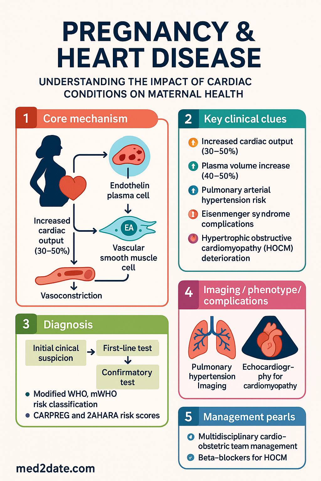
- Pregnancy imposes a 30–50% increase in cardiac output, 40–50% increase in plasma volume, and a physiological decline in systemic vascular resistance — these changes peak at 28–34 weeks gestation.
- Modified WHO (mWHO) risk classification (I–IV) is the standard tool for preconception counselling; mWHO IV carries a contraindication to pregnancy.
- Pulmonary arterial hypertension (PAH) carries a maternal mortality of 17–33% and is formally contraindicated (mWHO IV) — pregnancy should be avoided or terminated early.
- Eisenmenger syndrome mortality approaches 30–50% per pregnancy; multidisciplinary management in a tertiary centre with ICU access is mandatory.
- Severe symptomatic hypertrophic obstructive cardiomyopathy (HOCM) may deteriorate as venous return falls; beta-blockers are first-line therapy.
- Anticoagulation in pregnancy requires switching from warfarin to low-molecular-weight heparin (LMWH) — enoxaparin 1 mg/kg BD with anti-Xa monitoring — particularly between 6–12 weeks and from 36 weeks.
- Peripartum cardiomyopathy (PPCM) presents in the last month of pregnancy or within 5 months postpartum; early initiation of bromocriptine (2.5 mg BD for 2 weeks then 2.5 mg daily) plus standard HF therapy improves LV recovery.
- ACE inhibitors, ARBs, direct renin inhibitors, and spironolactone are teratogenic and contraindicated in pregnancy; hydralazine + nitrate or labetalol are safe antihypertensive alternatives.
- All pregnant women with known or suspected cardiac disease must be managed via a multidisciplinary cardio-obstetric team including obstetrician, cardiologist, anaesthetist, and neonatologist.
- Vaginal delivery with assisted second stage (forceps/vacuum) is preferred for most cardiac conditions; Caesarean section is reserved for obstetric indications or haemodynamic instability.
- Epidural analgesia reduces catecholamine surge and preload fluctuations — beneficial in most cardiac lesions except severe aortic stenosis or HOCM where a phenylephrine bolus strategy is preferred.
- Aboriginal and Torres Strait Islander women have higher rates of rheumatic heart disease and delayed cardiac diagnosis in pregnancy — proactive screening and culturally safe care pathways are essential.
Introduction & Australian Epidemiology
Cardiac disease is a leading cause of indirect maternal mortality in Australia, responsible for approximately 15–20% of maternal deaths reported in successive triennial MBRRACE-UK and Australian Maternal Deaths Committee reports. Outcomes are worst with pulmonary hypertension, severe mitral stenosis, and cardiomyopathy — conditions in which the haemodynamic demands of pregnancy, labour, and the immediate postpartum period can precipitate acute decompensation, arrhythmia, or death.
The incidence of cardiac disease in pregnancy is rising, driven by increasing maternal age, the obesity epidemic, improved survival of congenital heart disease (CHD) patients into reproductive years, and immigration from regions with high rheumatic heart disease (RHD) prevalence. In Australia, the estimated prevalence of significant cardiac disease in pregnancy is 1–3%, with congenital heart disease now exceeding acquired valvular disease in most metropolitan centres. However, in Northern Australia and among Aboriginal and Torres Strait Islander communities, RHD — particularly severe mitral valve disease — remains a dominant cause of cardiac morbidity in pregnancy.
The CARPREG (Cardiac Disease in Pregnancy) and ZAHARA risk scores, along with the modified WHO (mWHO) classification, provide evidence-based frameworks for risk stratification. The mWHO classification is now the most widely adopted system in Australian tertiary centres. Accurate preconception risk assessment and multidisciplinary planning are the cornerstones of safe management.
Physiological Cardiovascular Changes in Pregnancy
Understanding normal pregnancy physiology is essential for recognising pathological deterioration. The cardiovascular system undergoes profound adaptations beginning in the first trimester and peaking at 28–34 weeks of gestation.
| Parameter | Change | Magnitude | Peak Timing |
|---|---|---|---|
| Cardiac output | ↑ 30–50% | Up to 7 L/min | 28–34 weeks |
| Heart rate | ↑ 15–25 bpm | Resting HR 80–95 bpm | Third trimester |
| Stroke volume | ↑ 20–30% | - | Mid-pregnancy |
| Plasma volume | ↑ 40–50% | ~4.5–5.5 L | 32–34 weeks |
| Red cell mass | ↑ 20–30% | - | Term |
| Systemic vascular resistance | ↓ 20–30% | Nadir 70–80% baseline | Second trimester |
| Blood pressure | ↓ then ↑ | Nadir mid-2nd trimester | Returns to baseline by term |
| Oxygen consumption | ↑ 20–30% | ~300 mL/min | Term |
Intrapartum Haemodynamic Shifts
Labour imposes additional cardiovascular stress. Each uterine contraction produces an auto-transfusion of approximately 300–500 mL into the central circulation, increasing cardiac output by 15–25% above the already elevated third-trimester baseline. The immediate postpartum period is the most hazardous — cardiac output peaks at 60–80% above pre-pregnancy values within 10–15 minutes of delivery due to relief of inferior vena caval compression and uterine auto-transfusion. This is the critical window for pulmonary oedema and acute heart failure.
High-Risk Conditions: Pulmonary Hypertension, HOCM & Eisenmenger Syndrome
Pulmonary Arterial Hypertension (PAH)
Pulmonary arterial hypertension in pregnancy remains one of the most dangerous cardiovascular conditions, classified as mWHO IV — pregnancy is formally contraindicated. Maternal mortality rates range from 17–33% in contemporary series, with the highest risk in idiopathic PAH and connective tissue disease-associated PAH.
The pathophysiology centres on the fixed pulmonary vascular bed failing to accommodate the 30–50% increase in cardiac output. The normal pregnancy-induced decline in systemic vascular resistance, combined with the fixed high pulmonary vascular resistance, creates a haemodynamic mismatch that precipitates right ventricular failure and cardiovascular collapse.
Management of PAH in pregnancy involves continuation or initiation of pulmonary vasodilator therapy. Sildenafil (20–40 mg PO TDS; PBS Authority Required for PAH) and inhaled iloprost are the most commonly used agents in Australian practice. Intravenous epoprostenol (prostacyclin) is reserved for refractory cases and requires central venous access. Bosentan and macitentan are endothelin receptor antagonists with known teratogenicity and are generally avoided.
Eisenmenger Syndrome
Eisenmenger syndrome — irreversible pulmonary hypertension with reversal of shunt flow through a large intracardiac or great vessel defect — is associated with maternal mortality of 30–50%. Death typically occurs peripartum or in the early postpartum period from progressive right heart failure, hypoxaemia, thromboembolism, or paradoxical embolism. Pregnancy is formally contraindicated (mWHO IV).
If pregnancy proceeds despite counselling, management mandates admission to a tertiary centre from 20 weeks gestation. Therapeutic strategies include supplemental oxygen to maintain SpO₂ >90%, avoidance of dehydration and systemic vasodilatation, prophylactic anticoagulation (LMWH from second trimester), and planned early delivery by Caesarean section under epidural or combined spinal-epidural anaesthesia with invasive haemodynamic monitoring.
Hypertrophic Obstructive Cardiomyopathy (HOCM)
HOCM is the most common genetic cardiomyopathy, affecting approximately 1 in 500 individuals. Pregnancy is generally well tolerated in asymptomatic or mildly symptomatic patients (mWHO II), but women with significant left ventricular outflow tract (LVOT) obstruction (gradient >30 mmHg), NYHA III–IV symptoms, or prior arrhythmia are at higher risk (mWHO II–III).
The fall in systemic vascular resistance during pregnancy can worsen dynamic LVOT obstruction. Additionally, the increased venous return and heart rate in labour may trigger haemodynamic deterioration.
Key management principles:
- Beta-blockers are first-line: metoprolol 25–100 mg PO BD or propranolol 20–40 mg PO TDS (both Pregnancy Category C — generally acceptable with monitoring).
- Maintain adequate preload — avoid dehydration, vasodilatation, and Valsalva manoeuvre.
- Vaginal delivery with assisted second stage (forceps/vacuum) is preferred to avoid prolonged pushing.
- Epidural analgesia must be used cautiously — low-dose incremental technique with phenylephrine (100 mcg IV bolus) available to counteract hypotension. Avoid pure beta-agonists (e.g., ephedrine) which increase contractility and worsen LVOT obstruction.
- ICD or pacemaker in situ: no contraindication to vaginal delivery; magnet available at delivery for ICD deactivation during labour.
Other High-Risk Lesions
| Condition | mWHO Class | Key Risk | Management Focus |
|---|---|---|---|
| Severe mitral stenosis (area <1.0 cm²) | III–IV | Pulmonary oedema, AF | Beta-blocker, balloon valvuloplasty if feasible pre-pregnancy |
| Marfan syndrome (aortic root >40 mm) | III | Aortic dissection | Beta-blocker, serial echo, delivery by Caesarean if aorta >45 mm |
| Coarctation of aorta (repaired) | II–III | Aortic dissection, re-coarctation | MRI pre-pregnancy, BP control with labetalol |
| Mechanical valve | III | Valve thrombosis, warfarin embryopathy | Complex anticoagulation (see below) |
| Systemic RV (e.g., TGA post-Mustard/Senning) | III | RV failure, arrhythmia | Echo monitoring, low threshold for early delivery |
Management Principles & Anticoagulation in Pregnancy
General Management Principles
The foundation of managing cardiac disease in pregnancy is preconception risk assessment, shared decision-making, and structured multidisciplinary care. The following principles apply across all cardiac conditions:
- Preconception counselling: Discuss mWHO class, maternal and foetal risks, medication adjustments, and delivery planning before conception where possible.
- Medication review: Discontinue teratogens (ACE inhibitors, ARBs, statins, amiodarone, direct renin inhibitors). Substitute with pregnancy-safe alternatives (labetalol, hydralazine, nifedipine for hypertension; sotalol or flecainide for arrhythmias where indicated).
- Monitoring schedule: Clinical review every 2–4 weeks (mWHO I–II) or weekly (mWHO III–IV). Echocardiography each trimester at minimum — more frequent if valve disease, cardiomyopathy, or aortopathy.
- Delivery planning: Planned delivery at 37–39 weeks for most conditions; earlier if haemodynamic deterioration. Written delivery plan reviewed by all team members.
- Mode of delivery: Vaginal delivery is preferred for the majority of cardiac conditions. Indications for Caesarean section include obstetric indications, acute haemodynamic instability, severe aortopathy (aortic diameter >45 mm in Marfan), and some complex CHD.
- Intrapartum monitoring: Continuous telemetry, pulse oximetry, and invasive arterial monitoring for mWHO III–IV. Pulmonary artery catheterisation reserved for severe pulmonary hypertension or complex haemodynamics.
- Postpartum: Monitor in high-dependency for ≥48 hours (mWHO III–IV). Risk of decompensation persists for 3–6 months postpartum due to ongoing haemodynamic changes and fluid redistribution.
Teratogenic Medications to Discontinue
- ACE inhibitors (enalapril, perindopril, ramipril) — renal agenesis, oligohydramnios
- ARBs (valsartan, irbesartan, candesartan) — similar to ACEi
- Statins (atorvastatin, rosuvastatin) — skeletal anomalies (contraindicated in pregnancy)
- Amiodarone — foetal thyroid dysfunction, growth restriction
- Direct renin inhibitors (aliskiren) — contraindicated
- Eplerenone, spironolactone — anti-androgenic effects
- Warfarin — switch to LMWH between 6–12 weeks and from 36 weeks (see anticoagulation below)
Pregnancy-Safe Alternatives
Anticoagulation in Pregnancy
Pregnancy is a prothrombotic state with a 4–5-fold increased risk of venous thromboembolism (VTE). Anticoagulation management is particularly complex in women with mechanical heart valves, atrial fibrillation, previous VTE, or pulmonary hypertension.
| Indication | First-Line Agent | Dose & Monitoring | Key Considerations |
|---|---|---|---|
| Mechanical heart valve | Warfarin or LMWH | LMWH: enoxaparin 1 mg/kg BD + anti-Xa 4h post-dose (target 0.8–1.2 IU/mL) Warfarin: ≤5 mg/day if used in 2nd/3rd trimester |
Warfarin embryopathy risk if >5 mg/day in 1st trimester. Switch to LMWH 6–12 weeks then revert. Switch back to LMWH from 36 weeks. |
| Atrial fibrillation (non-valvular) | LMWH or aspirin | Enoxaparin 1 mg/kg OD–BD (risk-stratified) | DOACs (apixaban, rivaroxaban, edoxaban) contraindicated in pregnancy |
| VTE prophylaxis | LMWH | Enoxaparin 40 mg OD | Weight-adjust if BMI >40 or renal impairment |
| VTE treatment | LMWH | Enoxaparin 1 mg/kg BD | Continue for duration of pregnancy + 6 weeks postpartum |
Peripartum Cardiomyopathy (PPCM)
Peripartum cardiomyopathy (PPCM) is defined as heart failure occurring in the last month of pregnancy or within five months postpartum, with no identifiable cause and no pre-existing cardiac disease, and left ventricular ejection fraction (LVEF) <45% on echocardiography. The incidence in Australia is approximately 1 in 1,000–4,000 live births, with higher rates in Aboriginal and Torres Strait Islander women, women over 30 years, multiparous women, twin pregnancies, and those with pre-eclampsia or gestational hypertension.
Pathophysiology
The pathogenesis is multifactorial. Current evidence supports a "two-hit" hypothesis: (1) the haemodynamic stress of pregnancy on the myocardium, and (2) a prolactin-mediated vascular insult. Upregulated cathepsin D cleaves prolactin into a 16-kDa fragment (vasoinhibin) that is cardiotoxic and anti-angiogenic. Oxidative stress, inflammation, and autoimmune mechanisms also contribute. Selenoprotein deficiency has been implicated in some populations, notably in parts of Africa and potentially among nutritionally at-risk populations in Australia.
Clinical Presentation
- Dyspnoea, orthopnoea, paroxysmal nocturnal dyspnoea
- Peripheral oedema (may be confused with normal pregnancy changes)
- Palpitations, chest discomfort
- Cough, haemoptysis (severe cases)
- Thromboembolism — stroke, peripheral arterial embolism (up to 5–7% incidence due to low EF and hypercoagulable state)
Diagnostic Criteria
- Heart failure in the last month of pregnancy or within 5 months postpartum
- LVEF <45% on echocardiography (or fractional shortening <30%)
- No other identifiable cause of heart failure
- No pre-existing cardiac disease
Management of PPCM
Management follows standard heart failure principles with important modifications for pregnancy and lactation safety.
If presenting antenatally:
- Diuretics: Furosemide 20–40 mg PO/IV OD–BD (Category C, generally acceptable). Avoid excessive diuresis to maintain placental perfusion.
- Vasodilators: Hydralazine 25–50 mg PO TDS + isosorbide dinitrate 10–20 mg PO TDS (safe in pregnancy). Avoid ACEi/ARBs until after delivery.
- Beta-blockers: Carvedilol 3.125–25 mg PO BD or metoprolol 25–100 mg PO BD (Category C). Initiate cautiously; avoid in acute haemodynamic instability.
- Anticoagulation: LMWH if LVEF <35% due to high thromboembolic risk.
- Delivery: Expedite delivery if LVEF <30% or NYHA III–IV despite therapy. Vaginal delivery preferred.
If presenting postpartum:
- ACE inhibitor: Enalapril 2.5–20 mg OD or perindopril 2–8 mg OD (compatible with breastfeeding; preferred).
- Beta-blocker: Carvedilol or metoprolol as above.
- Diuretics: Furosemide as needed for congestion.
- Mineralocorticoid receptor antagonist: Spironolactone 25 mg OD if LVEF <35% (compatible with breastfeeding).
- Anticoagulation: Warfarin or LMWH if LVEF <35%.
- Bromocriptine: 2.5 mg PO BD for 2 weeks, then 2.5 mg daily for a further 4–6 weeks. Targets the prolactin-mediated pathway. The BRO-HF and another pilot trial showed improved LV recovery. Note: Bromocriptine suppresses lactation — combined with ACEi use, breastfeeding is generally not supported if bromocriptine is given.
Prognosis
Recovery (LVEF ≥50%) occurs in approximately 50–70% of women with PPCM, typically within 6–12 months. Factors associated with poor recovery include LVEF <25% at diagnosis, LV end-diastolic diameter >60 mm, African ancestry (higher incidence and worse outcomes), and delayed presentation. Relapse occurs in 20–30% of subsequent pregnancies, and subsequent pregnancy is contraindicated if LVEF has not recovered to ≥50%.
Risk Stratification: Modified WHO Classification
The modified WHO (mWHO) classification is the most widely validated risk assessment tool for cardiac disease in pregnancy and is recommended by the European Society of Cardiology (ESC) and endorsed by the Cardiac Society of Australia and New Zealand (CSANZ).
Special Populations
Aboriginal and Torres Strait Islander Health Considerations
Aboriginal and Torres Strait Islander women experience significantly higher rates of cardiac disease in pregnancy compared with non-Indigenous Australians, driven primarily by the burden of rheumatic heart disease (RHD). RHD remains the most common acquired heart disease in pregnancy among Indigenous Australians, particularly in Northern Territory, Western Australia, and Far North Queensland communities.
📚 References
- 1. Regitz-Zagrosek V, Roos-Hesselink JW, Bauersachs J, et al. 2018 ESC Guidelines for the management of cardiovascular diseases during pregnancy. Eur Heart J. 2018;39(34):3165–3241.
- 2. Cauldwell M, Steer PJ, Swan L, et al. Management of women with pulmonary arterial hypertension during pregnancy: a retrospective, case-matched study comparing outcomes in women treated with sildenafil. Open Heart. 2020;7(2):e001259.
- 3. Sliwa K, Hilfiker-Kleiner D, Petrie MC, et al. Current state of knowledge on aetiology, diagnosis, management, and therapy of peripartum cardiomyopathy: a position statement from the Heart Failure Association of the ESC Working Group on Peripartum Cardiomyopathy. Eur J Heart Fail. 2010;12(8):767–778.
- 4. Hoesli I, Strahl B, Buder T, et al. Bromocriptine in addition to standard heart failure therapy in peripartum cardiomyopathy: the BRO-HF pilot trial. ESC Heart Fail. 2021;8(6):4882–4890.
- 5. Siu SC, Sermer M, Colman JM, et al. Prospective multicenter study of pregnancy outcomes in women with heart disease (CARPREG). Circulation. 2001;104(5):515–521.
- 6. Drenthen W, Boersma E, Balci A, et al. Predictors of pregnancy complications in women with congenital heart disease (ZAHARA study). Eur Heart J. 2010;31(17):2124–2132.
- 7. RHDAustralia ARF/RHD writing group. The 2020 Australian guideline for prevention, diagnosis, and management of acute rheumatic fever and rheumatic heart disease. 3rd ed. Darwin: Menzies School of Health Research; 2020.
- 8. AIHW (Australian Institute of Health and Welfare). Maternal deaths in Australia 2018–2020. Cat. no. PER 113. Canberra: AIHW; 2023.
- 9. Gelson E, Curry R, Gatzoulis MA, et al. Effect of maternal heart disease on pregnancy outcomes in a low-income country. Heart. 2011;97(23):1927–1932.
- 10. European Society of Cardiology. 2021 ESC Guidelines on cardiovascular disease during pregnancy (executive summary). Eur Heart J. 2022;43(42):4333–4346.
- 11. National Blood Authority Australia. Clinical practice guidelines: use of anticoagulants in pregnancy. Canberra: NBA; 2023.
- 12. Australasian Society of Clinical Immunology and Allergy (ASCIA) & CSANZ. Consensus position statement: cardiac assessment in women planning pregnancy with known cardiovascular disease. Heart Lung Circ. 2020;29(9):1267–1275.
- 13. Parapia LA, Roberts A, Hussey J. The use of low-molecular-weight heparin in pregnancy: a review and meta-analysis. Thromb Haemost. 2022;122(10):1623–1633.
- 14. Carlin AJ, Alfirevic Z, Gyte GML. Interventions for treating persistent high blood pressure during pregnancy. Cochrane Database Syst Rev. 2020;(10):CD004281.
- 15. McGrady M, Krum H. Cardiovascular risk in women — the role of pregnancy and the menopause. Heart Lung Circ. 2019;28(12):1770–1777.