📋 Key Information Summary
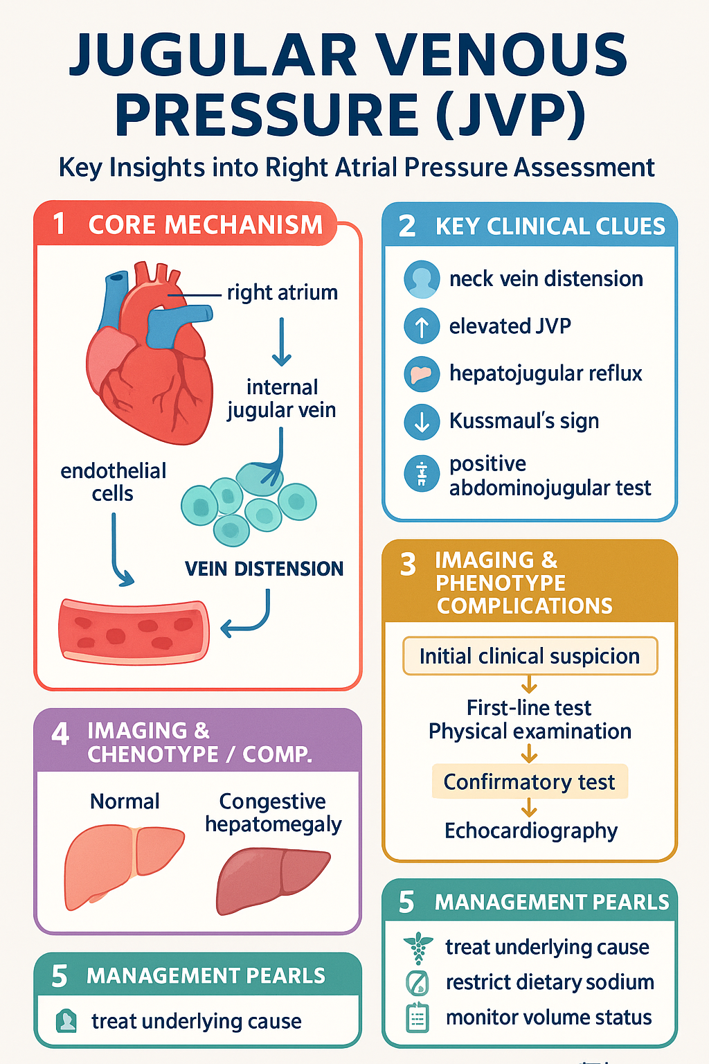
- Jugular venous pressure (JVP) estimation reflects right atrial (RA) pressure and is a cornerstone of bedside cardiovascular assessment in Australian clinical practice.
- The normal JVP is ≤3 cm vertical height above the sternal angle (right atrial centre ≈ 5 cm below sternal angle + 3 cm above = 8 cmH₂O; often quoted as <10 cm H₂O).
- The JVP waveform consists of five components: a-wave (atrial contraction), c-wave (tricuspid closure/bulging), x-descent (atrial relaxation), v-wave (atrial filling against closed tricuspid), and y-descent (rapid ventricular filling).
- An elevated JVP (>3 cm above sternal angle) indicates raised right-sided filling pressures: think heart failure, pericardial effusion, pulmonary hypertension, or tricuspid valve disease.
- Cannon a-waves occur when the right atrium contracts against a closed tricuspid valve — seen in complete heart block, ventricular tachycardia, and junctional rhythms.
- Giant v-waves (cv-waves) with rapid y-descent are the hallmark of tricuspid regurgitation.
- A blunted (slow) y-descent suggests constrictive pericarditis or restrictive cardiomyopathy when combined with elevated JVP and Kussmaul sign.
- Kussmaul sign (paradoxical rise in JVP with inspiration) is seen in constrictive pericarditis, cardiac tamponade (rarely), and right ventricular infarction.
- Technique: position patient at 45°, use internal jugular vein (preferred), locate the highest point of pulsation, measure vertical height above sternal angle with ruler.
- Distinguish venous pulsation (palpable but obliterated by light pressure, non-palpable above) from carotid artery pulsation (palpable, sustained, more lateral).
- Hepatojugular reflux (abdominojugular test) — sustained rise ≥3 cm for >15 s with firm RUQ pressure confirms elevated RA pressure.
- Aboriginal and Torres Strait Islander peoples experience rheumatic heart disease at disproportionately high rates; JVP assessment is critical for detecting tricuspid regurgitation secondary to RHD.
Introduction & Australian Epidemiology
The jugular venous pressure (JVP) is the vertical height of the venous column of blood above the sternal angle, reflecting right atrial (RA) pressure and providing a non-invasive window into right-heart haemodynamics. The JVP waveform — unlike the carotid pulse — transmits a distinctive oscillatory pattern that encodes information about atrial contraction, atrioventricular valve competence, and ventricular compliance.
Bedside JVP assessment remains a first-line investigation in Australian general practice, emergency departments, and inpatient medicine. Its diagnostic value extends to heart failure classification, pericardial disease, arrhythmia identification, and tricuspid valve pathology. In an era of echocardiography and invasive haemodynamics, the JVP remains a rapid, cost-free, and reproducible screening tool when performed correctly.
Heart failure affects approximately 500,000 Australians, with right-heart involvement common in chronic left-heart disease, pulmonary hypertension, and cor pulmonale. Rheumatic heart disease — particularly relevant in Aboriginal and Torres Strait Islander communities — frequently involves the tricuspid valve, making JVP waveform interpretation essential for early detection and management of tricuspid regurgitation. Pericardial disease, though less common, carries significant morbidity and is often first suspected at the bedside through JVP examination findings.
This guideline provides a comprehensive framework for JVP assessment, waveform interpretation, and clinical application in Australian practice, aligned with Heart Foundation, Cardiac Society of Australia and New Zealand (CSANZ), and RACGP standards.
JVP Waveform Components (a, c, x, v, y)
The normal JVP waveform is composed of three positive deflections (a-wave, c-wave, v-wave) and two negative descents (x-descent, y-descent). Understanding these components is essential for identifying pathological waveforms at the bedside.
| Component | Timing | Mechanism | Clinical Significance |
|---|---|---|---|
| a-wave | Late diastole / before S1 | Right atrial contraction against closed AV valve (atrial kick) | Absent in atrial fibrillation; cannon a-waves in heart block / VT |
| c-wave | Early systole / at S1 | Tricuspid valve closure bulging into RA; carotid transmission | Usually not visible at bedside; merges with a-wave |
| x-descent | Early–mid systole | Atrial relaxation and ventricular systolic descent of tricuspid annulus | Attenuated or absent in severe tricuspid regurgitation |
| v-wave | Late systole / before S2 | Venous filling of RA against closed tricuspid valve | Giant v-wave (cv-wave) in tricuspid regurgitation |
| y-descent | Early diastole / after S2 | Tricuspid valve opens; rapid passive emptying of RA into RV | Blunted in constrictive pericarditis; rapid in TR and constrictive physiology |
Normal Waveform Characteristics
- a-wave amplitude is approximately 3–5 mmHg above the v-wave trough.
- The y-descent is normally slow and gradual; a rapid y-descent suggests elevated RA pressure or tricuspid regurgitation.
- Normal mean RA pressure is 2–6 mmHg (equivalent to ~3 cm vertical column above sternal angle at 45°).
- Respiratory variation: JVP normally falls with inspiration (negative intrathoracic pressure increases venous return transiently but lowers the measured column).
Elevated JVP: Causes & Assessment
An elevated JVP — defined as venous pulsation visible >3 cm above the sternal angle with the patient reclined at 45° — indicates raised right atrial pressure. Systematic evaluation of the height, waveform morphology, and response to manoeuvres enables differentiation of underlying aetiologies.
Causes of Elevated JVP
| Category | Conditions | Associated Waveform |
|---|---|---|
| Right-heart failure | Cor pulmonale, right ventricular infarction, pulmonary hypertension, left-heart failure with secondary RV dysfunction | Prominent a-wave (PAH); rapid y-descent |
| Tricuspid valve disease | Tricuspid regurgitation (RHD, endocarditis, carcinoid), tricuspid stenosis (rare) | Giant v-wave (TR); slow y-descent, prominent a-wave (TS) |
| Pericardial disease | Cardiac tamponade, constrictive pericarditis, effusive–constrictive pericarditis | Blunted y-descent (tamponade); rapid y-descent + Kussmaul (constriction) |
| Volume overload | Renal failure, iatrogenic fluid overload, nephrotic syndrome | Normal waveform morphology, elevated height |
| Arrhythmias | Complete heart block, junctional tachycardia, VT | Cannon a-waves (AV dissociation) |
| Increased intrathoracic pressure | Tension pneumothorax, mechanical ventilation (high PEEP), severe COPD exacerbation | Elevated baseline, reduced respiratory variation |
| SVC obstruction | Malignancy (lung, lymphoma), mediastinal fibrosis | Non-pulsatile elevated venous pressure; may lose waveform |
Hepatojugular Reflux (Abdominojugular Test)
This bedside manoeuvre helps differentiate true elevation from venous pulsation artefact and unmasked elevated RA pressure:
- Position patient at 45° head-up.
- Identify the JVP level.
- Apply firm, sustained pressure to the right upper quadrant (RUQ) for 10–15 seconds.
- A sustained rise ≥3 cm that persists for >15 s is a positive test — indicating elevated RA pressure.
- A transient rise (<15 s) is normal (transient augmentation of venous return).
Clinical Grading of JVP Elevation
Abnormal Waveforms (Cannon a-waves, Absent x)
Specific waveform abnormalities at the JVP provide powerful diagnostic clues to underlying cardiac pathology. Recognition of these patterns at the bedside can accelerate diagnosis and guide management.
Cannon a-Waves
Cannon a-waves are large, intermittent, forceful pulsations in the JVP caused by right atrial contraction against a closed tricuspid valve during atrioventricular (AV) dissociation. They appear as periodic, exaggerated a-waves that tower above the normal waveform.
| Rhythm | Mechanism | JVP Pattern |
|---|---|---|
| Complete heart block (3rd degree) | Atrial contraction randomly coincides with ventricular systole | Intermittent cannon a-waves at irregular intervals |
| Ventricular tachycardia | Retrograde AV conduction or AV dissociation | Cannon a-waves at variable frequency; may be regular if 1:1 retrograde |
| Junctional tachycardia | AV node fires independently of SA node | Regular cannon a-waves if retrograde P waves coincide with systole |
| Premature ventricular contractions (PVCs) | Early ventricular beat → atrium contracts against closed tricuspid | Single cannon a-wave after each PVC |
Giant v-Waves (cv-Waves) — Tricuspid Regurgitation
In tricuspid regurgitation (TR), the x-descent is obliterated as blood regurgitates into the RA throughout systole. The v-wave merges with the absent x-descent to produce a prominent, continuous positive deflection termed a cv-wave. This produces a systolic pulsation visible in the neck that may be mistaken for a carotid pulse.
- Severe TR produces ventricularisation of the RA waveform — the RA pressure tracing mimics the RV waveform.
- Associated findings: pulsatile liver, pansystolic murmur at left lower sternal edge increasing with inspiration (Carvallo sign).
- Common causes in Australia: rheumatic heart disease (particularly in Aboriginal and Torres Strait Islander populations), infective endocarditis (IVDU), carcinoid heart disease, Ebstein anomaly.
Absent x-Descent
Loss of the normal x-descent indicates that atrial pressure does not fall during ventricular systole. This occurs in:
- Tricuspid regurgitation — blood regurgitates into the RA, preventing the normal systolic fall.
- Right atrial myxoma — obstructs RA emptying.
- Severe right-heart failure — RA pressure remains high throughout the cardiac cycle.
Blunted y-Descent
A slow or absent y-descent indicates impaired right ventricular filling:
- Cardiac tamponade — compressed RV resists filling; y-descent is virtually absent. Combined with elevated JVP, tachycardia, and hypotension, this constitutes Beck triad.
- Tricuspid stenosis — narrowed valve impedes passive RA emptying. Rare; seen in advanced rheumatic heart disease.
- Right atrial thrombus or tumour — mechanical obstruction to RA outflow.
Kussmaul Sign
Kussmaul sign is the paradoxical rise in JVP during inspiration, instead of the normal fall. It indicates impaired right ventricular filling:
- Constrictive pericarditis — classic association; the rigid pericardium prevents RV expansion during inspiration.
- Right ventricular infarction — non-compliant RV cannot accept increased venous return.
- Restrictive cardiomyopathy — similar physiology to constriction.
- Kussmaul sign is typically absent in cardiac tamponade — this is an important clinical distinction from constriction.
Clinical Assessment Technique
Accurate JVP assessment requires a systematic bedside technique. Common errors — confusing the JVP with the carotid pulse, incorrect patient positioning, or poor lighting — reduce diagnostic accuracy. The following method optimises reliability.
Step-by-Step Technique
Pitfalls & Troubleshooting
- Using the external jugular vein (valves confound measurement).
- Measuring from the clavicle instead of the sternal angle.
- Patient lying flat — venous pressure appears falsely normal.
- Confusing carotid pulsation with JVP (palpate above — venous disappears).
- Not adjusting for sternal angle elevation (pectus excavatum).
- Obesity — neck tissue obscures the vein; try left lateral decubitus position.
- Short, muscular neck — SCM heads may be close together.
- Very low JVP — recline further (30° or 15°) to increase venous filling.
- Patient anxiety / Valsalva — ask the patient to breathe normally and relax.
- Emphysema — hyperinflated chest may impede venous return and distort landmarks.
When to Refer for Echocardiography
- JVP >6 cm above sternal angle with no clear reversible cause (e.g., volume overload).
- Suspected constrictive pericarditis (elevated JVP + Kussmaul + rapid y-descent).
- Suspected cardiac tamponade (Beck triad: elevated JVP, hypotension, muffled heart sounds).
- Giant v-waves suggesting severe tricuspid regurgitation.
- New JVP elevation in the setting of right ventricular infarction (inferior STEMI).
- Aboriginal and Torres Strait Islander patients with suspected rheumatic heart disease and tricuspid involvement.
📚 References
- 1. Chaudhry GM, Fleg JL. Physical examination of the jugular venous pulse: a review and update. Am J Med. 2021;134(8):958–963.
- 2. Maisel AS, Atwood JE, Goldberger AL. Hepatojugular reflux: useful in the bedside diagnosis of heart failure. JAMA. 1987;257(8):1145–1148.
- 3. O'Brien F, O'Shea MJ. Bedside assessment of jugular venous pressure. BMJ. 2019;366:l5134.
- 4. Drazner MH, Rame JE, Stevenson LW, Dries DL. Prognostic importance of elevated jugular venous pressure and a third heart sound in patients with heart failure. N Engl J Med. 2001;345(8):574–581.
- 5. Heart Foundation. Guidelines for the prevention, detection, and management of heart failure in Australia. Melbourne: National Heart Foundation of Australia; 2018.
- 6. RHDAustralia (ARF/RHD writing group). The 2020 Australian guideline for prevention, diagnosis, and management of acute rheumatic fever and rheumatic heart disease (3rd edition). Darwin: Menzies School of Health Research; 2020.
- 7. AIHW. Rheumatic heart disease and acute rheumatic fever in Australia: 1996–2012. Cat. no. CVD 60. Canberra: Australian Institute of Health and Welfare; 2013.
- 8. Cardiac Society of Australia and New Zealand. Guidelines for the diagnosis and management of constrictive pericarditis. Heart Lung Circ. 2020;29(3):385–399.
- 9. Lange RA, Hillis LD. Cardiac tamponade. N Engl J Med. 2004;351(24):2562–2569.
- 10. RACGP. The RACGP curriculum for Australian general practice 2022: cardiovascular health. Melbourne: The Royal Australian College of General Practitioners; 2022.
- 11. McGee SR. Evidence-based physical diagnosis. 4th ed. Philadelphia: Elsevier; 2018:245–278.
- 12. Nishimura RA, Tajik AJ. Quantitative hemodynamics by Doppler echocardiography: a noninvasive alternative to cardiac catheterization. Prog Cardiovasc Dis. 1994;36(5):309–342.