📋 Key Information Summary
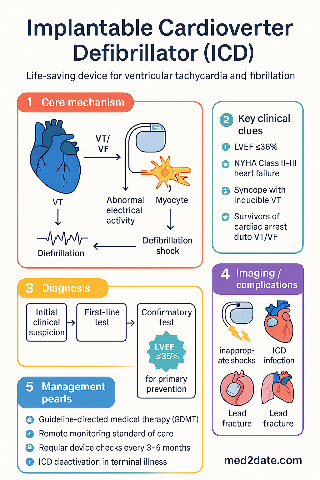
- ICDs are life-saving devices that detect and terminate ventricular tachycardia (VT) and ventricular fibrillation (VF) using anti-tachycardia pacing (ATP) or defibrillation shocks.
- Primary prevention ICD: Implant in patients at high risk of sudden cardiac death (SCD) who have not experienced sustained VT/VF — requires LVEF ≤35% and NYHA II–III heart failure on optimal medical therapy ≥3 months, or specific cardiomyopathy/genetic criteria.
- Secondary prevention ICD: Implant in survivors of cardiac arrest due to VT/VF, or patients with sustained haemodynamically significant VT, or syncope with inducible VT — no LVEF threshold required.
- MADIT-II and SCD-HeFT trials demonstrated 23–31% relative mortality reduction with primary prevention ICDs in ischaemic and non-ischaemic cardiomyopathy.
- All ICD patients must be on guideline-directed medical therapy (GDMT) for at least 3 months before primary prevention implantation to allow LVEF reassessment.
- CRT-D (cardiac resynchronisation therapy–defibrillator) is preferred when LVEF ≤35% with QRS ≥150 ms and LBBB morphology.
- Subcutaneous ICD (S-ICD) avoids transvenous lead complications and is an option for patients without pacing indications.
- Inappropriate shocks occur in 10–20% of patients — SVT is the most common cause; correct programming and beta-blockers reduce risk.
- ICD infection is a serious complication with 18–40% in-hospital mortality for endocarditis; complete device and lead extraction is mandatory.
- Remote monitoring (e.g., Medtronic CareLink, Abbott Merlin) is standard of care and reduces time to clinical intervention.
- Regular device checks at 3–6 monthly intervals (in-clinic) with continuous remote monitoring between visits.
- Generator replacement depends on battery status (ERI); typical longevity is 5–7 years for single-chamber, 4–6 years for dual-chamber/CRT-D.
- End-of-life discussions should include ICD deactivation to avoid painful shocks in patients with irreversible terminal illness.
Introduction & Australian Epidemiology
Implantable cardioverter defibrillators (ICDs) deliver life-saving shock therapy for ventricular fibrillation (VF) and ventricular tachycardia (VT) and are indicated for both primary and secondary prevention of sudden cardiac death (SCD) in high-risk patients. Since the first human implant by Dr Michel Mirowski in 1980, ICDs have become the single most effective intervention for prevention of arrhythmic death.
In Australia, approximately 5,000–6,000 new ICDs are implanted annually, with over 40,000 patients currently living with an active device. Implantation is performed at tertiary cardiac centres across all states. The Australian Institute of Health and Welfare (AIHW) data indicate that cardiac arrhythmias contribute to approximately 25,000 deaths per year, with SCD accounting for an estimated 3,000–4,000 of these.
ICDs are classified as Class I (strong) indications by the Cardiac Society of Australia and New Zealand (CSANZ), European Society of Cardiology (ESC), and American Heart Association/American College of Cardiology (AHA/ACC) guidelines for both primary and secondary prevention in appropriate patient populations. Device therapy is funded under Medicare Benefits Schedule (MBS) item numbers for implantation, generator changes, and lead revisions.
Contemporary ICDs offer tiered therapy — starting with painless anti-tachycardia pacing (ATP) before escalating to high-energy defibrillation. Modern devices incorporate remote monitoring, MRI-conditional designs, and extended battery longevity. The shift towards subcutaneous ICD (S-ICD) systems further reduces transvenous lead-related complications.
Indications: Primary vs Secondary Prevention
Primary Prevention
Primary prevention ICDs are implanted in patients at high risk of SCD who have not yet experienced a life-threatening ventricular arrhythmia. Landmark randomised trials — MADIT (1996), MUSTT (1999), MADIT-II (2002), and SCD-HeFT (2005) — established mortality benefit in selected populations.
| Indication | Criteria | Class | Key Evidence |
|---|---|---|---|
| Ischaemic cardiomyopathy | LVEF ≤35%, NYHA II–III, ≥40 days post-MI, ≥3 months GDMT | Class I | MADIT-II, SCD-HeFT |
| Non-ischaemic dilated cardiomyopathy (DCM) | LVEF ≤35%, NYHA II–III, ≥3 months GDMT | Class I | SCD-HeFT, DANISH* |
| LVEF ≤30%, NYHA I (ischaemic) | ≥40 days post-MI, ≥90 days post-revascularisation | Class I | MADIT-II |
| Hypertrophic cardiomyopathy (HCM) | ≥1 major risk factor (see below) | Class IIa | ESC HCM Guidelines 2020 |
| Arrhythmogenic right ventricular cardiomyopathy | ≥1 risk factor for SCD | Class IIa | Padua criteria |
| Long QT syndrome | Syncope/beta-blocker failure, QTc ≥500 ms | Class IIa | AHA/ACC 2017 |
| Brugada syndrome | Survived cardiac arrest, spontaneous VT with syncope | Class I (secondary prev.) | Brugada Consensus 2013 |
HCM Major Risk Factors for SCD
- Maximum left ventricular wall thickness ≥30 mm
- Family history of SCD in first-degree relative <40 years
- Unexplained syncope within preceding 6 months
- Non-sustained VT on Holter (≥3 beats at ≥120 bpm)
- Abnormal exercise blood pressure response (hypotensive or blunted rise)
- Late gadolinium enhancement on CMR (extensive fibrosis ≥15% LV mass)
Secondary Prevention
Secondary prevention ICDs are implanted in patients who have survived a cardiac arrest or experienced sustained VT. The AVID (1997), CIDS (2000), and CASH (2000) trials demonstrated 20–31% relative reduction in mortality compared with amiodarone.
| Indication | Class | Notes |
|---|---|---|
| Survived cardiac arrest (VF/VT) not due to reversible cause | Class I | Exclude acute MI <48 h, correctable electrolyte/ischaemic cause |
| Sustained VT with haemodynamic compromise | Class I | Regardless of LVEF |
| Syncope with inducible VT on EPS | Class I | EPS indicated when cause of syncope unclear |
| Sustained VT with near-normal LVEF | Class I | Consider underlying channelopathy or structural disease |
Device Components & Programming
System Components
ICD Programming — Rate Zones & Therapies
Modern ICDs use programmable rate-based detection zones and tiered therapy. Evidence from the ADVANCE III, MADIT-RIT, and PROVIDE trials demonstrates that long detection intervals and high rate thresholds significantly reduce inappropriate shocks.
| Zone | Rate Threshold | Detection | Therapy |
|---|---|---|---|
| VT-1 (slow VT) | 150–188 bpm | 30/40 intervals | ATP (burst, 8 pulses, 88% CL), then shock |
| VT-2 (fast VT) | 188–250 bpm | 18/24 intervals | ATP (burst), then shock 36 J → 36 J → 36 J |
| VF | >250 bpm | 12/16 intervals | Shock 36 J → 36 J → 36 J (max energy) |
Anti-Tachycardia Pacing (ATP)
ATP is a painless pacing therapy that terminates monomorphic VT by penetrating and resetting the re-entrant circuit. Up to 70–80% of VTs ≤200 bpm are terminated by ATP alone, avoiding painful shocks. ATP programming should include burst pacing at 84–88% of the VT cycle length.
Complications
Inappropriate Shocks
Inappropriate shocks are delivered for rhythms other than VT/VF, occurring in 10–20% of patients over 3–5 years. They cause significant psychological distress, reduce quality of life, and are associated with increased mortality.
ICD Infection
Pocket infection and CIED (cardiac implantable electronic device) endocarditis occur in 0.5–2.2% of initial implants and 1–4% of revision procedures. Staphylococcus aureus and coagulase-negative staphylococci (CoNS) account for >70% of cases.
Antibiotic Therapy for CIED Infection
Other Complications
| Complication | Incidence | Management |
|---|---|---|
| Pneumothorax | 0.5–2% | Aspiration or chest drain if symptomatic |
| Lead dislodgement | 1–2% | Repositioning within 6 weeks |
| Cardiac perforation/tamponade | 0.1–0.5% | Emergent pericardiocentesis; surgical repair |
| Venous thrombosis/occlusion | 2–5% | Anticoagulation; contralateral re-implant if severe |
| Lead fracture | 1–3% at 5 years | Lead revision or extraction |
| Twiddler syndrome | <1% | Generator pocket revision; suture anchoring |
| Electrical storm (≥3 shocks in 24 h) | 10–30% (lifetime) | IV amiodarone, sedation, beta-blockade, catheter ablation |
Device Checks & Follow-Up
Follow-Up Schedule
Post-ICD implantation follow-up is essential for safety, battery monitoring, arrhythmia review, and optimisation of device programming. Remote monitoring is now standard of care and is endorsed by CSANZ, ESC, and HRS/EHRS.
Remote Monitoring
At Each Device Check — Essential Components
Special Populations
End-of-Life & ICD Deactivation
ICD deactivation is an essential component of advance care planning for patients with terminal illness. Delivering shocks to dying patients causes suffering without altering outcomes. ICD deactivation should be discussed as part of palliative care planning, ideally well before end-stage disease.
Process: Device deactivation requires device interrogation by a cardiac physiologist, EP nurse, or cardiologist. The defibrillation function is programmed off while bradycardia pacing may be maintained if clinically appropriate. Written documentation of deactivation should be added to the patient's advance care directive.
Aboriginal and Torres Strait Islander Health Considerations
📚 References
- 1. Moss AJ, Hall WJ, Cannom DS, et al. Improved survival with an implanted defibrillator in patients with coronary disease at high risk for ventricular arrhythmia. N Engl J Med. 1996;335(26):1933–1940.
- 2. Moss AJ, Zareba W, Hall WJ, et al. Prophylactic implantation of a defibrillator in patients with myocardial infarction and reduced ejection fraction. N Engl J Med. 2002;346(12):877–883.
- 3. Bardy GH, Lee KL, Mark DB, et al. Amiodarone or an implantable cardioverter-defibrillator for congestive heart failure. N Engl J Med. 2005;352(3):225–237.
- 4. Køber L, Thune JJ, Nielsen JC, et al. Defibrillator implantation in patients with nonischemic systolic heart failure. N Engl J Med. 2016;375(13):1221–1230.
- 5. The Antiarrhythmics versus Implantable Defibrillators (AVID) Investigators. A comparison of antiarrhythmic-drug therapy with implantable defibrillators in patients resuscitated from near-fatal ventricular arrhythmias. N Engl J Med. 1997;337(22):1576–1583.
- 6. Gasparini M, Proclemer A, Klersy C, et al. Effect of long-detection interval vs standard-detection interval for implantable cardioverter-defibrillators on antitachycardia pacing and shock delivery: the ADVANCE III randomized clinical trial. JAMA. 2013;309(18):1903–1911.
- 7. Moss AJ, Schuger C, Beck CA, et al. Reduction in inappropriate therapy and mortality through ICD programming. N Engl J Med. 2012;367(24):2275–2283.
- 8. Hindricks G, Potpara T, Dagres N, et al. 2020 ESC Guidelines for the diagnosis and management of atrial fibrillation. Eur Heart J. 2021;42(5):373–498.
- 9. Writing Committee Members, Al-Khatib SM, Stevenson WG, et al. 2017 AHA/ACC/HRS guideline for management of patients with ventricular arrhythmias and the prevention of sudden cardiac death. Circulation. 2018;138(13):e272–e391.
- 10. Cardiac Society of Australia and New Zealand. Guidelines for the management of arrhythmias in heart failure. Heart Lung Circ. 2023;32(1):5–27.
- 11. Australian Institute of Health and Welfare. Cardiovascular disease in Aboriginal and Torres Strait Islander people. AIHW, Canberra. 2023.
- 12. Remenyi B, Carapetis J, Wyber R, et al. Position statement of the World Heart Federation on the diagnosis and management of rheumatic heart disease. Nat Rev Cardiol. 2013;10(11):668–670.
- 13. Sood N, Martin DT, Lampert R, et al. Incidence and predictors of cardiac implantable electronic device infection: results from the REPLACE registry. Pacing Clin Electrophysiol. 2018;41(11):1485–1493.
- 14. Slotwiner D, Varma N, Akar JG, et al. HRS Expert Consensus on remote interrogation and monitoring of cardiovascular implantable electronic devices. Heart Rhythm. 2015;12(7):e69–e100.
- 15. National Health and Medical Research Council. National Statement on Ethical Conduct in Human Research. NHMRC, Canberra. 2023 update.