📋 Key Information Summary
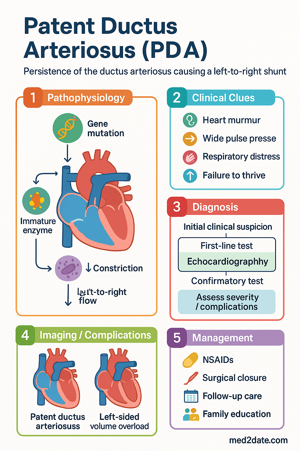
- Definition: Patent ductus arteriosus (PDA) is persistence of the ductus arteriosus beyond 72 hours of life in term neonates or beyond the first week in preterm neonates, causing a left-to-right shunt from aorta to pulmonary artery.
- Epidemiology: Affects 40–60 % of extremely preterm infants (<28 weeks) but <1 in 2 000 term births; is the third most common congenital heart lesion overall.
- Clinical features: Continuous 'machinery' murmur at the left upper sternal border, bounding pulses, widened pulse pressure, precordial hyperactivity; preterms may show only a new systolic murmur or be clinically silent.
- Complications: Left heart volume overload → pulmonary oedema, pulmonary hypertension, heart failure, necrotising enterocolitis (NEC), intraventricular haemorrhage (IVH), bronchopulmonary dysplasia (BPD), and death.
- Diagnosis: Echocardiography is the gold standard; assess PDA size (>1.5 mm = haemodynamically significant), flow pattern (restrictive vs. non-restrictive), LA:Ao ratio (>1.6 significant), and diastolic flow reversal in the descending aorta.
- Conservative management: Fluid restriction (120–140 mL/kg/day), optimise respiratory support, treat anaemia; appropriate for small, restrictive PDAs with minimal haemodynamic effect.
- Pharmacological closure — ibuprofen: First-line cyclooxygenase inhibitor; 10 mg/kg IV/PO day 1, then 5 mg/kg days 2 and 3 (PBS Authority Required). Equivalent efficacy to indomethacin with fewer renal side effects.
- Pharmacological closure — paracetamol: 15 mg/kg PO/IV 6-hourly for 5–7 days; evidence-based alternative when NSAIDs are contraindicated (renal impairment, NEC, active bleeding, thrombocytopaenia).
- Indomethacin: 0.1–0.25 mg/kg IV/PO q8–12 h for 3 doses; second-line in Australia due to higher risk of renal impairment and NEC; do not use if serum creatinine >120 µmol/L or urine output <0.6 mL/kg/h.
- Surgical ligation: Indicated when ≥2 courses of pharmacological therapy fail or are contraindicated; performed by a paediatric cardiothoracic surgeon. Post-ligation syndrome (haemodynamic instability, pulmonary hypertension) occurs in 5–10 %.
- Transcatheter device closure: Preferred for older infants (>5–6 kg, typically >6 months) and adults; Amplatzer™ duct occluder is the most widely used device. Performed in paediatric cardiology catheterisation suites (available in all tertiary children's hospitals).
- ATSI considerations: Congenital heart disease, including PDA, is more common in Aboriginal and Torres Strait Islander neonates; remote communities face delayed diagnosis and limited access to paediatric cardiology and interventional services.
- Monitoring: Serial echocardiography, daily clinical assessment (pulses, murmur, respiratory status, urine output), serum creatinine, electrolytes, platelet count, and bilirubin during pharmacological treatment.
🩺 Introduction & Australian Epidemiology
A patent ductus arteriosus (PDA) is the persistence of a fetal vascular channel — the ductus arteriosus — connecting the descending aorta to the main pulmonary artery beyond the normal postnatal closure period (typically within 24–72 hours of birth in term neonates). During fetal life, the ductus arteriosus is essential for diverting pulmonary blood flow to the systemic circulation. After birth, rising arterial oxygen tension and falling prostaglandin E₂ levels trigger functional closure, followed by anatomical obliteration over the subsequent 2–3 weeks.
Failure of this process results in a left-to-right shunt, the magnitude of which depends on the size of the PDA and the ratio of pulmonary to systemic vascular resistance. Haemodynamically significant PDAs cause volume overload of the left atrium and left ventricle, pulmonary overcirculation, and, if untreated, irreversible pulmonary hypertension (Eisenmenger syndrome).
Australian Epidemiology
- PDA affects approximately 40–60 % of extremely preterm neonates (<28 weeks' gestational age) and up to 70 % of neonates born at <26 weeks (AIHW, 2023).
- In term infants, isolated PDA accounts for 5–10 % of all congenital heart disease, with an estimated incidence of 1 in 2 000 live births in Australia.
- Female-to-male ratio is approximately 2:1 in term infants; the condition is more common at high altitude (>2 500 m) and in areas of low birth weight.
- PDA contributes to increased morbidity in Australian neonatal intensive care units (NICUs), including higher rates of bronchopulmonary dysplasia (BPD), necrotising enterocolitis (NEC), and intraventricular haemorrhage (IVH).
- The Australian & New Zealand Neonatal Network (ANZNN) reports that approximately 18 % of very-low-birth-weight (<1 500 g) infants undergo PDA treatment (pharmacological or surgical).
🔬 Pathophysiology & Aetiology
Normal Ductus Arteriosus Closure
The ductus arteriosus is a muscular artery that, in utero, shunts blood from the pulmonary artery to the descending aorta. Postnatal closure occurs in two phases:
- Functional closure (hours 1–72): Rising PaO₂ and declining prostaglandin E₂ (PGE₂) levels trigger smooth muscle contraction and intimal infolding, narrowing the lumen.
- Anatomical obliteration (weeks 2–8): Fibrosis and endothelial proliferation convert the ductus into the ligamentum arteriosum.
Why the Ductus Stays Patent
| Factor | Mechanism | Clinical Relevance |
|---|---|---|
| Prematurity | Immature ductal smooth muscle; elevated endogenous PGE₂; reduced sensitivity to oxygen-mediated constriction | Most common cause; inverse relationship with gestational age |
| Hypoxia / respiratory distress | Low PaO₂ maintains ductal patency via prostaglandin and nitric oxide pathways | Common in preterm infants with RDS |
| Genetic syndromes | Abnormal ductal musculature or elastic tissue | Trisomy 21, 18, 13; Rubella syndrome; Ehlers–Danlos |
| High altitude | Chronic hypoxaemia reduces oxygen-mediated constriction | Less relevant in Australia (populations largely <500 m) |
| Female sex | Oestrogen-related modulation of prostaglandin receptors | 2:1 female predominance in term PDA |
| Fluid overload | Increased preload raises left atrial pressure, preventing ductal constriction | Supports fluid-restriction strategies in NICU |
Haemodynamic Consequences
The direction and magnitude of shunting through a PDA depend on the relative resistances of the pulmonary and systemic vascular beds:
- Restrictive PDA (small, <1.5 mm): High resistance across the ductus limits shunt volume; often clinically insignificant.
- Non-restrictive PDA (large, >2.5 mm): Low ductal resistance permits significant left-to-right shunting, causing left atrial and left ventricular volume overload, pulmonary overcirculation, and systemic hypoperfusion (diastolic 'steal').
- In preterm infants, pulmonary vascular resistance is already low, amplifying shunt volume and accelerating pulmonary oedema.
- Chronic volume overload leads to left ventricular remodelling, dilated cardiomyopathy, and, if pulmonary hypertension develops, right heart failure.
🩺 Clinical Features & Complications
Clinical Presentation
Presentation varies dramatically with gestational age and PDA size:
Term Neonates & Older Children
- Continuous 'machinery' murmur: Best heard at the left upper sternal border (pulmonary area), radiating to the back; loudest in systole, continues through S2 into diastole.
- Bounding peripheral pulses: Widened pulse pressure (diastolic runoff through the PDA) produces a collapsing (Corrigan's) pulse.
- Precordial hyperactivity; displaced, hyperdynamic apex beat.
- Small PDAs may be asymptomatic and detected incidentally (murmur or echocardiography).
- Large PDAs: tachypnoea, poor feeding, failure to thrive, recurrent lower respiratory tract infections, exercise intolerance in older children.
Preterm Neonates
- Often presents without a continuous murmur — may have only a systolic murmur or no audible murmur at all.
- Signs of hsPDA: Systolic or continuous murmur, hyperdynamic precordium, bounding pulses, widened pulse pressure (>25 mmHg), ventilator dependence, increasing oxygen requirement, pulmonary haemorrhage, metabolic acidosis, oliguria.
- Clinical scores (e.g., McNamara score, Evens score) can assist in grading haemodynamic significance but echocardiography is definitive.
Complications
🔬 Investigations & Haemodynamic Assessment
Echocardiography — Gold Standard
Transthoracic echocardiography (TTE) is the definitive diagnostic and haemodynamic assessment tool. All tertiary and most secondary NICUs in Australia have access to neonatal echocardiography.
Laboratory Investigations
| Test | Purpose | MBS Item |
|---|---|---|
| Full blood count (FBC) | Anaemia exacerbates heart failure; thrombocytopaenia contraindicates NSAIDs | 65070 |
| Serum creatinine & electrolytes | Baseline renal function before NSAID therapy; monitor for NSAID nephrotoxicity | 66500 |
| Blood gas (capillary/arterial) | Metabolic acidosis suggests systemic hypoperfusion (diastolic steal) | 66500 |
| BNP / NT-proBNP | Correlates with shunt volume; guides treatment decisions and monitoring | 66844 |
| Blood culture | Rule out sepsis as a confounding diagnosis in preterm neonates | 69308 |
Echocardiographic Criteria for Haemodynamic Significance
| Parameter | Haemodynamically Significant | Non-significant |
|---|---|---|
| PDA diameter | >1.5 mm | ≤1.5 mm |
| LA:Ao ratio | >1.5–1.6 | ≤1.5 |
| Ductal flow pattern | Continuous (non-restrictive) or growing | Restrictive, low-velocity, closing |
| Diastolic flow reversal | Present in descending aorta | Absent |
| LV output | >300 mL/kg/min | Normal range |
💊 Management — Conservative, Pharmacological, & Interventional
Management of PDA depends on gestational age, PDA size, haemodynamic significance, and clinical context. The three arms of therapy are conservative (supportive), pharmacological (cyclooxygenase inhibitors or paracetamol), and interventional (surgical ligation or transcatheter device closure).
A. Conservative / Supportive Management
Appropriate for small, restrictive PDAs without haemodynamic significance, or as an adjunct to pharmacological therapy:
- Fluid restriction: 120–140 mL/kg/day (avoid excess free water; monitor serum sodium).
- Optimise respiratory support: Minimise mean airway pressure to reduce pulmonary blood flow; use CPAP/NIPPV as appropriate.
- Correct anaemia: Maintain haematocrit >35 % with packed red cell transfusion (anaemia increases cardiac output and shunt volume).
- Diuretics: Frusemide 1 mg/kg IV/PO 12–24-hourly for symptomatic pulmonary oedema (note: does not close the PDA; monitor electrolytes).
- Nutritional optimisation: Ensure adequate caloric intake (120–150 kcal/kg/day) to support growth.
B. Pharmacological Closure
Pharmacological therapy is the first-line intervention for hsPDA in preterm neonates. Two to three courses may be attempted before proceeding to interventional closure. The first course should be initiated within the first 2 weeks of life for maximum efficacy (early treatment).
C. Surgical Ligation
Surgical ligation via left thoracotomy remains the gold standard for failed pharmacological therapy. It is performed by paediatric cardiothoracic surgeons under general anaesthesia.
- Indications: Failure of ≥2 courses of pharmacological therapy; contraindication to all pharmacological agents; extremely large, non-restrictive PDA with severe haemodynamic compromise.
- Technique: Left posterolateral thoracotomy, dissection and ligation (or division) of the ductus with clips or suture.
- Success rate: >98 % complete closure.
- Complications: Post-ligation syndrome (hypotension, pulmonary hypertension, left ventricular dysfunction) in 5–10 %; recurrent laryngeal nerve injury (1–3 %); chylothorax; wound infection; scoliosis (long-term).
- Australian centres: All tertiary paediatric cardiac surgical units perform neonatal PDA ligation — The Children's Hospital at Westmead (Sydney), Royal Children's Hospital (Melbourne), Queensland Children's Hospital (Brisbane), Perth Children's Hospital, Women's & Children's Hospital (Adelaide).
- PBS/Insurance: Covered under public hospital surgical item numbers; no PBS listing required for surgery.
D. Transcatheter Device Closure
Transcatheter closure has become the preferred method for PDA closure in older infants (>5–6 kg), children, and adults. It avoids thoracotomy and is associated with shorter hospital stays and lower complication rates than surgery in suitable patients.
- Indications: Haemodynamically significant PDA in infants >5–6 kg (or >6 months of age, centre-dependent); any age child or adult with a suitable PDA anatomy; contraindication to or failure of surgical ligation.
- Devices: Amplatzer™ Duct Occluder (ADO) I and II (most common in Australia); coils (for small PDAs <2.5 mm); vascular plugs.
- Procedure: Percutaneous femoral venous (and sometimes arterial) access; catheter-directed deployment of occluder device under fluoroscopic and transoesophageal echocardiographic guidance.
- Success rate: 95–98 % complete closure at 6 months.
- Complications: Device embolisation (<1 %), residual shunt (2–5 %), haemolysis, femoral vessel injury, left pulmonary artery stenosis (device impingement), endocarditis.
- Australian centres: All tertiary paediatric cardiology centres with catheterisation facilities — primarily in Sydney (Westmead, Children's Hospital), Melbourne (RCH), Brisbane (QCH), Perth (PCH).
- Endocarditis prophylaxis: Required for 6 months post-device closure (or until complete endothelialisation confirmed on echo). Use amoxicillin 50 mg/kg (max 2 g) PO 1 h before dental or respiratory-tract procedures.
Treatment Algorithm Summary
👥 Special Populations
🏳️ Aboriginal and Torres Strait Islander Health
📚 References
- 1. Benitz WE; Committee on Fetus and Newborn, American Academy of Pediatrics. Patent ductus arteriosus in preterm infants. Pediatrics. 2016;137(1):e20153730.
- 2. Clyman RI, Couto J, Murphy GM. Patent ductus arteriosus: are current neonatal treatment options better or worse than no treatment at all? Semin Perinatol. 2012;36(2):123–129.
- 3. Australian Institute of Health and Welfare (AIHW). Congenital heart disease in Australia. Cat. no. CVD 89. Canberra: AIHW; 2023.
- 4. Australian & New Zealand Neonatal Network (ANZNN). Report of the Australian and New Zealand Neonatal Network 2021. Sydney: ANZNN; 2023.
- 5. Hundscheid T, Jansen ELS, Onland W, et al. Conservative management of patent ductus arteriosus in preterm infants — a systematic review and meta-analysis. Arch Dis Child Fetal Neonatal Ed. 2021;106(3):244–251.
- 6. Ohlsson A, Walia R, Shah SS. Ibuprofen for the treatment of patent ductus arteriosus in preterm or low birth weight (or both) infants. Cochrane Database Syst Rev. 2020;2:CD003481.
- 7. Terrin G, Conte F, Oncel MY, et al. Paracetamol for the treatment of patent ductus arteriosus in preterm neonates: a systematic review and meta-analysis. Arch Dis Child Fetal Neonatal Ed. 2016;101(2):F127–F136.
- 8. Backes CH, Rivera BK, Haque U, et al. Procedural closure of the patent ductus arteriosus in preterm infants: a review of the literature. J Perinatol. 2023;43(4):431–440.
- 9. Royal Australasian College of Physicians (RACP). Paediatric & Child Health Division. Position statement on neonatal cardiology services. Sydney: RACP; 2022.
- 10. National Health and Medical Research Council (NHMRC). Australian guidelines for the prevention and control of infection in healthcare. Canberra: NHMRC; 2019 (updated 2023). [Referenced for antimicrobial stewardship and endocarditis prophylaxis.]
- 11. Stout KK, Daniels CJ, Aboulhosn JA, et al. 2018 AHA/ACC guideline for the management of adults with congenital heart disease. Circulation. 2019;139(14):e698–e800.
- 12. Australian Commission on Safety and Quality in Health Care (ACSQHC). National Safety and Quality Health Service Standards. 2nd ed. Sydney: ACSQHC; 2021.