📋 Key Information Summary
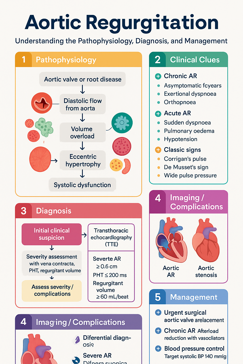
- Aortic regurgitation (AR) is characterised by diastolic flow of blood from the aorta back into the left ventricle due to aortic valve or root disease, leading to volume overload and progressive LV dilatation.
- Chronic AR is most commonly caused by degenerative aortic valve disease, bicuspid aortic valve, rheumatic heart disease, and aortic root dilatation (e.g., Marfan syndrome); it is typically well tolerated for years with compensatory LV dilatation.
- Acute AR is a cardiovascular emergency — most often due to infective endocarditis or aortic dissection — presenting with sudden haemodynamic collapse, pulmonary oedema, and cardiogenic shock.
- Acute AR requires urgent surgical aortic valve replacement in nearly all cases; medical stabilisation with vasodilators (sodium nitroprusside) and inotropes is a bridge to theatre, not definitive treatment.
- Transthoracic echocardiography (TTE) is the primary diagnostic tool; severity is graded using an integrated approach incorporating vena contracta width, pressure half-time (PHT), regurgitant volume/fraction, and LV dimensions.
- Severe AR is defined by vena contracta ≥ 0.6 cm, PHT ≤ 200 ms, regurgitant volume ≥ 60 mL/beat, regurgitant fraction ≥ 50%, and holodiastolic flow reversal in the descending aorta.
- Surgical indications (aortic valve replacement, ± root replacement) include: symptomatic severe AR (Class I), asymptomatic severe AR with LVEF ≤ 50%, asymptomatic severe AR with LV end-systolic dimension > 50 mm (or > 25 mm/m² BSA), and severe AR with aortic root dilatation ≥ 55 mm (≥ 50 mm in Marfan syndrome or bicuspid valve).
- Medical management of chronic AR centres on afterload reduction with vasodilators (ACE inhibitors/ARBs, dihydropyridine CCBs, long-acting nitrates) to slow LV remodelling; guideline-directed medical therapy for heart failure if LVEF declines.
- Blood pressure control is critical in chronic AR — target systolic BP < 140 mmHg to minimise regurgitant volume; avoid bradycardia (increases diastolic filling time and AR volume).
- Endocarditis prophylaxis is generally not recommended for native valve AR under current Australian (CSANZ) and international guidelines, except in specific high-risk prosthetic valve or post-surgical scenarios.
- Aboriginal and Torres Strait Islander Australians have higher rates of rheumatic heart disease–related AR, later presentation, and reduced access to specialist cardiac imaging and surgical services, particularly in remote communities.
- Serial echocardiographic surveillance every 6–12 months is essential for moderate-to-severe AR to detect early LV decompensation before symptoms develop.
Introduction & Australian Epidemiology
Aortic regurgitation (AR) is a valvular heart lesion in which blood flows retrograde from the aorta into the left ventricle (LV) during diastole, due to incompetence of the aortic valve cusps, aortic root dilatation, or both. The resultant volume overload leads to progressive LV eccentric hypertrophy, increased end-diastolic volume, and ultimately, systolic dysfunction and heart failure if left untreated.
In Australia, valvular heart disease affects approximately 2.5% of the population, with degenerative aortic valve disease being the most common aetiology in older adults. Bicuspid aortic valve — the most common congenital cardiac anomaly, affecting 1–2% of the population — predisposes to both aortic stenosis and regurgitation. Rheumatic heart disease (RHD) remains an important cause of AR in Aboriginal and Torres Strait Islander Australians, with incidence rates 20–60 times higher than in non-Indigenous Australians, particularly in the Northern Territory, Queensland, and Western Australia.
The Australian Institute of Health and Welfare (AIHW) reports that valvular heart disease contributed to over 2,300 deaths in Australia in 2021, and aortic valve surgery (including replacement and repair) accounts for a significant proportion of cardiac surgical caseloads nationally. Transcatheter aortic valve replacement (TAVR), originally developed for severe aortic stenosis, is increasingly being explored for high-risk AR patients at selected Australian centres.
This guideline provides an evidence-based, Australian-contextualised approach to the diagnosis, severity assessment, surgical decision-making, and medical management of aortic regurgitation in adults.
Acute vs Chronic Aortic Regurgitation
The distinction between acute and chronic AR is fundamental, as the haemodynamic consequences, clinical presentation, and management strategies differ profoundly. Acute AR is a cardiovascular emergency, whereas chronic AR is often tolerated for decades with compensatory LV remodelling.
Haemodynamic Differences
| Parameter | Acute AR | Chronic AR |
|---|---|---|
| Onset | Minutes to hours | Months to years |
| LV adaptation | No time for compensatory hypertrophy or dilatation | Eccentric hypertrophy with increased end-diastolic volume; Frank–Starling compensation maintains stroke volume |
| LV end-diastolic pressure (LVEDP) | Markedly elevated (rapid rise due to non-compliant ventricle) | Normal or mildly elevated initially; rises with decompensation |
| Diastolic BP | Very low (often < 50 mmHg); wide pulse pressure | Low; progressive widening of pulse pressure |
| Cardiac output | Markedly reduced (forward failure) | Preserved until late decompensation |
| Pulmonary oedema | Early and severe (LVEDP transmitted to pulmonary circulation) | Late finding |
| Mitral valve timing | Premature mitral closure (protective) and diastolic mitral regurgitation | Normal mitral valve timing |
| Peripheral signs | Often absent (no time for compensatory vasodilation) | Classic: Corrigan's pulse, de Musset's sign, wide pulse pressure, Duroziez's sign, Quincke's pulse |
Acute AR — Emergencies
Common causes of acute AR:
- Infective endocarditis (most common): Destruction or perforation of aortic valve cusps by vegetations; may also cause aortic root abscess with valve ring disruption. Community-acquired Staphylococcus aureus is the most virulent pathogen; Streptococcus species remain the most common overall in Australia.
- Type A aortic dissection: Dissection flap can involve the aortic root, causing acute aortic valve incompetence by preventing cusp coaptation. Stanford Type A dissection with AR requires emergency surgical repair (Bentall procedure or valve-sparing root replacement).
- Traumatic aortic injury: Blunt thoracic trauma can disrupt the aortic valve annulus or cusps, often in combination with aortic root injury.
- Iatrogenic: Catheter or guidewire injury to the aortic valve during coronary or structural heart interventions.
- Spontaneous cusp perforation or flail: Rare; can occur with myxomatous degeneration or Marfan syndrome.
Emergency management of acute AR:
- Rapid haemodynamic assessment and invasive monitoring (arterial line, central venous access).
- Intravenous vasodilator therapy: sodium nitroprusside 0.5–5 mcg/kg/min IV titrated to reduce afterload and forward cardiac output (requires intra-arterial BP monitoring).
- Inotropic support if cardiogenic shock: dobutamine 2.5–20 mcg/kg/min IV or noradrenaline if hypotension predominates.
- Avoid beta-blockers — slowing the heart rate increases diastolic time and worsens regurgitant volume.
- Avoid intra-aortic balloon pump (IABP) — contraindicated in AR (increases afterload during systole, worsening regurgitation).
- Urgent cardiothoracic surgical consultation and transfer to a cardiac surgical centre if not already at one.
- If endocarditis-related: initiate empiric antibiotics immediately per eTG Antibiotic guidelines (vancomycin + gentamicin ± rifampicin for prosthetic valve; see Infectious Disease guidelines), but surgery should not be delayed for antibiotic response alone.
Endocarditis-Related AR
Infective endocarditis (IE) is the most common cause of acute severe AR in Australia. AR occurs in 25–40% of left-sided IE cases and may result from leaflet destruction, perforation, prolapse, or perivalvular extension (abscess, pseudoaneurysm, fistula).
Key Australian considerations for endocarditis-related AR:
- Intravenous drug use (IVDU)-associated IE is increasing in urban centres (Melbourne, Sydney), often involving S. aureus and tricuspid involvement, but aortic valve disease occurs with left-sided involvement.
- RHD-related valve disease increases endocarditis risk in Aboriginal and Torres Strait Islander populations.
- Staphylococcus aureus bacteraemia (SAB) — a notifiable condition in Australia — should prompt urgent echocardiography in all cases to assess for IE-related AR.
Clinical Presentation
Acute AR presentation: Sudden dyspnoea, orthopnoea, pulmonary oedema, hypotension, tachycardia, peripheral vasoconstriction (cool peripheries). Auscultation: early diastolic murmur may be short or absent; S3 gallop common; soft S1 due to premature mitral closure.
Chronic AR presentation: Often asymptomatic for decades. Symptoms develop insidiously: exertional dyspnoea → orthopnoea → paroxysmal nocturnal dyspnoea → fatigue and exercise intolerance. Auscultation: early diastolic decrescendo murmur (left sternal edge, sitting forward, end-expiration), Austin Flint murmur (mid-diastolic low-pitched rumble at the apex), wide pulse pressure with collapsing pulse.
Severity Assessment — Echocardiographic Grading
An integrated, multi-parametric approach is recommended for AR severity grading, as no single echocardiographic parameter is sufficient in isolation. The 2020 ACC/AHA and 2021 ESC/EACTS guidelines endorse a comprehensive assessment incorporating qualitative, semi-quantitative, and quantitative measures.
Integrated Echocardiographic Grading
| Parameter | Mild | Moderate | Severe |
|---|---|---|---|
| Vena contracta width | < 0.3 cm | 0.3–0.59 cm | ≥ 0.6 cm |
| Pressure half-time (PHT) | > 500 ms | 200–500 ms | ≤ 200 ms |
| Regurgitant volume (RVol) | < 30 mL/beat | 30–59 mL/beat | ≥ 60 mL/beat |
| Regurgitant fraction (RF) | < 30% | 30–49% | ≥ 50% |
| Effective regurgitant orifice area (EROA) | < 0.10 cm² | 0.10–0.29 cm² | ≥ 0.30 cm² |
| Descending aorta diastolic flow reversal | Brief early diastolic | Intermediate | Holodiastolic (pandastolic) reversal |
| Jet width / LVOT width ratio | < 25% | 25–64% | ≥ 65% |
| AR jet density (CW Doppler) | Faint/incomplete | Dense | Dense, early termination (triangular CW profile) |
Key Echocardiographic Parameters — Detailed Explanation
Pressure Half-Time (PHT)
PHT is the time for the peak aortic–LV diastolic pressure gradient to fall by half. A short PHT (≤ 200 ms) reflects rapid pressure equalisation between the aorta and LV, indicating severe AR. PHT may be shortened by elevated LVEDP (heart failure, acute AR) and prolonged by vasodilator therapy, aortic compliance changes, or low-cardiac-output states. Therefore, PHT should be interpreted alongside other parameters.
Vena Contracta
The vena contracta is the narrowest width of the regurgitant jet at the level of the aortic valve, measured in the parasternal long-axis view with colour Doppler. It correlates closely with EROA and is relatively load-independent. A vena contracta ≥ 0.6 cm has high specificity for severe AR. Measurement requires careful image optimisation and may be technically challenging in eccentric jets.
Regurgitant Volume and Fraction (PISA Method)
Quantification using the proximal isovelocity surface area (PISA) method allows calculation of EROA and regurgitant volume. Regurgitant volume is calculated as EROA × velocity-time integral (VTI) of the AR jet. The regurgitant fraction is regurgitant volume divided by total (forward) stroke volume. The PISA method requires aliasing velocity optimisation and may underestimate EROA in eccentric jets or irregular orifices.
LV Dimensions and Function
LV end-diastolic dimension (LVEDD), LV end-systolic dimension (LVESD), LVEF, and indexed LV volumes are essential for determining timing of intervention in chronic AR. Progressive LV dilatation or declining LVEF (below 55–60%) in the setting of severe AR is an indication for surgery even in asymptomatic patients.
Additional Imaging
- Transoesophageal echocardiography (TOE): Superior for identifying the mechanism of AR (cusp perforation, prolapse, vegetation, aortic root anatomy), particularly pre-operatively and in suspected endocarditis. Available at all major Australian cardiac surgical centres.
- Cardiac MRI (CMR): Gold standard for quantification of regurgitant volume and fraction using phase-contrast flow mapping; also provides accurate LV volumes and LVEF. Indicated when echocardiographic assessment is suboptimal or discrepant. Available at major tertiary centres (MBS item 63000 series).
- Cardiac CT: Valuable for aortic root and ascending aorta assessment, particularly in bicuspid aortic valve, Marfan syndrome, or aortic dissection. Useful for procedural planning if surgery or TAVR is considered.
Surgical Indications
Aortic valve replacement (AVR) is the definitive treatment for severe AR, restoring valve competence and allowing reverse LV remodelling. Surgical timing is critical: intervention before irreversible LV systolic dysfunction yields the best long-term outcomes.
1. Symptomatic severe AR (exertional dyspnoea, angina, NYHA II–IV)
2. Asymptomatic severe AR with resting LVEF ≤ 50%
3. Asymptomatic severe AR undergoing other cardiac surgery (e.g., CABG, ascending aortic surgery, other valve surgery)
All carry Level of Evidence B–R.
Complete Indication Summary
| Indication | Class | Level |
|---|---|---|
| Symptomatic severe AR (NYHA II–IV or CCS angina class II–IV) | I | B |
| Asymptomatic severe AR with resting LVEF ≤ 50% | I | B |
| Severe AR undergoing CABG, ascending aortic surgery, or other valve surgery | I | C |
| Asymptomatic severe AR with normal LVEF and progressive LV dilatation: LVEDD > 65 mm or LVESD > 50 mm (or LVESD indexed > 25 mm/m² BSA) — Class IIa | IIa | B |
| Asymptomatic severe AR with normal LVEF, LVESD 45–49 mm (indexed 22–24.9 mm/m²) and declining exercise tolerance or progressive LV dilatation on serial imaging | IIb | C |
| Severe AR with bicuspid aortic valve and ascending aorta ≥ 50 mm (≥ 45 mm with additional risk factors) | IIa | C |
| Severe AR with Marfan syndrome and aortic root ≥ 50 mm (or ≥ 45 mm with risk factors) | IIa | C |
| Acute severe AR (endocarditis, dissection, trauma) — emergency surgery | I | B |
Surgical Approaches
Aortic valve replacement (AVR):
- Mechanical prosthesis: Durable (20–30+ years); requires lifelong anticoagulation with warfarin (target INR 2.0–3.0). Preferred in patients aged < 50–55 years who can reliably maintain anticoagulation.
- Bioprosthetic (tissue) valve: No long-term anticoagulation required; limited durability (10–20 years depending on age). Preferred in patients aged > 65 years or those with contraindications to anticoagulation. May be placed via surgical or transcatheter approach (TAVR-in-SAVR for valve-in-valve).
- Ross procedure (pulmonary autograft): Patient's own pulmonary valve replaces the aortic valve; homograft in pulmonary position. Excellent haemodynamics and no anticoagulation; performed at select Australian centres for younger patients (especially women of childbearing age). Complex surgery requiring specialist expertise.
- Aortic valve repair: Preserves native valve tissue; suitable for selected patients with cusp prolapse or fenestration, particularly with experienced surgical teams. Growing role in bicuspid aortic valve repair.
Aortic root surgery:
- Bentall procedure: Composite valve–conduit graft (mechanical or bioprosthetic valve within a Dacron tube graft) with reimplantation of coronary ostia. Standard for aortic root aneurysm with AR.
- Valve-sparing aortic root replacement (David procedure): Reimplants the native aortic valve within a graft; avoids prosthetic valve but requires skilled surgical team. Appropriate for aortic root dilatation with pliable, repairable valve cusps.
- Ascending aortic replacement: For ascending aortic aneurysm ≥ 55 mm (or ≥ 50 mm in bicuspid valve with risk factors, or ≥ 45 mm in Marfan syndrome with risk factors).
Post-Surgical Outcomes and Australian Data
Australian cardiac surgical outcomes are reported through the Australian and New Zealand Society of Cardiac and Thoracic Surgeons (ANZSCTS) Cardiac Surgery Database. In-hospital mortality for isolated AVR in Australia is approximately 2–3% for elective cases, rising to 6–10% for urgent/emergency operations (e.g., endocarditis, dissection). Long-term survival after successful AVR for severe AR is favourable, particularly when surgery is performed before the onset of irreversible LV dysfunction.
Referral Pathway in Australia
Patients meeting surgical criteria should be referred promptly to a cardiothoracic surgical unit. Cardiac surgical services are available at major tertiary hospitals in each Australian state and territory. Regional and remote patients should be referred via local cardiologist or through state-based cardiac telehealth pathways (e.g., NT Cardiac, Queensland Health telecardiology). Indigenous cardiac outreach programmes (e.g., RHDAustralia, Heart of Australia) facilitate access in remote communities.

Medical Management
Medical therapy in AR is primarily indicated for chronic AR and serves as bridging therapy in acute AR pending surgery. The goals of medical management are to reduce afterload, decrease regurgitant volume, preserve LV function, control blood pressure, and manage symptoms of heart failure when present.
Vasodilator Therapy in Chronic AR
Vasodilators reduce aortic impedance and diastolic blood pressure, thereby decreasing the pressure gradient driving regurgitant flow and reducing LV wall stress. This can slow the rate of LV dilatation and potentially delay the need for surgery. However, vasodilator therapy is not a substitute for surgery when surgical indications are met.
Class I: Symptomatic severe AR when surgery is not feasible (contraindication, patient preference) — use ACE inhibitor/ARB or hydralazine.
Class IIa: Asymptomatic severe AR with hypertension — use ACE inhibitor/ARB, dihydropyridine CCB, or long-acting nitrate.
Class IIb: Asymptomatic severe AR with normal blood pressure and normal LVEF — vasodilator therapy may be considered to delay surgical referral (evidence is limited).
Pharmacotherapy — Drug Cards
Heart Failure Management in Chronic AR with LV Dysfunction
When chronic AR progresses to LV systolic dysfunction (LVEF < 50%), guideline-directed medical therapy (GDMT) for heart failure should be initiated in addition to afterload reduction and surgical referral:
Blood Pressure Control
Endocarditis Prophylaxis
Current Australian (CSANZ), ESC, AHA, and Australian Dental Association guidelines do not recommend routine antibiotic prophylaxis for infective endocarditis in patients with native valve AR, including bicuspid aortic valve. The consensus is that the risk of adverse reactions to prophylactic antibiotics outweighs the uncertain benefit in most native valve disease.
High-risk scenarios where endocarditis prophylaxis MAY be considered (discuss with cardiology):
- Prosthetic heart valve (mechanical or bioprosthetic), including transcatheter valves
- Previous infective endocarditis
- Congenital heart disease with prosthetic material or repaired defect with residual shunt
- Cardiac transplant recipients with valvulopathy
For eligible patients undergoing dental procedures involving manipulation of gingival tissue or periapical region of teeth:
Dental and procedural advice for all patients with AR:
- Maintain excellent oral hygiene and regular dental reviews.
- Prompt treatment of skin infections, soft tissue infections, and any source of bacteraemia.
- Advise against cosmetic piercing (tongue, body) due to bacteraemia risk.
- Patients with prosthetic valves should carry a medical alert card and inform all treating health professionals.
Surveillance and Follow-Up Schedule
Exercise advice: Asymptomatic patients with mild-to-moderate AR and normal LV function may participate in competitive sports and vigorous exercise. Symptomatic patients, those with severe AR, or those with LV dysfunction should avoid isometric (heavy lifting) exercise and high-intensity competitive sport until reviewed by a cardiologist.
Special Populations
Pregnancy
Paediatrics
Elderly
Renal Impairment
Hepatic Impairment
Immunocompromised
Aboriginal and Torres Strait Islander Australians experience a disproportionate burden of valvular heart disease, including aortic regurgitation, driven primarily by rheumatic heart disease (RHD). These disparities reflect the broader social determinants of health including housing overcrowding, limited access to primary healthcare, geographic remoteness, and systemic disadvantage.
📚 References
- 1. Otto CM, Nishimura RA, Bonow RO, et al. 2020 ACC/AHA Guideline for the Management of Patients With Valvular Heart Disease: A Report of the American College of Cardiology/American Heart Association Joint Committee on Clinical Practice Guidelines. Circulation. 2021;143(5):e72–e227.
- 2. Vahanian A, Beyersdorf F, Praz F, et al. 2021 ESC/EACTS Guidelines for the management of valvular heart disease. Eur Heart J. 2022;43(7):561–632.
- 3. Zoghbi WA, Adams D, Bonow RO, et al. Recommendations for Noninvasive Evaluation of Native Valvular Regurgitation: A Report from the American Society of Echocardiography. J Am Soc Echocardiogr. 2017;30(4):303–371.
- 4. National Heart Foundation of Australia and Cardiac Society of Australia and New Zealand. Diagnosis and management of heart failure in Australia: a position statement from the National Heart Foundation of Australia and Cardiac Society of Australia and New Zealand. Med J Aust. 2018;209(1):35–42.
- 5. RHDAustralia (RHD Australia), Menzies School of Health Research. 2020 Australian Guideline for Prevention, Diagnosis and Management of Acute Rheumatic Fever and Rheumatic Heart Disease. 3rd ed. Darwin: RHDAustralia; 2020.
- 6. Australian Institute of Health and Welfare (AIHW). Rheumatic heart disease and acute rheumatic fever in Australia: 2017–2018 update. AIHW; 2020.
- 7. Remenyi B, ElGuindy A, Smith SC Jr, Yacoub M, Holmes DR Jr. Valvular aspects of rheumatic heart disease. Lancet. 2016;387(10025):1314–1324.
- 8. Chambers JB, Garbi M, Nieman K, et al. Appropriateness criteria for the use of cardiovascular imaging in heart valve disease in adults: a European Association of Cardiovascular Imaging report of literature review and current practice. Eur Heart J Cardiovasc Imaging. 2017;18(3):248–256.
- 9. Yeo TC, Dujardin KS, Tei C, Mahoney DW, McGoon MD, Seward JB. Value of a Doppler-derived index combining systolic and diastolic time intervals in predicting outcome in primary pulmonary hypertension. Am J Cardiol. 1998;81(9):1157–1161. [Reference for pressure half-time methodology in regurgitant lesions.]
- 10. Habib G, Lancellotti P, Antunes MJ, et al. 2015 ESC Guidelines for the management of infective endocarditis. Eur Heart J. 2015;36(44):3075–3128.
- 11. Stout KK, Verrier ED, Borer JS, et al. ACC/AHA Guideline Update on Valvular Heart Disease: Valvular Aortic Stenosis and Aortic Regurgitation. Circulation. 2008;118(15):e523–e661.
- 12. Australian and New Zealand Society of Cardiac and Thoracic Surgeons (ANZSCTS). ANZSCTS Cardiac Surgery Database Annual Report. Melbourne: ANZSCTS; 2023.
- 13. D'Arcy JL, Coffey S, Loudon MA, et al. Large-scale community echocardiographic screening for aortic stenosis using a hand-held device in the UK. Eur Heart J. 2021;42(16):1611–1621.
- 14. Carabello BA. Aortic regurgitation: a lesion with similarities to both aortic stenosis and mitral regurgitation. Circulation. 2013;127(12):1259–1263.
- 15. RHDAustralia. Rheumatic Heart Disease Endgame Strategy 2021–2031. Darwin: Menzies School of Health Research; 2021.