📋 Key Information Summary
- The cardiac cycle comprises systole (contraction, ejection) and diastole (relaxation, filling); at resting heart rate (~70 bpm) one cycle lasts approximately 0.85 seconds.
- Wiggers' diagram convention divides the cycle into five phases: isovolumetric contraction, rapid ejection, reduced ejection, isovolumetric relaxation, and rapid ventricular filling.
- The pressure-volume (PV) loop is the gold-standard framework for assessing myocardial performance — the loop's width represents stroke volume (SV) and its area represents external work per beat.
- End-systolic pressure-volume relationship (ESPVR) defines contractility; a steeper slope indicates increased inotropy independent of loading conditions.
- End-diastolic pressure-volume relationship (EDPVR) defines passive ventricular stiffness; the slope shift leftward indicates diastolic dysfunction (HFpEF).
- The first heart sound (S1) occurs at mitral and tricuspid valve closure (onset of systole); the second heart sound (S2) occurs at aortic and pulmonic valve closure (onset of diastole).
- Splitting of S2 is physiological during inspiration (increased venous return delays pulmonic closure); fixed splitting suggests atrial septal defect (ASD).
- Third heart sound (S3) in adults over 40 years suggests volume overload or heart failure; fourth heart sound (S4) indicates reduced ventricular compliance (e.g. hypertensive heart disease).
- Haemodynamic changes in heart failure include elevated LVEDP (>16 mmHg), reduced cardiac index (<2.2 L/min/m²), and elevated pulmonary capillary wedge pressure (PCWP >18 mmHg).
- Aortic stenosis narrows the PV loop width (reduced SV), prolongs isovolumetric contraction, and produces a slow-rising arterial pressure waveform.
- Mitral regurgitation shifts the PV loop rightward (volume overload), reduces effective forward SV, and increases left atrial pressure.
- Australian cardiology services utilise transthoracic echocardiography (TTE, MBS item 55121), cardiac catheterisation (MBS item 38217), and cardiac MRI for PV loop–derived haemodynamic assessment.
- Aboriginal and Torres Strait Islander peoples experience rheumatic heart disease (RHD) at rates 5–20 times higher than non-Indigenous Australians, significantly altering cardiac cycle haemodynamics through chronic valvular disease.
Introduction & Australian Epidemiology
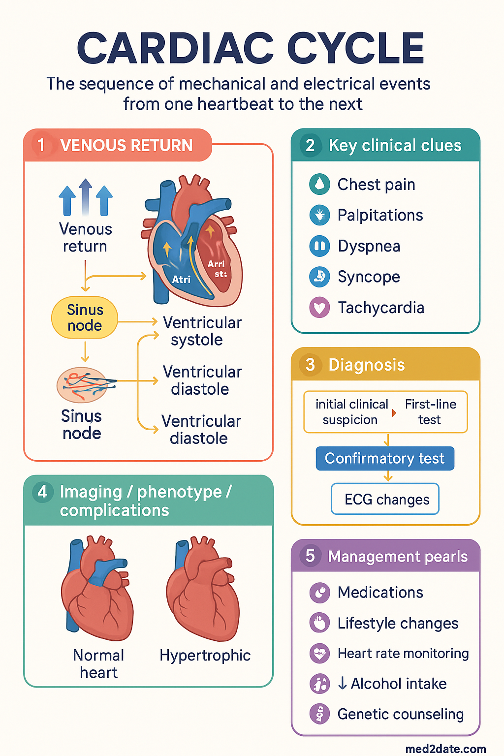
The cardiac cycle describes the sequence of mechanical and electrical events that occur from the onset of one heartbeat to the next. Mastery of the cardiac cycle — including its pressure–volume relationships, valve timing, and auscultatory findings — is fundamental to haemodynamic assessment in both primary care and specialist settings across Australia.
Haemodynamic monitoring is central to the management of heart failure, valvular heart disease, and cardiogenic shock. Approximately 480,000 Australians live with heart failure (AIHW, 2023), and cardiac valvular disease affects an estimated 3–5% of adults over 65 years. Rheumatic heart disease (RHD) remains a significant burden in Aboriginal and Torres Strait Islander communities, with notification rates exceeding 100 per 100,000 in the Northern Territory compared with <2 per 100,000 in non-Indigenous populations (RHDAustralia, 2023).
This guideline provides a comprehensive overview of the cardiac cycle, pressure–volume loop physiology, valve events and heart sounds, and the haemodynamic derangements encountered in common disease states seen in Australian clinical practice. It is intended for physicians, advanced trainees, general practitioners, and critical care staff involved in cardiac assessment.
Phases of the Cardiac Cycle
The cardiac cycle is conventionally divided into two major periods — systole (contraction and ejection) and diastole (relaxation and filling). At a resting heart rate of 70 bpm, systole occupies approximately 0.3 seconds and diastole approximately 0.55 seconds. As heart rate increases, diastole shortens disproportionately, limiting ventricular filling time.
Systolic Phases
| Phase | Duration (70 bpm) | Key Events | Valve Status |
|---|---|---|---|
| Isovolumetric contraction | ~50 ms | Ventricles contract with all valves closed; pressure rises rapidly without volume change | AV valves closed, semilunar valves closed |
| Rapid ejection | ~100 ms | Semilunar valves open; ~70% of SV ejected; ventricular pressure peaks | Semilunar valves open |
| Reduced ejection | ~150 ms | Ventricular pressure falls below arterial pressure; remaining ~30% of SV ejected; coronary perfusion occurs | Semilunar valves closing |
Diastolic Phases
| Phase | Duration (70 bpm) | Key Events | Valve Status |
|---|---|---|---|
| Isovolumetric relaxation | ~60 ms | Ventricles relax with all valves closed; pressure falls rapidly at constant volume | All valves closed |
| Rapid ventricular filling | ~200 ms | AV valves open; ~70–80% of ventricular filling occurs passively; S3 may be normal in young adults | AV valves open |
| Diastasis (reduced filling) | Variable | Atrial and ventricular pressures equalise; minimal flow; shortens at high heart rates | AV valves open |
| Atrial contraction (a-wave) | ~100 ms | Atrial systole contributes ~20–25% of ventricular filling ("atrial kick"); loss (e.g. AF) reduces SV by ~15–25% | AV valves open, closing at end |
Electrical–Mechanical Correlation
The P wave precedes atrial contraction by ~40–60 ms. The QRS complex precedes ventricular contraction; the interval between QRS onset and the rise in ventricular pressure (pre-ejection period, PEP) is approximately 50–80 ms. The T wave corresponds to ventricular repolarisation and overlaps with isovolumetric relaxation.
QT interval = electrical systole. Normal corrected QT (QTc) is <450 ms (males) and <470 ms (females). Prolonged QTc increases risk of torsades de pointes — relevant to drug safety monitoring in Australian practice (e.g. sotalol, amiodarone, erythromycin).
At higher heart rates, diastole shortens more than systole. At 150 bpm, diastole may be only ~200 ms, limiting coronary perfusion (which occurs primarily in diastole) and ventricular filling — important in tachycardia-mediated cardiomyopathy.
Pressure-Volume Loop
The left ventricular pressure–volume (PV) loop, plotted with LV volume on the x-axis and LV pressure on the y-axis, is the most informative single-graph representation of cardiac mechanics. Each loop represents one cardiac cycle and encodes information about preload, afterload, contractility, and diastolic function.
Loop Corners and Segments
Key Relationships
| Parameter | PV Loop Representation | Clinical Significance |
|---|---|---|
| Stroke volume (SV) | Width of the loop (EDV − ESV) | Normal 60–100 mL; reduced in systolic HF, aortic stenosis |
| Cardiac output (CO) | SV × HR | Normal 4–8 L/min; CI 2.5–4.0 L/min/m² |
| Stroke work | Area enclosed by the loop | External work per beat; oxygen consumption correlate |
| Contractility (ESPVR) | Slope through end-systolic points | Steeper = ↑ inotropy; independent of preload/afterload |
| Diastolic stiffness (EDPVR) | Curve along which filling occurs | Left-shifted/upward = HFpEF, hypertrophy, fibrosis |
| Elastance (Ees) | End-systolic pressure ÷ ESV | Load-independent index of contractility; normal 1.5–3.0 mmHg/mL |
Effects of Loading Conditions
Valve Events & Heart Sounds
Heart sounds are generated by valve closure and, in abnormal states, by turbulent flow through stenotic or regurgitant valves. Careful auscultation — using the bell (low-frequency) and diaphragm (high-frequency) — remains a cornerstone of cardiac examination in Australian general practice and specialist clinics.
Normal Heart Sounds
| Sound | Timing | Mechanism | Best Heard |
|---|---|---|---|
| S1 | Onset of systole | Closure of mitral (M1) and tricuspid (T1) valves; M1 precedes T1 by ~20 ms | Apex (mitral area), left lower sternal border (tricuspid) |
| S2 | Onset of diastole | Closure of aortic (A2) and pulmonic (P2) valves; A2 normally precedes P2 | Aortic area (2nd right ICS), pulmonary area (2nd left ICS) |
Splitting of S2
During inspiration, increased venous return delays P2 closure (by ~30–40 ms) while A2 remains unchanged. Splitting is heard best at the pulmonary area and disappears during expiration. Normal in young adults.
Fixed splitting: No respiratory variation — hallmark of atrial septal defect (ASD). Wide splitting: Delayed P2 — RBBB, pulmonary stenosis. Paradoxical splitting: A2 delayed after P2 — LBBB, severe aortic stenosis; splits during expiration, disappears during inspiration.
Extra Heart Sounds
Murmurs — Timing and Significance
| Murmur | Timing | Character | Causes |
|---|---|---|---|
| Aortic stenosis | Systolic ejection (crescendo–decrescendo) | Harsh, radiates to carotids | Bicuspid AV, degenerative calcification, RHD |
| Mitral regurgitation | Pansystolic (holosystolic) | Blowing, radiates to axilla | MV prolapse, ischaemic MR, RHD, endocarditis |
| Aortic regurgitation | Early diastolic (decrescendo) | High-pitched, blowing; best heard sitting forward, end-expiration | Bicuspid AV, aortic root dilation, endocarditis, RHD |
| Mitral stenosis | Mid-diastolic (low-pitched rumble) | Opening snap → rumble; best at apex, left lateral decubitus | RHD (most common cause in Australia, especially in ATSI populations) |
| Flow murmur | Systolic ejection | Soft, no radiation; disappears with Valsalva | Anaemia, pregnancy, fever, thyrotoxicosis — benign |
Clicks and Other Sounds
Ejection click: Early systolic, high-pitched; heard in bicuspid aortic valve, pulmonary stenosis. Mid-systolic click: Pathognomonic of mitral valve prolapse (MVP); timing varies with manoeuvres (earlier with standing/ Valsalva, later with squatting). Opening snap: Early diastolic sound in mitral stenosis due to rigid valve leaflets snapping open; shorter A2–OS interval indicates more severe stenosis.
Haemodynamic Changes in Disease States
Disease states profoundly alter the morphology of the cardiac cycle and PV loop. Understanding these alterations enables targeted therapy and haemodynamic optimisation.
Heart Failure with Reduced Ejection Fraction (HFrEF)
- PV loop shifts rightward (increased ESV and EDV) and narrows (reduced SV).
- ESPVR slope flattens — reduced contractility.
- LVEDP elevated (>16 mmHg); PCWP often >18 mmHg.
- Cardiac index reduced (<2.2 L/min/m²).
- S3 gallop common; displaced apex beat.
- Australian guideline-directed medical therapy (GDMT): ACE inhibitor or ARNI (sacubitril/valsartan — Entresto®, PBS Authority Required), beta-blocker (bisoprolol, carvedilol, metoprolol succinate), mineralocorticoid receptor antagonist (spironolactone or eplerenone), SGLT2 inhibitor (dapagliflozin — Forxiga®, PBS Authority Required for HFrEF).
Heart Failure with Preserved Ejection Fraction (HFpEF)
- EDPVR shifted leftward and upward — increased passive stiffness.
- Normal or near-normal EF (≥50%) but impaired diastolic filling.
- Elevated LA pressure → pulmonary congestion despite normal systolic function.
- S4 gallop common; left atrial enlargement on echocardiography.
- Dapagliflozin (Forxiga®) now PBS-listed for HFpEF (EF >40%); evidence from DELIVER trial.
- Management: diuretics for congestion, blood pressure control, weight loss, management of comorbidities (AF, obesity, diabetes).
- Narrowed PV loop (reduced SV due to fixed obstruction).
- Prolonged isovolumetric contraction phase; slow-rising carotid pulse (pulsus parvus et tardus).
- Concentric LV hypertrophy → elevated LVEDP despite normal or reduced chamber volume.
- Peak gradient >40 mmHg (or velocity >4 m/s) with valve area <1.0 cm² defines severe AS.
- Late-peaking crescendo–decrescendo systolic murmur at right upper sternal border, radiating to carotids.
- Australian management: Surgical aortic valve replacement (SAVR) for low surgical risk; transcatheter aortic valve implantation (TAVI) available at major public and private centres (MBS item 38600 series) for intermediate–high risk patients.
- PV loop shifted rightward — volume-overloaded ventricle (increased EDV).
- Effective forward SV reduced; total SV = forward SV + regurgitant volume.
- Elevated LA pressure and V-wave on pulmonary capillary wedge tracing.
- Dilated LV with eccentric hypertrophy (volume overload pattern).
- Pansystolic murmur at apex radiating to axilla; S3 if acute/severe.
- Primary MR: surgical repair preferred over replacement when feasible (mitral repair, MBS item 38652). Secondary MR: GDMT for HF ± cardiac resynchronisation therapy (CRT).
- Pericardial pressure equilibrates with intracardiac pressures — all four chamber pressures equalise.
- PV loop narrows markedly due to reduced ventricular filling (restricted preload).
- Pulsus paradoxus: systolic BP drop >10 mmHg during inspiration (exaggerated ventricular interdependence).
- Elevated JVP, muffled heart sounds, tachycardia — Beck's triad.
- Emergency pericardiocentesis (MBS item 38400) — often ultrasound-guided in Australian centres.
Aortic Stenosis
Mitral Regurgitation
Cardiac Tamponade
Restrictive Cardiomyopathy vs. Constrictive Pericarditis
Both conditions produce diastolic dysfunction with elevated filling pressures. Key differentiating haemodynamic features:
| Feature | Restrictive CM | Constrictive Pericarditis |
|---|---|---|
| LV/RV pressure dissociation | Present | Absent (ventricles move in parallel) |
| Ventricular interdependence | Minimal | Marked (enhanced with inspiration) |
| Pericardial thickness | Normal | Increased (CT/MRI findings) |
| Square root sign | May be present | Characteristic ("dip-and-plateau") |
| Treatment | Diuretics, treat underlying cause (amyloid, sarcoid) | Pericardiectomy (MBS item 38450) |
Special Populations
Aboriginal and Torres Strait Islander Health Considerations
📚 References
- 1. Klabunde RE. Cardiovascular Physiology Concepts. 3rd ed. Philadelphia: Wolters Kluwer; 2021.
- 2. Opie LH, Gersh BJ. Heart Physiology: From Cell to Circulation. 5th ed. Philadelphia: Lippincott Williams & Wilkins; 2023.
- 3. NHFA CSANZ. Guidelines for the prevention, detection, and management of heart failure in Australia 2018. National Heart Foundation of Australia and Cardiac Society of Australia and New Zealand. Heart Lung Circ. 2018;27(10):1123–1208.
- 4. RHDAustralia (ARF/RHD writing group). The 2020 Australian guideline for prevention, diagnosis, and management of acute rheumatic fever and rheumatic heart disease. 3rd ed. Menzies School of Health Research; 2020.
- 5. Australian Institute of Health and Welfare (AIHW). Heart, stroke, and vascular diseases — Australian facts 2023. Cat. no. CVD 87. Canberra: AIHW; 2023.
- 6. Baumgartner H, et al. 2017 ESC/EACTS Guidelines for the management of valvular heart disease. Eur Heart J. 2017;38(36):2739–2791.
- 7. McDonagh TA, et al. 2021 ESC Guidelines for the diagnosis and treatment of acute and chronic heart failure. Eur Heart J. 2021;42(36):3599–3726.
- 8. Burkhoff D, Mirsky I, Suga H. Assessment of systolic and diastolic ventricular properties via pressure-volume analysis: a guide for clinical, translational, and basic researchers. Am J Physiol Heart Circ Physiol. 2005;289(2):H501–H512.
- 9. Nishimura RA, Otto CM, Bonow RO, et al. 2014 AHA/ACC Guideline for the management of patients with valvular heart disease. J Am Coll Cardiol. 2014;63(22):e57–e185.
- 10. McMurray JJV, et al. Dapagliflozin in patients with heart failure and reduced ejection fraction. N Engl J Med. 2019;381(21):1995–2008 (DAPA-HF trial).
- 11. Solomon SD, et al. Dapagliflozin in heart failure with mildly reduced or preserved ejection fraction. N Engl J Med. 2022;387(12):1089–1098 (DELIVER trial).
- 12. Australian Government Department of Health. Pharmaceutical Benefits Schedule (PBS). Available at: pbs.gov.au. Accessed 2024.
- 13. Services for Australian Rural and Remote Allied Health (SARRAH). Access to cardiology services in rural and remote Australia. Position paper. 2022.
- 14. Australian Commission on Safety and Quality in Health Care (ACSQHC). National Safety and Quality Health Service Standards. 2nd ed. Sydney: ACSQHC; 2021.