📋 Key Information Summary
- Cardiac myocytes exhibit four intrinsic electrophysiological properties — automaticity, excitability (conductivity in some texts), conductivity, and contractility — which together sustain rhythmic, coordinated ventricular contraction.
- The ventricular action potential has five phases (0–4); Phase 4 in working myocytes is normally stable, while pacemaker cells exhibit spontaneous diastolic depolarisation driven by the funny current (If).
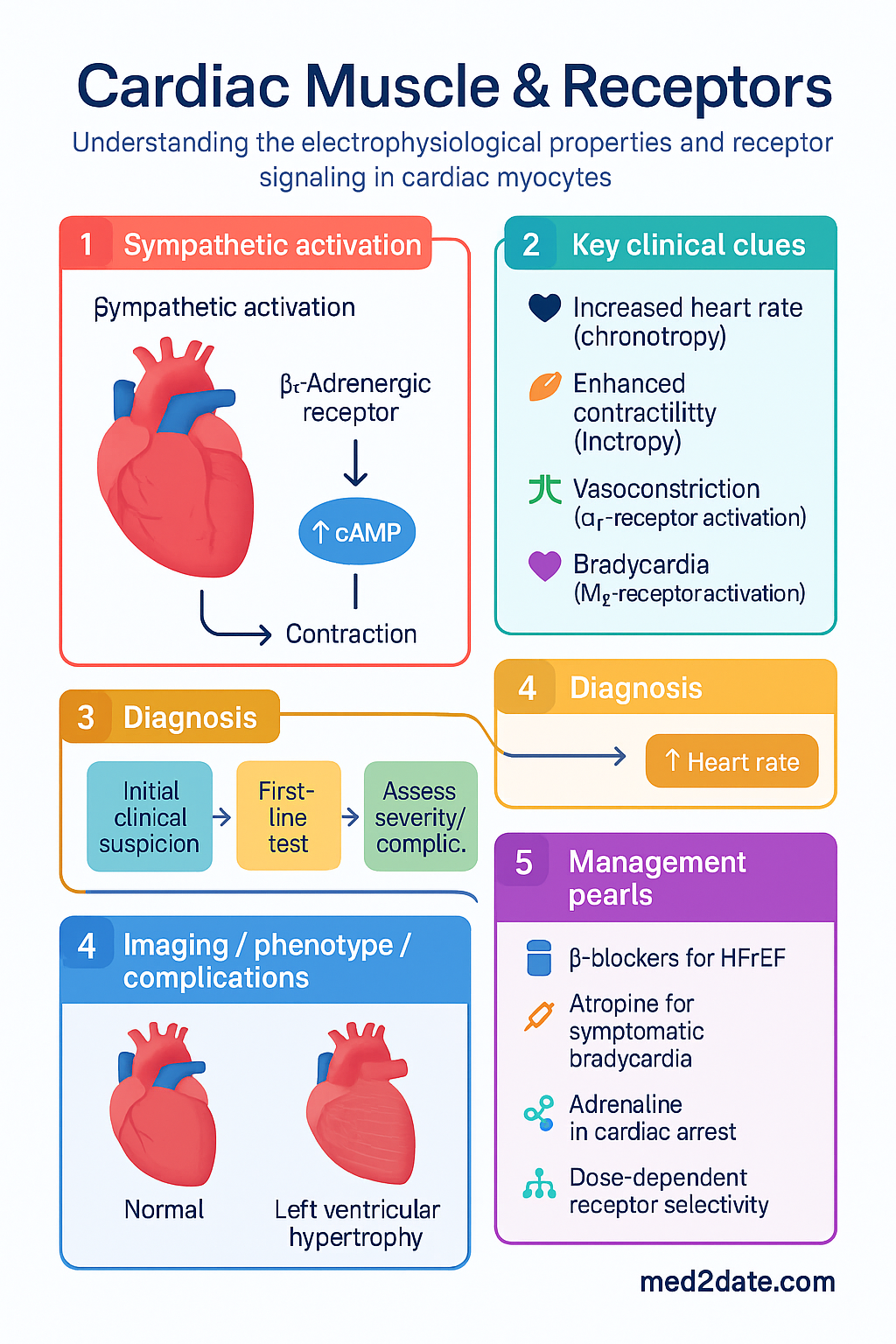
- Sympathetic activation via β₁-adrenergic receptors increases heart rate (chronotropy), contractility (inotropy), conduction velocity (dromotropy), and relaxation rate (lusitropy) through Gs–cAMP–PKA signalling.
- β₂-adrenergic receptors are present in human myocardium but at lower density than β₁ (ratio ≈ 70:30); they mediate vasodilatation, bronchodilatation, and contribute to inotropy at high catecholamine concentrations.
- α₁-adrenergic receptors activate Gq–PLC–IP₃/DAG signalling causing vasoconstriction, modest positive inotropy (especially in failing myocardium), and myocardial hypertrophy when chronically stimulated.
- Parasympathetic (vagal) influence acts via M₂ muscarinic receptors coupled to Gi, reducing cAMP and activating IKACh to produce bradycardia, reduced atrial contractility, and attenuated AV-nodal conduction.
- β-blockers (metoprolol, bisoprolol, carvedilol) reduce mortality in heart failure with reduced ejection fraction (HFrEF) by reversing chronic β₁-receptor downregulation and desensitisation.
- Atropine (M₂ antagonist) is first-line for symptomatic bradycardia; adrenaline (mixed adrenergic agonist) is first-line in cardiac arrest per Australian Resuscitation Council (ARC) guidelines.
- Pharmacological receptor selectivity is dose-dependent — high-dose adrenaline activates α₁, β₁, and β₂ receptors, whereas low-dose infusion is predominantly β-mediated.
- Understanding receptor physiology is essential for rational use of inotropes, vasopressors, chronotropes, and antiarrhythmics in Australian emergency and critical-care settings.
- Chronic sympathetic overdrive leads to β₁-receptor downregulation and G-protein uncoupling — the rationale for initiating β-blockers at low dose in HFrEF and up-titrating slowly over weeks.
- Aboriginal and Torres Strait Islander Australians experience cardiovascular disease at 1.7× the rate of non-Indigenous Australians; receptor-targeted therapies must be accessible in rural and remote settings.
Introduction & Australian Epidemiology
Cardiac muscle possesses four fundamental electrophysiological properties that distinguish it from skeletal and smooth muscle: automaticity (spontaneous impulse generation by pacemaker cells), excitability (the ability to respond to a stimulus), conductivity (propagation of the action potential through the syncytium), and contractility (force generation in response to depolarisation). The interplay of these properties, modulated by the autonomic nervous system through adrenergic and muscarinic receptors, underpins virtually every pharmacological intervention used in contemporary Australian cardiology practice.
Cardiovascular disease remains the leading cause of death in Australia, accounting for approximately 26% of all deaths in 2022 (Australian Bureau of Statistics). Heart failure alone affects an estimated 480 000 Australians, with prevalence rising sharply after age 65 years (AIHW, 2023). Ischaemic heart disease, hypertensive heart disease, and cardiomyopathies all converge on alterations in cardiac muscle electrophysiology and receptor signalling, making a thorough understanding of these systems essential for evidence-based prescribing.
This guideline reviews cardiac muscle physiology, the major adrenergic receptor subtypes (α₁, β₁, β₂), muscarinic receptor (M₂) autonomic control, and the pharmacological implications relevant to Australian primary-care and specialist practice. All drug references align with the Pharmaceutical Benefits Scheme (PBS) and current Australian therapeutic standards.
Cardiac Muscle Physiology (Action Potential)
The cardiac action potential differs markedly between pacemaker cells (SA node, AV node) and working ventricular myocytes. Understanding both is essential because the receptor-mediated modulation of each phase determines the clinical effect of adrenergic and muscarinic agonists and antagonists.
Ventricular Myocyte Action Potential (Fast-Response)
| Phase | Name | Ionic Basis | Receptor Modulation |
|---|---|---|---|
| Phase 0 | Rapid depolarisation | Na⁺ influx via voltage-gated INa channels | β₁-agonism ↑ Na⁺ channel availability; Class I antiarrhythmics block INa |
| Phase 1 | Early repolarisation | Transient K⁺ efflux (Ito) | Modest adrenergic modulation of Ito |
| Phase 2 | Plateau | Balance between Ca²⁺ influx (ICaL) and K⁺ efflux | β₁ ↑ ICaL via PKA phosphorylation → ↑ contractility; β-blockers attenuate this |
| Phase 3 | Repolarisation | K⁺ efflux via IKr and IKs | β₁ ↑ IKs → shortened APD at high HR; M₂ ↑ IKACh in atria |
| Phase 4 | Resting membrane potential | K⁺ equilibrium (≈ −90 mV); Na⁺/K⁺-ATPase | Stable in working myocytes; β₁ increases Ca²⁺ leak → arrhythmia risk |
Pacemaker Cell Action Potential (Slow-Response, SA Node)
Pacemaker cells lack fast Na⁺ channels. Their action potential depends on:
- Phase 4 — Spontaneous diastolic depolarisation: Driven primarily by the funny current (If, a mixed Na⁺/K⁺ current activated by hyperpolarisation), T-type Ca²⁺ channels, and decay of outward K⁺ current. This is the basis of automaticity.
- Phase 0 — Upstroke: Ca²⁺ influx through L-type (ICaL) and T-type (ICaT) calcium channels — slower and of lower amplitude than ventricular Phase 0.
- Phase 3 — Repolarisation: K⁺ efflux via IK channels.
Excitation–Contraction Coupling
Depolarisation opens voltage-gated L-type Ca²⁺ channels (Phase 2), admitting a small Ca²⁺ influx that triggers a much larger release of Ca²⁺ from the sarcoplasmic reticulum (SR) via ryanodine receptors (RyR2) — a process termed calcium-induced calcium release (CICR). Cytoplasmic Ca²⁺ binds troponin C, displacing tropomyosin and permitting actin–myosin cross-bridge cycling. β₁-adrenergic stimulation enhances this cascade at multiple points:
- ↑ ICaL → more trigger Ca²⁺
- Phosphorylation of phospholamban → disinhibition of SERCA2a → faster SR Ca²⁺ reuptake → positive lusitropy (enhanced relaxation)
- Phosphorylation of RyR2 → increased SR Ca²⁺ release sensitivity
- Phosphorylation of troponin I → reduced myofilament Ca²⁺ sensitivity → faster cross-bridge detachment
Adrenergic Receptors (Alpha, Beta-1, Beta-2)
Adrenergic receptors are G-protein-coupled receptors (GPCRs) activated by endogenous catecholamines (noradrenaline, adrenaline, dopamine) and by exogenous sympathomimetic drugs. Three principal subtypes are clinically relevant in cardiovascular medicine.
| Receptor | G-Protein | Second Messenger | Primary Cardiac / Vascular Effects | Endogenous Affinity |
|---|---|---|---|---|
| α₁ | Gq | ↑ IP₃ / DAG → ↑ intracellular Ca²⁺ | Vasoconstriction (arteriolar smooth muscle); modest positive inotropy; myocardial hypertrophy with chronic stimulation | Adrenaline ≥ Noradrenaline |
| β₁ | Gs | ↑ cAMP → ↑ PKA | ↑ Heart rate (chronotropy); ↑ contractility (inotropy); ↑ conduction velocity (dromotropy); ↑ relaxation rate (lusitropy); ↑ renin release | Noradrenaline ≈ Adrenaline |
| β₂ | Gs (also Gi at high conc.) | ↑ cAMP; vasodilatation, bronchodilatation | Vascular smooth-muscle relaxation; bronchodilatation; hepatic glycogenolysis; mild positive inotropy in ventricle; hypokalaemia (skeletal muscle K⁺ uptake) | Adrenaline >> Noradrenaline |
β₁-Adrenergic Receptors — The Dominant Cardiac Receptor
Approximately 70–80% of β-receptors in the healthy human ventricle are β₁-subtype, localised predominantly at the T-tubule junction near L-type Ca²⁺ channels and at the Z-line adjacent to RyR2 clusters. This spatial coupling ensures that cAMP/PKA signalling is delivered precisely to excitation–contraction coupling machinery.
In chronic heart failure, persistent sympathetic activation (plasma noradrenaline is typically elevated 2–3-fold) leads to β₁-receptor downregulation (reduced receptor density) and desensitisation (GRK-mediated phosphorylation → β-arrestin recruitment → uncoupling from Gs). This is the pathophysiological rationale for β-blocker therapy in HFrEF — by reducing chronic catecholamine drive, β-blockers permit receptor density to recover, restoring physiological responsiveness.
β₂-Adrenergic Receptors
Although comprising only ~20–30% of cardiac β-receptors, β₂-subtypes gain functional importance in heart failure (where β₁ is downregulated) and during high-dose catecholamine infusion. β₂ receptors couple to both Gs and Gi; the latter can activate cardioprotective pathways (PI3K–Akt) but also promote arrhythmogenesis via spatially restricted cAMP microdomains. In clinical practice, β₂ effects are exploited therapeutically by salbutamol (bronchodilatation) and must be recognised as a cause of tachycardia and hypokalaemia.
α₁-Adrenergic Receptors
α₁-receptors are the dominant vasoconstrictor adrenergic receptors in arteriolar smooth muscle, mediating the pressor response to noradrenaline and phenylephrine. In the myocardium, α₁-receptors (predominantly α₁A and α₁B subtypes) couple to Gq–PLC, generating IP₃ (sarcoplasmic reticulum Ca²⁺ release) and DAG (PKC activation). The net effect is a modest positive inotropic response that is independent of cAMP and therefore additive to β₁-mediated inotropy. Chronic α₁-stimulation activates foetal gene programmes (ANP, β-MHC) contributing to pathological hypertrophy.
Receptor Selectivity Is Dose-Dependent
A critical pharmacological principle is that receptor selectivity diminishes at high doses:
- Adrenaline: Low dose (0.01–0.03 µg/kg/min) → predominantly β₁/β₂ (↑ HR, ↑ CO, vasodilatation). High dose (>0.15 µg/kg/min) → α₁ predominates (vasoconstriction, ↑ SVR).
- Noradrenaline: Predominantly α₁ and β₁ across all doses; minimal β₂ activity — hence causes vasoconstriction without significant bronchodilatation.
- Dobutamine: Predominantly β₁ with some β₂ and α₁; net effect is ↑ contractility with modest ↓ SVR.
Muscarinic Receptors & Autonomic Control
The parasympathetic nervous system modulates cardiac function primarily through acetylcholine (ACh) acting on M₂ muscarinic receptors located on SA-node pacemaker cells, atrial myocytes, and AV-nodal conduction tissue. The ventricles have sparse vagal innervation, so parasympathetic effects on ventricular contractility are indirect (mediated via sympathetic withdrawal and presynaptic inhibition of noradrenaline release).
M₂ Receptor Signalling Cascade
- G-protein coupling: Gi (inhibitory) — directly inhibits adenylyl cyclase → ↓ cAMP → ↓ PKA activity.
- Gβγ subunit: Opens IKACh (G-protein-gated inward-rectifier K⁺ channels) → membrane hyperpolarisation → slowed Phase 4 diastolic depolarisation → negative chronotropy.
- AV node: ↓ cAMP and ↑ IKACh slow conduction velocity and prolong refractory period → negative dromotropy (slowed AV conduction, risk of AV block at high vagal tone).
- Atria: ↓ ICaL and ↑ IKACh reduce atrial contractility and shorten atrial action potential duration.
- Presynaptic M₂/M₄: Inhibit noradrenaline release from sympathetic nerve terminals — a key mechanism of vagal–sympathetic interaction.
Vagal Tone and Heart Rate Variability
Resting heart rate in healthy adults (60–80 bpm) reflects tonic vagal dominance over sympathetic drive — a phenomenon termed vagal tone. High vagal tone (e.g., in trained athletes) produces resting bradycardia. Loss of vagal tone — as in autonomic neuropathy (diabetes), post-cardiac transplant, or with anticholinergic drugs — results in an elevated resting heart rate with reduced heart rate variability (HRV), an independent predictor of cardiovascular mortality.
Other Muscarinic Subtypes (Brief Overview)
| Subtype | Location | Cardiovascular Relevance |
|---|---|---|
| M₁ | Autonomic ganglia, CNS | Minimal direct cardiac role; relevant to cognitive side-effects of anticholinergics |
| M₂ | SA node, AV node, atria | Primary cardiac parasympathetic receptor — negative chronotropy, dromotropy |
| M₃ | Vascular endothelium, smooth muscle | Endothelial M₃ → NO release → vasodilatation; smooth muscle M₃ → vasoconstriction |
| M₄, M₅ | CNS | No direct cardiovascular role |
Pharmacological Implications
Knowledge of receptor subtypes, G-protein coupling, and downstream signalling directly informs prescribing decisions across the spectrum of cardiovascular medicine. Below are clinically important drug classes mapped to their receptor targets.
Sympathomimetics (Adrenergic Agonists)
β-Blockers (Adrenergic Antagonists)
Anticholinergics (Muscarinic Antagonists)
Receptor-Targeted Therapy Quick Reference
Investigations
While receptor physiology is primarily an applied-pharmacology topic, several investigations are relevant when assessing autonomic function or titrating receptor-targeted therapies.
Special Populations
Aboriginal and Torres Strait Islander Health Considerations
Aboriginal and Torres Strait Islander peoples experience cardiovascular disease at approximately 1.7 times the rate of non-Indigenous Australians, with premature onset (10–20 years earlier) and higher case-fatality rates (AIHW, 2023). Rheumatic heart disease (RHD) remains a significant burden, particularly in Northern Australia, and directly impacts cardiac muscle function and receptor-mediated physiology through chronic valvular pathology and myocardial remodelling.
📚 References
- 1. Australian Institute of Health and Welfare (AIHW). Heart, stroke and vascular disease — Australian facts 2023. Canberra: AIHW; 2023.
- 2. McMurray JJV, Packer M, Desai AS, et al. Angiotensin–neprilysin inhibition versus enalapril in heart failure. N Engl J Med. 2014;371(11):993–1004.
- 3. MERIT-HF Study Group. Effect of metoprolol CR/XL in chronic heart failure: Metoprolol CR/XL Randomised Intervention Trial in Congestive Heart Failure (MERIT-HF). Lancet. 1999;353(9169):2001–2007.
- 4. Packer M, Coats AJS, Fowler MB, et al. Effect of carvedilol on survival in severe chronic heart failure. N Engl J Med. 2001;344(22):1651–1658.
- 5. Australian Resuscitation Council (ARC). Guideline 11.2 – Protocols for adult advanced life support. ARC; 2024. Available from: https://resus.org.au
- 6. Brodde O-E, Michel MC. Adrenergic and muscarinic receptors in the human heart. Pharmacol Rev. 1999;51(4):651–690.
- 7. Bristow MR, Ginsburg R, Umans V, et al. β₁- and β₂-adrenergic-receptor subpopulations in nonfailing and failing human ventricular myocardium: coupling of both receptor subtypes to muscle contraction and selective β₁-receptor downregulation in heart failure. Circ Res. 1986;59(3):297–309.
- 8. National Heart Foundation of Australia and Cardiac Society of Australia and New Zealand. Guidelines for the prevention, detection, and management of heart failure in Australia 2018. Updated 2024.
- 9. RHDAustralia (a program of Menzies School of Health Research). The 2020 Australian guideline for prevention, diagnosis, and management of acute rheumatic fever and rheumatic heart disease. 3rd ed. Darwin: RHDAustralia; 2020.
- 10. Rhee EP, Berk BC. Adrenergic receptors and the failing heart. In: Heart Failure: A Companion to Braunwald's Heart Disease. 4th ed. Elsevier; 2020:141–157.
- 11. Australian Commission on Safety and Quality in Health Care (ACSQHC). National Safety and Quality Health Service Standards. 2nd ed. Sydney: ACSQHC; 2021.
- 12. Pharmaceutical Benefits Scheme (PBS). Australian Government Department of Health and Aged Care. Available from: https://www.pbs.gov.au. Accessed 2024.