Home Endocrinology Vitamin D Deficiency & Osteomalacia

Vitamin D Deficiency & Osteomalacia

📋 Key Information Summary

📋
  • Vitamin D deficiency (25-OH vitamin D <50 nmol/L) is extremely common in Australia, affecting approximately 23–31% of adults, with higher prevalence in southern states, veiled populations, the elderly, and Aboriginal and Torres Strait Islander peoples.
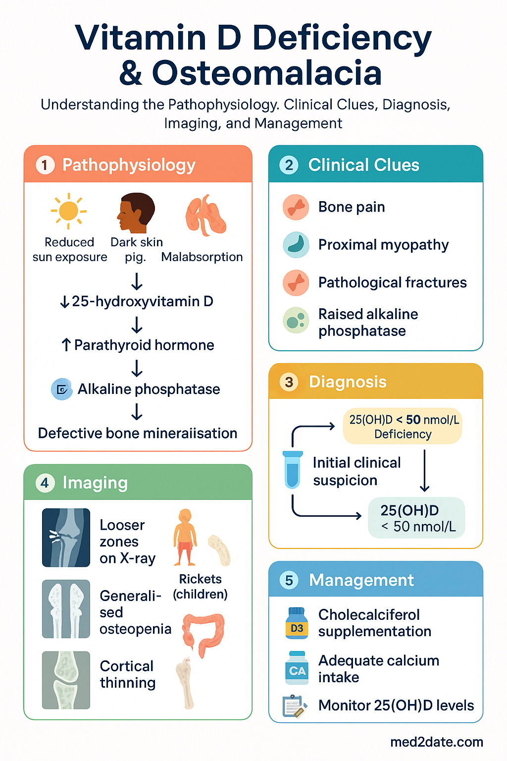
  • Osteomalacia results from severe, prolonged vitamin D deficiency causing defective bone mineralisation — characterised by bone pain, proximal myopathy, pathological fractures, and raised alkaline phosphatase (ALP).
  • Rickets is the paediatric equivalent, with growth-plate involvement, skeletal deformities (bowed legs, rachitic rosary, frontal bossing), and delayed milestones.
  • 25-hydroxyvitamin D [25(OH)D] is the recommended screening test; levels <50 nmol/L define deficiency, 50–74 nmol/L insufficiency, and ≥75 nmol/L adequacy.
  • Causes include reduced sun exposure (indoor lifestyle, veiling), dark skin pigmentation, malabsorption (coeliac disease, bariatric surgery), chronic kidney/hepatic disease, anticonvulsants, and obesity.
  • Dietary sources alone are insufficient; most Australians require sunlight exposure and/or supplementation to maintain adequate levels.
  • First-line supplementation: cholecalciferol (vitamin D₃) — loading dose followed by maintenance; ergocalciferol (D₂) is a less effective alternative.
  • Calcitriol is reserved for patients with severe renal impairment or hepatic failure who cannot hydroxylate cholecalciferol.
  • Concurrent calcium intake must be assessed and corrected (aim ≥1000–1200 mg/day in adults with osteomalacia).
  • Monitoring: repeat 25(OH)D at 3–6 months after initiation; ALP normalises over 6–12 months with adequate treatment.
  • Aboriginal and Torres Strait Islander populations have significantly higher prevalence of deficiency; culturally safe supplementation programmes and dietary education are essential.
  • Hypervitaminosis D (>250 nmol/L) can cause hypercalcaemia — avoid empiric mega-dosing without monitoring.

🎧 Audio Brief

Why sunny Australia lacks Vitamin D

A short clinical audio briefing generated from this article — perfect for the commute or ward round.

Introduction & Australian Epidemiology

Vitamin D deficiency is one of the most prevalent nutritional deficiencies worldwide and is remarkably common in sun-rich Australia. It causes defective bone mineralisation — osteomalacia in adults and rickets in children — and is independently associated with musculoskeletal weakness, falls, fractures, and impaired quality of life. Emerging evidence also links low vitamin D status to adverse cardiovascular, immune, and metabolic outcomes, though causality remains under investigation.

Australia's paradoxically high prevalence is driven by sun-safe messaging (slip, slop, slap), indoor working environments, multicultural demographics with darker skin pigmentation and cultural veiling, an ageing population, and high rates of obesity. The Australian Bureau of Statistics National Health Measures Survey (2011–12) found that 23% of adults were vitamin D-deficient (25-OH D <50 nmol/L), rising to 31% during winter–spring. Prevalence is highest in Victoria, Tasmania, and the ACT, and among Aboriginal and Torres Strait Islander peoples, where rates exceed 50–60% in some remote communities.

Despite increasing public and clinical awareness, osteomalacia remains underdiagnosed in Australian primary care, often presenting late with non-specific musculoskeletal symptoms, pathological fractures, or incidental biochemical findings. Early recognition and appropriate supplementation can reverse the condition in most patients.

Vitamin D Deficiency & Osteomalacia clinical infographic — pathophysiology, clinical clues, diagnosis, imaging, and management
Tap or click image to enlarge — Vitamin D Deficiency & Osteomalacia: pathophysiology, clinical clues, diagnosis, imaging, and management.
Vitamin D Deficiency & Osteomalacia infographic, full size

Vitamin D Metabolism & Causes of Deficiency

Physiology

Vitamin D₃ (cholecalciferol) is synthesised in the skin from 7-dehydrocholesterol upon exposure to ultraviolet B radiation (wavelength 290–315 nm). It can also be obtained from dietary sources (oily fish, fortified foods, egg yolks, liver). Vitamin D₂ (ergocalciferol) is derived from plant sterols and fungi. Both forms undergo hepatic 25-hydroxylation (CYP2R1, CYP27A1) to produce 25-hydroxyvitamin D [25(OH)D], the major circulating form measured clinically. Subsequent renal 1α-hydroxylation (CYP27B1), regulated by parathyroid hormone (PTH), calcium, and phosphate, produces the active hormone 1,25-dihydroxyvitamin D [1,25(OH)₂D or calcitriol]. Calcitriol promotes intestinal calcium and phosphate absorption, bone mineralisation, and modulates immune function.

Causes of Deficiency

Category Examples Mechanism
Reduced synthesis Indoor lifestyle, institutionalised elderly, cultural veiling/burqa, high-latitude residence (southern Australia/Tasmania), winter season Inadequate UVB exposure
Skin pigmentation Darkly pigmented skin (Aboriginal, African, South Asian, Middle Eastern backgrounds) Melanin absorbs UVB, reducing synthesis by up to 90%
Malabsorption Coeliac disease, inflammatory bowel disease, bariatric surgery, cholestatic liver disease, cystic fibrosis, pancreatic insufficiency Fat-soluble vitamin malabsorption; reduced bile salts
Impaired metabolism Chronic kidney disease (CKD stages 3–5), hepatic failure Reduced 1α-hydroxylation
Increased catabolism Anticonvulsants (phenytoin, carbamazepine, phenobarbitone), rifampicin, glucocorticoids Induction of CYP enzymes (CYP3A4) accelerating 25(OH)D degradation
Obesity BMI ≥30 kg/m² Vitamin D sequestration in adipose tissue; volumetric dilution
Inadequate intake Strict veganism, exclusive breastfeeding (without supplementation), anorexia nervosa Low dietary vitamin D; breast milk contains only ~25 IU/L
⚠️
Clinical pearl: Patients on anticonvulsants, rifampicin, or long-term corticosteroids should have 25(OH)D checked annually and supplementation considered proactively, as drug-induced catabolism can produce severe deficiency despite adequate intake.

Clinical Features

Osteomalacia (Adults)

Osteomalacia results from failure of osteoid mineralisation. Onset is typically insidious.

  • Bone pain: Diffuse, deep, aching — most commonly affecting the pelvis, lumbar spine, ribs, and lower limbs. Often worse with weight-bearing and palpation of bone (tibial tenderness is a classic sign).
  • Proximal myopathy: Difficulty rising from a chair, climbing stairs, or walking. Waddling gait pattern. Reflects both vitamin D receptor effects on skeletal muscle and hypophosphataemia.
  • Fatigue and weakness: Non-specific but common; may be the presenting complaint.
  • Pathological fractures: Insufficiency fractures, particularly of the femoral neck, pubic rami, and vertebrae (pseudofractures/Looser zones on X-ray are pathognomonic).
  • Tetany and paraesthesiae: In severe cases with hypocalcaemia — Chvostek's and Trousseau's signs may be positive.
  • Waddling gait: Due to combined proximal myopathy and pelvic bone pain.

Rickets (Children)

  • Skeletal deformities: Bowed legs (genu varum) or knock knees (genu valgum), frontal bossing, craniotabes (soft occipital skull), rachitic rosary (costochondral beading).
  • Delayed milestones: Late walking, fontanelle closure delay.
  • Growth retardation: Short stature, widened wrists and ankles.
  • Dental abnormalities: Enamel hypoplasia, delayed eruption.
  • Hypocalcaemic seizures: May be the presenting feature, especially in infants of dark-skinned mothers with low vitamin D status.
🚨
Red flag: Infant hypocalcaemic seizure in a breastfed baby of a veiled or darkly pigmented mother should prompt urgent assessment for vitamin D deficiency and rickets. This is a potentially preventable cause of morbidity.

Investigations

Biochemical Investigations

Essential
Serum 25-hydroxyvitamin D [25(OH)D]
The gold-standard screening test. Reflects total vitamin D stores (cutaneous + dietary). MBS item 66830 (assay of 25-hydroxyvitamin D). Interpretation: <25 nmol/L — severe deficiency; 25–49 nmol/L — deficiency; 50–74 nmol/L — insufficiency; ≥75 nmol/L — sufficient.
Essential
Serum alkaline phosphatase (ALP)
Markedly elevated in osteomalacia (often >2× ULN) due to increased osteoblast activity attempting to mineralise unmineralised osteoid. Bone-specific ALP or GGT can help confirm a skeletal origin. MBS item 66500.
Available
Serum calcium (corrected for albumin)
May be low or low-normal in osteomalacia. Hypocalcaemia indicates severe, long-standing deficiency. MBS item 66500.
Available
Serum phosphate
Low or low-normal. Combined with elevated ALP and low/normal calcium, the triad is highly suggestive. MBS item 66500.
Available
Parathyroid hormone (PTH)
Secondary hyperparathyroidism is common in vitamin D deficiency — PTH elevated as a compensatory response to maintain serum calcium. Useful to distinguish from primary hyperparathyroidism. MBS item 66837.
Available
Renal function (eGFR) + urine calcium:creatinine ratio
To assess renal contribution and monitor for hypercalciuria during supplementation. MBS items 66500, 66515.
Referral
1,25-dihydroxyvitamin D [1,25(OH)₂D]
Not routinely needed. May be useful in suspected vitamin D-dependent rickets types I and II or CKD. Often normal or low in deficiency. Request via specialist referral.

Imaging

Modality Findings in Osteomalacia/Rickets MBS Item
X-ray (long bones) Looser zones (pseudofractures) — radiolucent lines perpendicular to cortex, typically at pubic rami, femoral neck, scapula, ribs. Generalised osteopenia, cortical thinning. In rickets: widened, frayed metaphyses, cupped growth plates. 57508 (limb), 57708 (pelvis)
DXA scan Low bone mineral density (T-score ≤ −1.0) — often significantly reduced. Repeat after 12–24 months of treatment to document recovery. Not diagnostic of osteomalacia per se. 12320
Bone biopsy (gold standard) Increased osteoid volume, wide osteoid seams (>12.5 µm), reduced mineralisation rate on tetracycline labelling. Rarely performed — diagnosis is usually clinical + biochemical. Specialist only
ℹ️
MBS note: 25(OH)D testing is covered by Medicare (MBS item 66830) but is subject to RACGP-aligned testing indications — deficiency risk factors (dark skin, veiling, CKD, malabsorption, anticonvulsant use, institutionalisation, obesity) must be documented.

Management (Supplementation)

Treatment Principles

  • Correct vitamin D deficiency with a loading (repletion) dose followed by a long-term maintenance dose.
  • Ensure adequate concurrent calcium intake (diet ± supplements).
  • Treat the underlying cause where possible (e.g., coeliac disease, medication review).
  • Monitor response biochemically (25-OH D, ALP, calcium, PTH) and clinically.

Cholecalciferol (Vitamin D₃) — First-Line

💊
Cholecalciferol
Ostelin® · Various generics · Vitamin D₃
Indication Vitamin D deficiency and osteomalacia — first-line agent
Loading dose (adults) 300,000–500,000 IU orally over 6–10 weeks (e.g., 50,000 IU weekly for 6–10 weeks)
Maintenance (adults) 1,000–2,000 IU daily (or 7,000–14,000 IU weekly)
Paediatric dose Rickets: 3,000–5,000 IU/day (or 150,000 IU stat then monthly) for 3–6 months, then 400–1,000 IU/day maintenance
Route Oral
Obesity adjustment 2–3× standard dose required (BMI ≥30 kg/m²)
Renal adjustment CKD stages 1–4: cholecalciferol adequate; CKD stage 5/dialysis: consider calcitriol
Caution Monitor calcium if on thiazides or calcium supplements. Avoid mega-dose (>500,000 IU stat) — linked to increased falls in elderly.
PBS status ✔ PBS General Benefit Ostelin® OTC available

Ergocalciferol (Vitamin D₂) — Alternative

💊
Ergocalciferol
Ostelin® D₂ (discontinued) · Compounded · Vitamin D₂
Role Alternative if D₃ unavailable or vegan preference; less effective at raising 25(OH)D and shorter half-life
Adult dose 50,000 IU weekly for 8 weeks then monthly maintenance, or 1,000–2,000 IU daily
PBS status ✘ Not PBS listed (compounded)

Calcitriol (1,25-Dihydroxyvitamin D₃) — Specialist Use

💊
Calcitriol
Rocaltrol® · Calcitriol-Teva
Indication CKD stage 5/dialysis, hepatic failure, vitamin D-dependent rickets — patients unable to hydroxylate cholecalciferol
Adult dose 0.25–1.0 µg daily, titrate to calcium/PTH targets
Caution Higher risk of hypercalcaemia than cholecalciferol — requires close monitoring (calcium every 1–2 weeks during titration)
PBS status ✔ PBS Restricted Benefit

Calcium Supplementation

💊
Calcium carbonate
Caltrate® · Calsource®
Adult target intake ≥1,000–1,200 mg elemental calcium/day (diet + supplements) during active osteomalacia treatment
Supplement dose 500–600 mg elemental calcium PO BD (with food for absorption)
Paediatric 500–1,000 mg/day depending on age and dietary intake
Renal adjustment CKD: avoid calcium-based phosphate binders; use sevelamer/calcium acetate under nephrology
PBS status ✔ PBS General Benefit

Monitoring Schedule

Baseline
25(OH)D, corrected calcium, phosphate, ALP, PTH, renal function, urine calcium:creatinine ratio.
4–6 weeks
Serum calcium — to detect hypercalcaemia from rapid repletion, especially if on calcium supplements.
3–6 months
Repeat 25(OH)D — confirm target ≥75 nmol/L. ALP — expect to be falling.
6–12 months
ALP — should normalise in osteomalacia. If persistently elevated, reassess for alternative pathology (Paget's, biliary obstruction, malignancy).
Annually (ongoing)
25(OH)D, calcium. Discontinue supplements if levels >125 nmol/L and adjust.
Treatment response: With adequate supplementation, bone pain improves within weeks, ALP normalises by 6–12 months, and pseudofractures heal radiographically within 3–6 months. DXA improvement is seen at 1–2 years.

Special Populations

🤰 Pregnancy & Breastfeeding
Vitamin D All pregnant women should take ≥600 IU/day (NHMRC). Women at risk of deficiency (dark skin, veiling, limited sun) should take 1,000–2,000 IU/day. Deficiency increases risk of pre-eclampsia, gestational diabetes, low birth weight, and neonatal hypocalcaemia.
Breastfed infants All exclusively breastfed infants require 400 IU/day vitamin D supplementation from birth (RACP/ASCIA). Breast milk alone provides insufficient vitamin D.
👶 Paediatrics
Rickets treatment Cholecalciferol 3,000–5,000 IU/day for 3–6 months (or 150,000 IU stat oral dose followed by monthly doses). Calcium 500–1,000 mg/day. Maintenance 400–1,000 IU/day. Severe hypocalcaemia requires IV calcium gluconate 0.5–1 mL/kg of 10% solution (max 20 mL) over 10–20 min with cardiac monitoring.
Infants <12 months High risk groups: dark skin, veiled mothers, exclusively breastfed, preterm. Consider prophylactic 400 IU/day.
👴 Elderly
Cholecalciferol Higher prevalence of deficiency (reduced skin synthesis, indoor, frailty). Fall prevention: 800–1,000 IU/day + calcium 1,000 mg reduces fracture risk by 15–25%. Avoid mega-dose bolus (>500,000 IU stat) — associated with increased falls (Bischoff-Ferrari 2010).
🩺 Chronic Kidney Disease
CKD 1–4 Cholecalciferol 1,000–2,000 IU/day for deficiency. Treat secondary hyperparathyroidism with cholecalciferol first. Monitor calcium, phosphate, PTH (KDIGO targets).
CKD 5 / dialysis Use calcitriol (Rocaltrol®) 0.25–1.0 µg/day or alfacalcidol under nephrology supervision. Cholecalciferol may still be used as adjunct. Monitor calcium-phosphate product closely.
🫁 Hepatic Impairment
Cholecalciferol Cholecalciferol remains effective in most hepatic disease (25-hydroxylation is preserved except in severe cirrhosis). In severe cholestatic liver disease or cirrhosis with impaired hydroxylation, use calcitriol.
🛡️ Immunocompromised / Malabsorption
Post-bariatric surgery Roux-en-Y and biliopancreatic diversion cause fat malabsorption. Use liquid/water-miscible cholecalciferol. Doses of 3,000–6,000 IU/day may be required. Monitor 25(OH)D, calcium every 3–6 months.
Coeliac disease Strict gluten-free diet may restore absorption. Supplementation until 25(OH)D ≥75 nmol/L, then reassess.

Aboriginal and Torres Strait Islander Health Considerations

Aboriginal and Torres Strait Islander Health
Prevalence
Vitamin D deficiency is significantly more prevalent in Aboriginal and Torres Strait Islander peoples — estimated 50–60% in some remote communities. Higher rates of rickets reported in Northern Territory and Far North Queensland.
Risk factors
Darker skin pigmentation (reduced UVB synthesis), indoor lifestyle shifts, limited dietary diversity in remote communities, higher rates of chronic disease (CKD, diabetes), and socioeconomic barriers to healthcare access.
Nutritional status
Food insecurity in remote communities limits access to vitamin D–rich foods (fortified dairy, oily fish). Traditional diets historically contained adequate vitamin D from bush foods and sun exposure; dietary transition has reduced intake.
Healthcare access
Limited specialist availability in remote and very remote areas. Point-of-care 25(OH)D testing may improve screening rates. Aboriginal Community Controlled Health Organisations (ACCHOs) play a vital role in screening, supplementation, and follow-up.
Paediatric rickets
Aboriginal children have higher rates of nutritional rickets, particularly in Top End communities. Prophylactic vitamin D supplementation for at-risk infants (breastfed, dark-skinned, limited sun) should be strongly encouraged at child health checks.
Recommendations
Proactive screening at health assessments (MBS item 715), community-based supplementation programmes, culturally appropriate health education about safe sun exposure and nutrition, integration with Closing the Gap PBS co-payment initiatives for affordable supplementation.

📚 References

  1. 1. Nowson CA, McGrath JJ, Ebeling PR, et al. Vitamin D and health in adults in Australia and New Zealand: a position statement. Med J Aust. 2012;196(11):686–687.
  2. 2. Daly RM, Gagnon C, Lu ZX, et al. Prevalence of vitamin D deficiency and its determinants in Australian adults aged 25 years and older: a national, population-based study. Clin Endocrinol (Oxf). 2012;77(1):26–35.
  3. 3. Australian Institute of Health and Welfare (AIHW). Australia's health 2022: data insights. Canberra: AIHW; 2022.
  4. 4. Munns CF, Shaw N, Kiely M, et al. Global consensus recommendations on prevention and management of nutritional rickets. J Clin Endocrinol Metab. 2016;101(2):394–415.
  5. 5. Royal Australian College of General Practitioners (RACGP). RACGP position statement: Vitamin D and adult bone health in Australia. Melbourne: RACGP; 2012.
  6. 6. Bischoff-Ferrari HA, Willett WC, Orav EJ, et al. A pooled analysis of vitamin D dose requirements for fracture prevention. N Engl J Med. 2012;367(1):40–49.
  7. 7. Sanders KM, Stuart AL, Williamson EJ, et al. Annual high-dose oral vitamin D and falls and fractures in older women: a randomized controlled trial. JAMA. 2010;303(18):1815–1822.
  8. 8. Holick MF, Binkley NC, Bischoff-Ferrari HA, et al. Evaluation, treatment, and prevention of vitamin D deficiency: an Endocrine Society clinical practice guideline. J Clin Endocrinol Metab. 2011;96(7):1911–1930.
  9. 9. National Health and Medical Research Council (NHMRC). Nutrient reference values for Australia and New Zealand including recommended dietary intakes. Canberra: NHMRC; 2006.
  10. 10. Royal Australasian College of Physicians (RACP). Position statement: Vitamin D and health in pregnancy, infancy and childhood. Sydney: RACP; 2013.
  11. 11. KDIGO 2017 Clinical Practice Guideline Update for the Diagnosis, Evaluation, Prevention, and Treatment of Chronic Kidney Disease–Mineral and Bone Disorder (CKD-MBD). Kidney Int Suppl. 2017;7(1):1–59.
  12. 12. Gutiérrez OM, Farwell WR, Kermah D, Taylor EN. Racial differences in the relationship between vitamin D, bone mineral density, and parathyroid hormone in the National Health and Nutrition Examination Survey. Osteoporos Int. 2011;22(6):1745–1753.
  13. 13. Maple-Brown LJ, Hughes JT, Lu ZX, et al. Vitamin D in Aboriginal and Torres Strait Islander peoples of the Northern Territory. Aust N Z J Public Health. 2019;43(2):180–184.
  14. 14. Winzenberg T, Jones G. Vitamin D and bone health in childhood and adolescence. Calcif Tissue Int. 2013;92(2):140–150.
co-pay for eligible patients).
Pregnancy & maternal health
Antenatal screening for thyroid disease should be integrated into Aboriginal Community Controlled Health Organisation (ACCHO) maternal health programmes. Untreated hypothyroidism in pregnancy disproportionately impacts communities with limited access to early antenatal care.
Comorbidity burden
Higher rates of diabetes, cardiovascular disease, and chronic kidney disease in Aboriginal and Torres Strait Islander communities mean hypothyroid-related dyslipidaemia and cardiovascular risk require particularly active management. Integrating thyroid function testing into chronic disease management plans (MBS Item 721) is recommended.
Iodine status
Although Australia-wide mandatory iodisation has improved status, some Aboriginal and Torres Strait Islander communities — particularly in very remote areas — may have borderline iodine adequacy. Urinary iodine monitoring in these communities should be maintained.

📚 References

  1. 1. Chaker L, Bianco AC, Jonklaas J, Peeters RP. Hypothyroidism. Lancet. 2017;390(10101):1550–1562.
  2. 2. Garber JR, Cobin RH, Gharib H, et al. Clinical practice guidelines for hypothyroidism in adults: cosponsored by the American Association of Clinical Endocrinologists and the American Thyroid Association. Endocr Pract. 2012;18(6):988–1028.
  3. 3. Pearce SH, Brabant G, Duntas LH, et al. 2013 ETA guideline: management of subclinical hypothyroidism. Eur Thyroid J. 2013;2(4):215–228.
  4. 4. Alexander EK, Pearce EN, Brent GA, et al. 2017 guidelines of the American Thyroid Association for the diagnosis and management of thyroid disease during pregnancy and the postpartum. Thyroid. 2017;27(3):315–389.
  5. 5. RACGP. Red Book: Guidelines for preventive activities in general practice. 9th ed. East Melbourne: RACGP; 2018.
  6. 6. Australian Institute of Health and Welfare (AIHW). Aboriginal and Torres Strait Islander health performance framework. Canberra: AIHW; 2023.
  7. 7. Li Y, Teng D, Shi X, et al. Prevalence of diabetes recorded in mainland China using 2018 diagnostic criteria from the American Diabetes Association: national cross sectional study. BMJ. 2020;369:m997. [TSH population reference data]
  8. 8. Ross DS. Diagnosis of and screening for hypothyroidism. In: UpToDate, Cooper DS (Ed). Wolters Kluwer; 2024. Accessed June 2024.
  9. 9. NHMRC. National evidence-based guideline: diagnosis, management and prevention of congenital hypothyroidism. Canberra: NHMRC; 2019.
  10. 10. Wiersinga WM, Duntas L, Fadeyev V, Nygaard B, Vanderpump MP. 2012 ETA guidelines: the use of L-T4 + L-T3 in the treatment of hypothyroidism. Eur Thyroid J. 2012;1(2):55–71.
  11. 11. Pharmaceuticals Benefits Scheme (PBS). Levothyroxine sodium. Australian Government Department of Health. Available at: pbs.gov.au. Accessed June 2024.
  12. 12. Australian Government Department of Health. National Newborn Bloodspot Screening — Congenital Hypothyroidism. Available at: www.newbornscreening.gov.au. Accessed June 2024.