📋 Key Information Summary
- Hypopituitarism is partial or complete deficiency of anterior pituitary hormones (± posterior pituitary); ACTH and TSH deficiencies are most life-threatening if untreated.
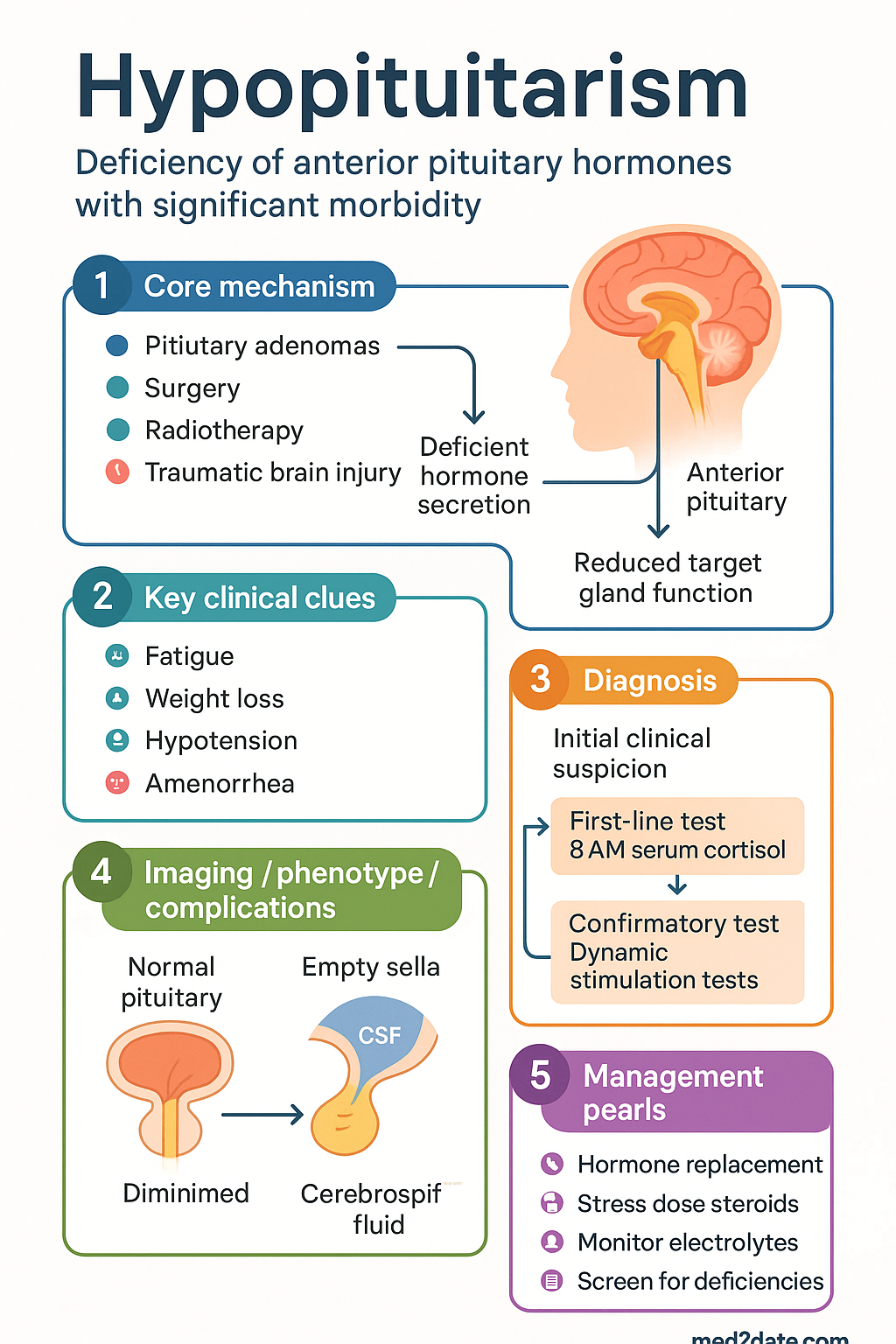
- Common causes in Australia include pituitary adenomas (functioning or non-functioning), transsphenoidal surgery, cranial radiotherapy ≥30 Gy, traumatic brain injury (TBI), and Sheehan syndrome.
- Loss of hormones follows a characteristic order: GH → LH/FSH → ACTH → TSH; GH is most vulnerable and ACTH deficiency is the most dangerous.
- Cortisol must be replaced BEFORE levothyroxine — initiating thyroxine in undiagnosed ACTH deficiency precipitates adrenal crisis.
- Acute adrenal crisis (pituitary apoplexy or undiagnosed deficiency) presents with hypotension, hyponatraemia, hypoglycaemia; treat IV hydrocortisone 100 mg bolus then 50 mg 6–8 hourly.
- Dynamic testing confirms axis failure: short Synacthen test (ACTH), GnRH stimulation test (LH/FSH), insulin tolerance test or glucagon stimulation test (GH), TRH stimulation test (TSH).
- Hydrocortisone replacement 15–25 mg/day in divided doses (10 mg / 5 mg / 5 mg) for secondary adrenal insufficiency; no mineralocorticoid replacement required.
- Levothyroxine 1.6 µg/kg/day for central hypothyroidism; monitor free T4 (not TSH) as TSH is unreliable.
- Gonadal replacement: testosterone enantate 250 mg IM every 2–4 weeks or transdermal gel for males; cyclical oestrogen–progestogen HRT for premenopausal females.
- GH replacement (somatropin) requires Authority PBS approval; retest in adulthood if childhood-onset; initiation under endocrinologist supervision.
- All patients need a crisis plan, MedicAlert identification, and education on stress dosing of hydrocortisone (double dose for minor illness; triple dose + seek medical review for febrile illness).
- Aboriginal and Torres Strait Islander peoples have higher rates of TBI and may face barriers to specialist endocrinology access; culturally safe outreach and education are essential.
🎧 Audio Brief
Introduction & Australian Epidemiology
Hypopituitarism is the partial or complete deficiency of one or more anterior pituitary hormones, with or without posterior pituitary involvement (diabetes insipidus). It may present insidiously with non-specific fatigue and weight gain, or acutely with life-threatening adrenal crisis. The condition carries significant morbidity from untreated hormone deficiencies and excess mortality — primarily driven by untreated ACTH deficiency and cardiovascular disease.
| Epidemiological Parameter | Data |
|---|---|
| Prevalence | 45.5 per 100,000 population (European data; Australian estimates similar) |
| Incidence | 4.2 per 100,000 per year |
| Commonest cause | Pituitary adenoma (including post-surgical/radiotherapy) |
| Most vulnerable axis | GH → Gonadotrophin → ACTH → TSH (in order of susceptibility) |
| Excess mortality | SIR 1.2–2.2; higher in females, younger patients, and those with craniopharyngioma |
| TBI-related | Up to 30% of moderate–severe TBI develop some pituitary dysfunction |
In Australia, the most common aetiologies encountered in specialist practice are non-functioning pituitary macroadenomas managed with transsphenoidal surgery, prolactinomas treated medically, and craniopharyngiomas. Post-radiotherapy hypopituitarism is increasingly recognised as a late effect of cranial irradiation in survivors of childhood and adult cancers. Traumatic brain injury is an emerging cause, with Australian trauma registries documenting significant rates of post-TBI pituitary dysfunction.
Aetiology & Pathophysiology
Hypopituitarism results from destruction or dysfunction of the anterior pituitary gland (secondary) or impairment of hypothalamic releasing factors (tertiary). The anterior pituitary is particularly susceptible to compression, ischaemia, and radiation damage due to its confined anatomical position within the sella turcica.
| Aetiological Category | Examples | Mechanism |
|---|---|---|
| Pituitary tumours | Non-functioning adenoma, craniopharyngioma, Rathke cleft cyst, meningioma, metastasis | Compression and destruction of normal pituitary tissue |
| Post-surgical | Transsphenoidal surgery, craniotomy | Direct surgical trauma, stalk damage, vascular compromise |
| Radiotherapy | Cranial irradiation ≥30 Gy (brain tumours, nasopharyngeal CA, ALL) | Progressive vascular fibrosis; GH most vulnerable (onset 2–5 yr post-RT) |
| Vascular | Pituitary apoplexy, Sheehan syndrome, subarachnoid haemorrhage, DIC | Haemorrhagic infarction of pituitary; Sheehan: postpartum haemorrhage |
| Infiltrative / Inflammatory | Lymphocytic hypophysitis, sarcoidosis, haemochromatosis, Langerhans cell histiocytosis, IgG4 disease | Immune-mediated destruction or iron deposition |
| Traumatic brain injury | Closed head injury, blast injury | Stalk transection, hypothalamic contusion, vascular spasm |
| Infections | Tuberculosis, fungal (aspergillosis), abscess | Granulomatous destruction or abscess formation |
| Genetic / Developmental | Combined pituitary hormone deficiency (PROP1, PIT1, HESX1 mutations), septo-optic dysplasia | Defective transcription factor signalling |
| Empty sella | Primary or secondary empty sella syndrome | Arachnoid herniation into sella with pituitary compression |
Pathophysiological Cascade
Anterior pituitary cell types have differential sensitivity to insult. Somatotrophs (GH) are the most vulnerable, followed by gonadotrophs (LH/FSH), then thyrotrophs (TSH), and finally corticotrophs (ACTH) — the most resistant. This hierarchy explains why GH deficiency typically appears first, and ACTH deficiency last, in progressive pituitary destruction. However, in acute insults (pituitary apoplexy, surgery), multiple axes may fail simultaneously.
The posterior pituitary may also be affected in craniopharyngioma, surgery near the stalk, and traumatic brain injury, leading to central diabetes insipidus (ADH deficiency). Posterior pituitary function is generally preserved in adenoma-related hypopituitarism and after radiotherapy.
Clinical Features by Hormone Deficiency
Presentation depends on the number and type of hormone deficiencies, speed of onset, and underlying aetiology. Chronic deficiencies present insidiously; acute pituitary apoplexy presents as an emergency.
| Hormone Deficiency | Key Clinical Features | Distinguishing Features |
|---|---|---|
| ACTH deficiency (secondary adrenal insufficiency) | Fatigue, weakness, anorexia, nausea, weight loss, postural hypotension, hyponatraemia, hypoglycaemia | Hyperpigmentation ABSENT (unlike primary adrenal insufficiency); aldosterone secretion preserved (no hyperkalaemia, no salt-wasting) |
| TSH deficiency (central hypothyroidism) | Fatigue, cold intolerance, constipation, weight gain, dry skin, bradycardia | TSH may be low, normal, or mildly elevated (inappropriately low for the fT4 level); normal TSH does NOT exclude central hypothyroidism |
| LH/FSH deficiency (hypogonadotropic hypogonadism) | Males: erectile dysfunction, decreased libido, reduced shaving frequency, muscle wasting, gynaecomastia. Females: amenorrhoea or oligomenorrhoea, infertility, vaginal dryness, osteoporosis | In premenopausal women, amenorrhoea is an early and prominent sign; in males, symptoms may be attributed to ageing |
| GH deficiency | Fatigue, reduced exercise capacity, central adiposity, decreased lean mass, reduced bone mineral density, impaired quality of life, dyslipidaemia | Most non-specific symptoms; often the last axis tested but the first to fail; significant impact on cardiovascular risk profile |
| Prolactin deficiency | Inability to lactate postpartum (Sheehan syndrome) | Rarely clinically significant outside lactation; prolactin is usually the last anterior hormone to be lost |
| ADH deficiency (central diabetes insipidus) | Polyuria (>3 L/day), polydipsia, nocturia, dilute urine (USG <1.005), hypernatraemia | Usually post-surgical or craniopharyngioma; transient DI may occur post-operatively with triphasic response |
Investigations (Dynamic Testing)
Diagnosis of hypopituitarism requires demonstration of low target gland hormones in the setting of inappropriately low or normal pituitary trophic hormones (TSH, ACTH, LH/FSH). Dynamic stimulation tests are required to confirm ACTH and GH reserve, as basal levels may be misleading.
Baseline Investigations
Dynamic Stimulation Tests
| Test | Protocol | Diagnostic Threshold | Notes |
|---|---|---|---|
| Short Synacthen test (ACTH reserve) | Synacthen 250 µg IV/IM; measure cortisol at 0 and 30 min | Peak cortisol <450 nmol/L = insufficient | Most widely used in Australia; adequate screening for secondary adrenal insufficiency. MBS item 66727 |
| Insulin tolerance test (ITT — gold standard for GH & ACTH) | Insulin 0.1–0.15 U/kg IV bolus; target symptomatic hypoglycaemia (glucose <2.2 mmol/L); measure cortisol and GH at 0, 30, 60, 90, 120 min | Peak GH <3 µg/L = GH deficiency; Peak cortisol <500 nmol/L = ACTH deficiency | Requires inpatient setting, continuous monitoring; contraindicated in epilepsy, ischaemic heart disease, elderly |
| Glucagon stimulation test (alternative to ITT) | Glucagon 1 mg IM (or 1.5 mg if >90 kg); measure GH and cortisol at 0, 90, 120, 150, 180 min | Peak GH <3 µg/L = GH deficiency; Peak cortisol <500 nmol/L = ACTH deficiency | Preferred outpatient alternative when ITT contraindicated; nausea is common |
| GnRH stimulation test | GnRH 100 µg IV; measure LH and FSH at 0, 30, 60 min | Inadequate LH rise (<2× baseline) with low sex steroids = hypothalamic/pituitary gonadotrophin failure | Rarely required clinically; basal gonadotrophins + sex steroids usually sufficient |
| TRH stimulation test | TRH 200 µg IV; measure TSH at 0, 20, 60 min | Absent or delayed TSH rise with low fT4 = central hypothyroidism | Largely superseded by fT4 + TSH interpretation; TRH availability in Australia may be limited |
| Water deprivation test (diabetes insipidus) | 8-hour fluid deprivation; measure urine osmolality, serum osmolality, body weight hourly; desmopressin 2 µg SC at end | Urine osmolality <300 mOsm/kg after dehydration, rising >50% after desmopressin = complete central DI | Requires specialist supervision; plasma copeptin (if available) is emerging as an alternative |
Replacement Therapy
Hormone replacement must be initiated in a physiologically logical order: cortisol first, then thyroid, then sex hormones and GH. The goal is to mimic normal physiology as closely as possible using the minimum effective doses.
Cortisol Replacement (ACTH Deficiency)
Secondary adrenal insufficiency does NOT require mineralocorticoid replacement — aldosterone secretion is primarily regulated by the renin–angiotensin system, which remains intact. Only glucocorticoid replacement is needed.
- IV hydrocortisone 100 mg bolus immediately
- Then 50 mg IV every 6–8 hours
- IV normal saline 0.9% — aggressive fluid resuscitation (1 L over 30–60 min, then titrate)
- Monitor glucose; dextrose infusion if hypoglycaemic
- Taper to oral hydrocortisone over 2–3 days when stable
Stress Dosing (Sick Day Rules)
| Situation | Hydrocortisone Adjustment |
|---|---|
| Minor illness (cold, low-grade fever) | Double oral dose until recovered (usually 2–3 days) |
| Moderate illness (fever >38.5°C, gastroenteritis, UTI) | Triple oral dose; if unable to take orally → IM hydrocortisone 100 mg and present to ED |
| Major illness / surgery | IV hydrocortisone 50–100 mg stat then 50 mg 6–8 hourly; taper over 2–3 days post-op |
| Minor dental procedure | Extra 20–25 mg PO on day of procedure |
| Vomiting / unable to take oral meds | IM hydrocortisone 100 mg (self-administered emergency injection) → attend ED |
Thyroid Replacement (Central Hypothyroidism)
Sex Hormone Replacement
Males — Testosterone Replacement
Females — Oestrogen–Progestogen Replacement
For premenopausal women with hypogonadotropic hypogonadism, physiological oestradiol replacement is essential to maintain bone density, cardiovascular health, and quality of life. Combined oral contraceptive pill (COCP) is often used as a convenient alternative to physiological HRT, though it provides supraphysiological oestradiol.
Fertility — Gonadotrophin Therapy
When fertility is desired, sex steroid replacement must be stopped and replaced with gonadotrophin therapy under specialist reproductive endocrinology supervision:
- Males: FSH (recombinant or hMG) + hCG to stimulate spermatogenesis; treatment duration 6–18 months; MBS authority required
- Females: FSH ± LH (recombinant gonadotrophins) with ultrasound monitoring for ovulation induction; high multiple pregnancy risk
Growth Hormone Replacement
Desmopressin (Central Diabetes Insipidus)
Monitoring
Lifelong monitoring is essential as replacement doses may change over time, and the underlying condition (e.g., adenoma) may progress. All patients should have an individualised management plan and emergency action plan.
| Parameter | Frequency | Target / Notes |
|---|---|---|
| Clinical review | Every 6–12 months (endocrinologist) | Symptoms, weight, BP, well-being assessment, QoL questionnaire |
| 9 AM cortisol or day-curve | Annually or after dose change | If on prednisolone; hydrocortisone day curve (3-point) if symptoms of under/over-replacement |
| Free T4 | Every 6–12 months | Aim mid-upper reference range; do NOT use TSH for dose titration |
| Testosterone (males) | Mid-cycle if IM; trough if gel | Aim 15–25 nmol/L; also monitor haematocrit (polycythaemia risk) |
| Oestradiol / withdrawal bleed (females) | Every 6–12 months | Confirm physiological replacement; ensure regular withdrawal bleeds if on cyclical HRT |
| IGF-1 | Every 4–6 weeks during titration; every 6–12 months once stable | Aim mid-normal for age and sex; avoid supraphysiological levels |
| Fasting glucose / HbA1c | Annually | GH replacement may impair glucose tolerance |
| Lipid profile | Annually | GH and sex steroid deficiency contribute to dyslipidaemia |
| DEXA bone density | At diagnosis; then every 2 years | Osteoporosis risk in GH and sex hormone deficiency |
| MRI pituitary | Per underlying pathology (e.g., 3–6 months post-surgery; annually for residual adenoma) | Assess tumour recurrence/residual mass; visual fields if chiasmal compression |
| Sodium (if on desmopressin) | Within 1 week of dose change; then 3–6 monthly | Hyponatraemia risk |
| PSA (males on testosterone) | Annually (age >50 or >40 if FHx) | Prostate screening per RACGP guidelines |
Patient Education & Crisis Plan
- All patients with ACTH deficiency must carry a MedicAlert bracelet/necklace
- Provide a written steroid emergency card and individualised adrenal crisis action plan
- Patients (and family) should be trained in IM hydrocortisone self-injection
- Supply an emergency hydrocortisone kit (Hydrocortisone 100 mg powder for injection or prefilled syringe)
- Educate on sick day rules (see stress dosing table above)
- Advise all healthcare providers of adrenal insufficiency before any procedure or illness
Special Populations
Aboriginal and Torres Strait Islander Health Considerations
📚 References
- 1. Higham CE, Johannsson G, Shalet SM. Hypopituitarism. Lancet. 2016;388(10058):2403–2415.
- 2. Fleseriu M, Hashim IA, Engel T, et al. Hypothalamic-pituitary-adrenal axis and related disorders. In: Melmed S, ed. Williams Textbook of Endocrinology. 14th ed. Elsevier; 2020:195–244.
- 3. Fleseriu M, Auchus R, Bancos I, et al. Consensus on diagnosis and management of Cushing's disease: a guideline update. Lancet Diabetes Endocrinol. 2021;9(12):847–875.
- 4. The Endocrine Society. Hormonal replacement in hypopituitarism: an Endocrine Society clinical practice guideline. J Clin Endocrinol Metab. 2016;101(11):3888–3921.
- 5. Australian Institute of Health and Welfare (AIHW). Head Injury in Aboriginal and Torres Strait Islander Peoples. Cat. no. INJCAT 125. Canberra: AIHW; 2019.
- 6. Royal Australasian College of Physicians (RACP). Australian Adult Growth Hormone Deficiency Consensus Guidelines. Sydney: RACP; 2019.
- 7. Schneider HJ, Kreitschmann-Andermahr I, Ghigo E, Stalla GK, Agha A. Hypothalamopituitary dysfunction following traumatic brain injury and aneurysmal subarachnoid hemorrhage: a systematic review. JAMA. 2007;298(12):1429–1438.
- 8. Grossman AB, Johannsson G, Quinkler M, Zelissen P. Therapy of endocrine disease: perspectives on the management of adrenal insufficiency: clinical insights from across Europe. Eur J Endocrinol. 2013;169(6):R165–R175.
- 9. Department of Health and Aged Care, Australian Government. Pharmaceutical Benefits Schedule (PBS). Available at: pbs.gov.au. Accessed 2024.
- 10. Puar TH, Stiksma J, Engelen M, et al. Pituitary apoplexy: an often forgotten medical emergency. Lancet. 2022;400(10353):709–721.
- 11. Australian Commission on Safety and Quality in Health Care (ACSQHC). National Safety and Quality Health Service Standards. 2nd ed. Sydney: ACSQHC; 2021.
- 12. Wass JAH, Reddy N, Gurnell M, et al. Oxford Textbook of Endocrinology and Diabetes. 3rd ed. Oxford University Press; 2022.
- 13. Gurnell M, Chatterjee VK, et al. Central hypothyroidism. In: Feingold KR, ed. Endotext. South Dartmouth (MA): MDText.com, Inc.; 2000–2024.
- 14. Royal Australian College of General Practitioners (RACGP). RACGP Red Book: Guidelines for Preventive Activities in General Practice. 9th ed. East Melbourne: RACGP; 2016.