Home Endocrinology Pituitary Apoplexy

Pituitary Apoplexy

๐Ÿ“‹ Key Information Summary

๐Ÿ“‹
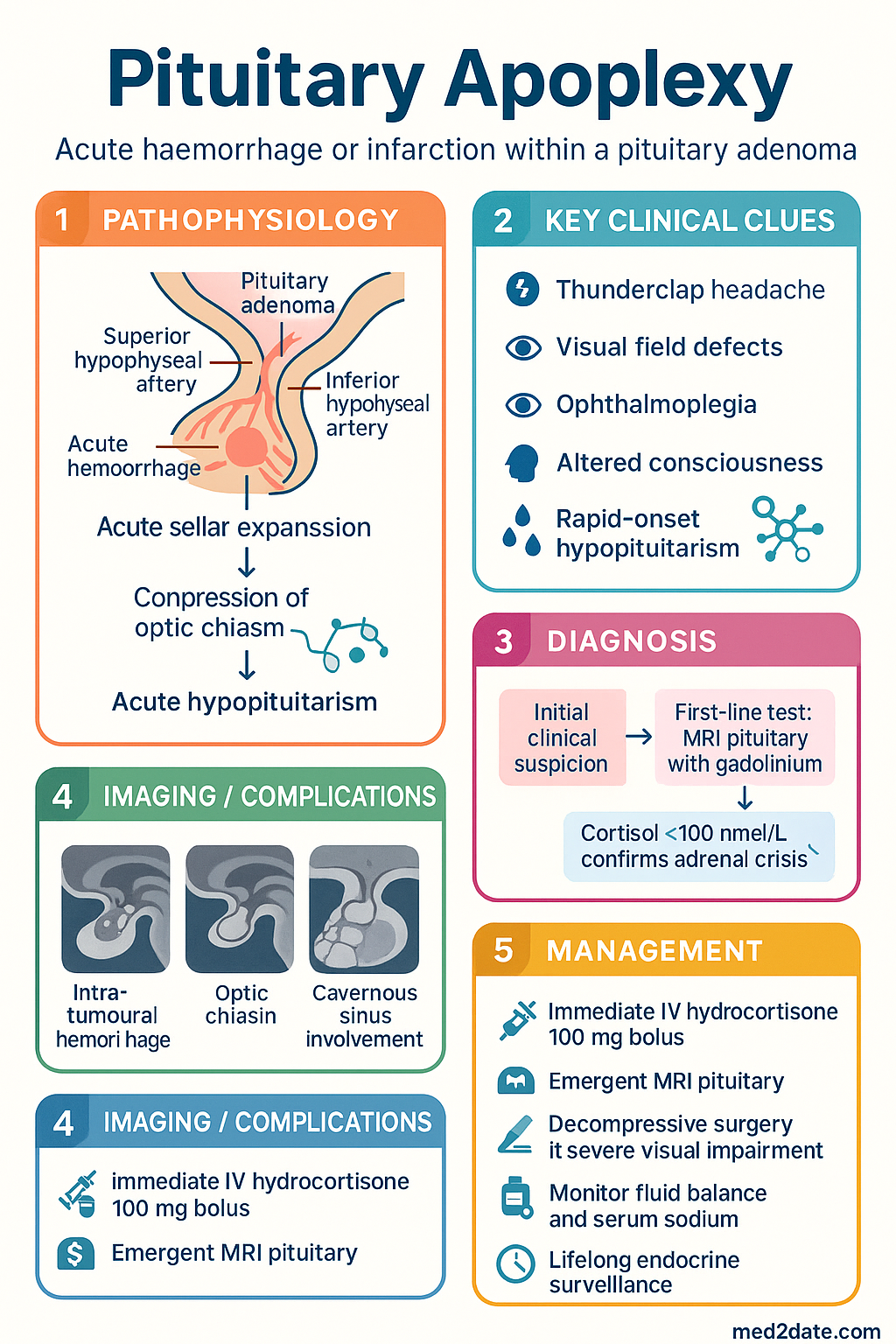
  • Pituitary apoplexy is a neuroendocrine emergency caused by acute haemorrhage or infarction within a pre-existing pituitary adenoma, occurring in approximately 0.6โ€“10% of all pituitary adenomas.
  • Severe sudden-onset headache ("thunderclap headache") is the cardinal symptom, often accompanied by visual field defects, ophthalmoplegia, altered consciousness, and rapid-onset hypopituitarism.
  • Immediate IV hydrocortisone 100 mg bolus is lifesaving and must be administered before diagnostic confirmation โ€” do not delay steroids for MRI.
  • Cortisol <100 nmol/L in the acute setting confirms adrenal crisis and mandates urgent glucocorticoid replacement.
  • Emergent MRI pituitary with gadolinium is the investigation of choice; CT may show haemorrhage but has lower sensitivity for infarction.
  • Decompressive transsphenoidal surgery should be performed within 7 days for patients with severe visual impairment, progressive visual deterioration, or declining consciousness.
  • Mild cases (no visual compromise, intact consciousness) may be managed conservatively with close monitoring in an HDU setting.
  • All patients require complete anterior pituitary hormone panel (TSH/fT4, cortisol, LH/FSH, testosterone/oestradiol, IGF-1, prolactin) and posterior pituitary function assessment post-event.
  • Precipitants include dynamic pituitary function testing (GnRH analogue stimulation), anticoagulation, pregnancy, and raised intracranial pressure.
  • Diabetes insipidus may develop acutely or 5โ€“10 days post-surgery; monitor strict fluid balance and serum sodium in all patients.
  • Aboriginal and Torres Strait Islander patients may have reduced access to specialist neurosurgical and endocrinological care, particularly in remote regions; initiate early retrieval coordination.
  • Lifelong endocrine surveillance and hormone replacement therapy are required for most patients following pituitary apoplexy.

๐ŸŽง Audio Brief

The Pituitary Apoplexy Emergency

A short clinical audio briefing generated from this article โ€” perfect for the commute or ward round.

Introduction & Australian Epidemiology

Pituitary apoplexy is a clinical emergency caused by acute haemorrhage or infarction within a pituitary adenoma, resulting in rapid expansion of the sellar mass, compression of adjacent neural structures, and abrupt pituitary hormone deficiency. The syndrome may also rarely occur in non-adenomatous pituitary tissue, during pregnancy (Sheehan syndrome), or as a consequence of pharmacological or radiological interventions.

The estimated incidence is 6.2 per 100,000 person-years among patients with known pituitary adenomas. Clinically apparent apoplexy occurs in 2โ€“12% of all pituitary adenomas, though histopathological evidence of haemorrhage or infarction is found in up to 25% of surgical specimens. The true incidence in the Australian population is not well established; however, data from the Australian Pituitary Tumour Registries and tertiary referral centres (Royal Melbourne Hospital, Westmead Hospital, St Vincent's Hospital Sydney) suggest approximately 80โ€“120 clinically significant presentations per year nationally.

The condition has a male predominance (M:F โ‰ˆ 2:1), likely reflecting the higher prevalence of non-functioning macroadenomas in men. Presentation is most common in the fifth and sixth decades. Non-functioning adenomas account for the majority of cases, though prolactinomas, GH-secreting, and ACTH-secreting adenomas may also undergo apoplexy.

โš ๏ธ
Time-critical diagnosis: Pituitary apoplexy is frequently misdiagnosed as subarachnoid haemorrhage, meningitis, or hypertensive emergency. A high index of suspicion is required in any patient presenting with thunderclap headache and visual disturbance, particularly in those with known pituitary adenomas.
Pituitary Apoplexy clinical infographic โ€” pathophysiology, clinical clues, diagnosis, imaging, and management
Tap or click image to enlarge โ€” Pituitary Apoplexy: pathophysiology, clinical clues, diagnosis, imaging, and management.
Pituitary Apoplexy infographic, full size

Pathophysiology & Precipitants

Mechanism

Pituitary adenomas are particularly vulnerable to haemorrhage and infarction due to their unique vascular architecture. The pituitary gland is supplied by the superior and inferior hypophyseal arteries, with adenomas relying on a fragile, sinusoidal capillary network. Rapid tumour expansion outstrips blood supply, creating zones of ischaemia that may progress to infarction or haemorrhagic transformation.

Two pathological processes underlie apoplexy:

  • Haemorrhagic apoplexy: Intra-tumoural haemorrhage causing rapid sellar expansion, compression of the optic chiasm, cavernous sinus, and hypothalamus. Gross haemorrhage is identified in approximately 50% of cases.
  • Ischaemic (infarctive) apoplexy: Thrombosis or vasospasm within the tumour vasculature leading to coagulative necrosis, oedema, and secondary mass effect. This variant may have a more insidious onset.

Recognised Precipitants

Precipitant Mechanism Clinical Context
Dynamic pituitary function testing GnRH analogue stimulation (e.g., triptorelin) causes rapid tumour enlargement Growth hormone stimulation testing in children with pituitary adenoma
Anticoagulation Haemorrhagic transformation of pre-existing ischaemic focus Heparin, warfarin, DOACs; post-cardiac surgery
Pregnancy Oestrogen-mediated lactotroph hyperplasia; haemodynamic changes Peripartum period; Sheehan syndrome (postpartum pituitary necrosis)
Raised intracranial pressure Venous congestion of pituitary sinusoidal system Lumbar puncture, Valsalva, mechanical ventilation with high PEEP
Radiotherapy Vascular endothelial damage; delayed vasculopathy Months to years after stereotactic or fractionated radiotherapy
Dopamine agonist initiation/withdrawal Rapid tumour shrinkage with infarction, or rebound expansion on withdrawal Cabergoline initiation in macroprolactinoma
Diabetes mellitus Microvascular disease of pituitary vasculature Independent risk factor in observational studies
Head trauma Direct vascular injury or shearing of stalk vessels Road traffic accidents, falls

Sequence of Pathological Events

The acute expansion of the pituitary fossa contents occurs over minutes to hours, producing a cascade of compressive effects: first on the optic chiasm (bitemporal hemianopia), then the cavernous sinus (cranial nerve III, IV, VI palsies), and finally the hypothalamus and third ventricle (altered consciousness, autonomic instability, hydrocephalus). Concurrently, destruction of functioning pituitary tissue causes acute hypopituitarism, with ACTH/cortisol deficiency being the most immediately life-threatening.

Clinical Features

Classic Triad

  • Severe sudden-onset headache (85โ€“97%)
  • Visual impairment (60โ€“80%)
  • Ophthalmoplegia / cranial nerve palsy (40โ€“70%)

Severe Headache

The headache of pituitary apoplexy is typically retro-orbital or frontal, of sudden onset ("thunderclap"), severe, and unremitting. It may be bilateral or unilateral and is often described as the "worst headache of my life." Nausea and vomiting occur in approximately 70% of cases due to meningeal irritation from subarachnoid haemorrhage or raised intracranial pressure. Neck stiffness may be present, mimicking meningitis or subarachnoid haemorrhage.

๐Ÿšจ
Red flag: Thunderclap headache with visual loss in a patient with a known pituitary adenoma is pituitary apoplexy until proven otherwise. Immediate administration of IV hydrocortisone is indicated before further investigation.

Visual Loss & Field Defects

Visual impairment results from compression of the optic chiasm and/or optic nerves. Classical findings include:

  • Bitemporal hemianopia: The hallmark visual field defect, occurring in approximately 50โ€“60% of cases
  • Unilateral or bilateral visual acuity loss: Ranging from mild blurring to complete blindness
  • Afferent pupillary defect (RAPD): Indicates significant unilateral optic nerve compression
  • Papilloedema: May be present with raised intracranial pressure

Patients with pre-existing bitemporal hemianopia from a known macroadenoma may not report new visual symptoms; therefore, formal bedside confrontation testing and pupillary reflex assessment are essential in all presentations.

Ophthalmoplegia & Cranial Nerve Palsies

Lateral extension of haemorrhage or oedema into the cavernous sinus compresses cranial nerves traversing this structure:

  • CN III (oculomotor): Ptosis, dilated pupil, "down and out" eye โ€” most common
  • CN IV (trochlear): Diplopia on downward and inward gaze
  • CN VI (abducens): Lateral rectus palsy, horizontal diplopia
  • CN Vโ‚ (ophthalmic): Reduced corneal sensation, forehead numbness

Hypopituitarism

Acute anterior pituitary failure develops rapidly, with the following deficiency pattern and approximate frequency:

Hormone Deficient Frequency Acute Consequences
ACTH โ†’ Cortisol ~70% Adrenal crisis: hypotension, shock, hyponatraemia, hypoglycaemia โ€” potentially fatal
TSH โ†’ Thyroid ~55% Secondary hypothyroidism (rarely acute; do not replace until cortisol replaced)
LH/FSH โ†’ Gonadal ~75% Hypogonadism: rarely acutely life-threatening
GH ~80% Hypoglycaemia (especially in children); rarely acute concern in adults
ADH (posterior pituitary) ~3โ€“5% acutely Diabetes insipidus: polyuria, polydipsia, hypernatraemia
Prolactin ~50% (or โ†‘ from stalk effect) Lactation failure in postpartum women
โš ๏ธ
ACTH/cortisol deficiency is the most immediately dangerous. Clinical features include hypotension refractory to fluids, hyponatraemia (dilutional), hypoglycaemia, altered consciousness, and cardiovascular collapse. Suspect adrenal crisis in any hypotensive patient with a pituitary mass.

Investigations

Immediate Bedside & Laboratory

ESSENTIAL
Serum cortisol (random)
Cortisol <100 nmol/L in acute presentation confirms adrenal crisis. Cortisol 100โ€“400 nmol/L is equivocal โ€” treat empirically. MBS Item 66549.
ESSENTIAL
Full anterior pituitary panel
TSH, fT4, fT3, LH, FSH, testosterone (men) / oestradiol (women), prolactin, IGF-1. May be drawn concurrently with cortisol but should not delay treatment. MBS Items 66630, 66634.
ESSENTIAL
Urgent biochemistry
Serum sodium, potassium, glucose, urea, creatinine, osmolality. Urine osmolality if polyuric. Hyponatraemia (dilutional, <130 mmol/L) common in cortisol deficiency. MBS Item 66506.
AVAILABLE
Full blood count
May show haemoconcentration in dehydration. Leucocytosis non-specific.
AVAILABLE
Coagulation studies
If patient is anticoagulated โ€” may need urgent reversal. INR, APTT, fibrinogen. MBS Item 65070.

Neuroimaging

ESSENTIAL
MRI pituitary with gadolinium (sella protocol)
Investigation of choice. Demonstrates intra-tumoural haemorrhage (T1 hyperintense in acute haemorrhage), infarction (T2 hyperintense), sellar expansion, suprasellar extension, and optic chiasm compression. Available at all Australian tertiary centres and most regional centres. MBS Item 63042.
AVAILABLE
CT head (non-contrast)
May demonstrate acute haemorrhage (hyperdense lesion in sella) but has lower sensitivity for infarction and early oedema. Suitable as initial screening if MRI not immediately available. MBS Item 56800.
REFERRAL
CT angiography
May be indicated to exclude aneurysmal subarachnoid haemorrhage or internal carotid artery involvement. Available at tertiary centres.

Formal Visual Assessment

Urgent ophthalmological assessment with formal visual field testing (Humphrey or Goldmann perimetry) should be arranged, though bedside confrontation testing and pupillary assessment must not be delayed. Documentation of baseline visual function is critical for surgical decision-making.

Lumbar Puncture

โš ๏ธ
Caution: Lumbar puncture is generally contraindicated in the acute setting as it may worsen herniation if there is significant suprasellar extension or hydrocephalus. CSF analysis may show xanthochromia and elevated protein but should only be performed after neurosurgical consultation.

Management

Immediate Management (First 0โ€“2 Hours)

The following steps should be initiated simultaneously in the emergency department:

1
IV Hydrocortisone โ€” STAT
Hydrocortisone 100 mg IV bolus immediately, then 50 mg IV 6โ€“8 hourly. Do not delay for cortisol results or MRI. This is the single most important intervention. Do NOT give dexamethasone โ€” hydrocortisone provides both glucocorticoid and mineralocorticoid activity needed in adrenal crisis.
2
Haemodynamic Stabilisation
IV 0.9% sodium chloride resuscitation (1โ€“2 L bolus if hypotensive). Monitor in HDU or ICU. Arterial line for continuous BP monitoring. Treat hypoglycaemia with IV glucose 50 mL 50% if glucose <4 mmol/L.
3
Bloods & Imaging
Draw cortisol, pituitary panel, biochemistry, FBC, coagulation simultaneously with steroid administration. Arrange emergent MRI pituitary (transfer to scanner with nursing escort and resuscitation equipment).
4
Neurosurgical Referral
Contact on-call neurosurgery at the nearest tertiary centre early. Telehealth consultation available for rural/remote sites via the Australian Telestroke Network.

Steroid Therapy โ€” Detailed Regimen

๐Ÿ’Š
Hydrocortisone (Solu-Cortefยฎ)
Hydrocortisone sodium succinate ยท Corticosteroid (glucocorticoid + mineralocorticoid)
Adult dose 100 mg IV bolus STAT, then 50 mg IV 6โ€“8 hourly for 24โ€“48 hours; taper to oral 20 mg mane / 10 mg nocte as acute phase resolves
Paediatric dose Infants: 25 mg IV bolus, then 25 mg/mยฒ/day IV divided 6-hourly. Children: 50 mg IV bolus, then 50 mg/mยฒ/day IV divided 6-hourly
Route / Frequency IV bolus โ†’ IV bolus or infusion 6โ€“8 hourly
Renal adjustment No dose adjustment required
Hepatic adjustment No dose adjustment; monitor for fluid retention
PBS status โœ” PBS General Benefit
๐Ÿ’Š
Dexamethasone
Dexamethasone sodium phosphate ยท Glucocorticoid (no mineralocorticoid activity)
Adult dose 4 mg IV if hydrocortisone unavailable. Not preferred โ€” lacks mineralocorticoid activity; cannot be used for adrenal crisis confirmation testing
Paediatric dose 0.15 mg/kg IV (max 4 mg) if hydrocortisone unavailable
Route / Frequency IV bolus 6โ€“8 hourly
PBS status โœ” PBS General Benefit
๐Ÿšจ
Safety critical: Never delay hydrocortisone for imaging. If pituitary apoplexy is clinically suspected, steroids must be given immediately. Untreated acute adrenal crisis carries a mortality of up to 50%.

Surgical Management

Transsphenoidal decompression (TSS) is the standard surgical approach. Surgical timing and indications remain debated, but the following evidence-based thresholds are used in Australian neurosurgical centres:

Surgery Indicated
Visual Compromise
Visual acuity deterioration, new or worsening visual field defect, progressive ophthalmoplegia
Timing: Within 24โ€“48 hours (ideally <7 days from onset)
Surgery Indicated
Neurological Deterioration
Declining GCS, signs of hydrocephalus, brainstem compression, worsening consciousness
Timing: Emergent โ€” within hours
Conservative
Mild Presentation
Headache only, no visual deficit, stable neurological status, improving with steroids
Setting: HDU monitoring with serial visual assessment and repeat MRI at 1 week

Key surgical considerations in Australia:

  • Surgery is performed at designated pituitary neurosurgical centres (Royal Melbourne, Westmead, St Vincent's Sydney, Royal Adelaide, Royal Brisbane & Women's, Sir Charles Gairdner Perth)
  • Endoscopic endonasal transsphenoidal approach is standard; craniotomy reserved for unusual anatomy or lateral extension
  • Pre-operative steroid therapy must be optimised โ€” hydrocortisone 100 mg IV at induction
  • Intra-operative CSF leak may require lumbar drain placement
  • Inter-hospital retrieval via RFDS or state retrieval services for rural/remote patients

Management of Diabetes Insipidus

Diabetes insipidus occurs in approximately 3โ€“5% acutely but may develop 5โ€“10 days post-surgery in up to 20% of cases. Monitor strict fluid balance and serum sodium daily.

๐Ÿ’Š
Desmopressin (DDAVP)
Minirinยฎ ยท Synthetic ADH analogue
Adult dose 1โ€“2 ยตg SC/IV 8โ€“12 hourly; or 100โ€“400 ยตg intranasal BD; or 0.1โ€“0.4 mg oral TDS
Paediatric dose 0.05โ€“0.1 mg oral BDโ€“TDS; or 5โ€“10 ยตg intranasal daily
Key monitoring Serum sodium 4โ€“6 hourly โ€” risk of hyponatraemia with over-replacement
PBS status โœ” PBS General Benefit

Long-Term Hormone Replacement

Following acute stabilisation, formal pituitary function reassessment at 6โ€“12 weeks determines long-term replacement needs. Approximately 80% of patients require lifelong glucocorticoid replacement; 60โ€“70% require thyroid hormone replacement; 50โ€“60% require gonadal hormone replacement; and GH replacement may be indicated in confirmed severe GH deficiency.

๐Ÿ’Š
Hydrocortisone (oral)
Hydrocortisone ยท Cortisol replacement
Adult dose 15โ€“25 mg/day PO in divided doses (e.g., 10 mg mane, 5 mg midday, 5 mg 1600h)
Sick-day rules Double dose for febrile illness; triple for severe illness/vomiting; IM/IV if unable to take orally
PBS status โœ” PBS General Benefit
๐Ÿ’Š
Levothyroxine (Oroxineยฎ)
Eutroxsigยฎ ยท Thyroid hormone replacement
Adult dose 50โ€“100 ยตg PO daily on empty stomach; titrate to fT4 mid-normal range
Critical rule Never start before cortisol is replaced โ€” risk of precipitating adrenal crisis
PBS status โœ” PBS General Benefit
โš ๏ธ
Thyroid replacement sequence: Always ensure adequate glucocorticoid replacement BEFORE commencing levothyroxine. Starting T4 in a cortisol-deficient patient can precipitate fatal adrenal crisis.

Supportive & Adjunctive Measures

  • VTE prophylaxis: Mechanical prophylaxis (TED stockings, intermittent pneumatic compression) immediately; pharmacological prophylaxis (enoxaparin 40 mg SC daily) once haemostasis confirmed โ€” usually 24โ€“48 hours post-surgery
  • Analgesia: Paracetamol 1 g QDS PO/IV; avoid NSAIDs if coagulopathy or recent haemorrhage
  • Anti-emetics: Ondansetron 4 mg IV PRN
  • Glucose monitoring: BSL 4โ€“6 hourly during acute phase โ€” hypoglycaemia is a feature of cortisol deficiency
  • Steroid emergency card: Issue MedicAlert bracelet and adrenal crisis information card prior to discharge

Monitoring

Acute Phase (0โ€“7 Days)

0โ€“24 hours
Continuous cardiac monitoring in HDU/ICU. Hourly GCS and pupillary checks. Strict fluid balance with hourly urine output. Serum sodium, glucose, and potassium 4โ€“6 hourly. Visual assessment q4h.
24โ€“72 hours
Repeat MRI at 48โ€“72 hours if conservative management. Formal ophthalmological assessment. Commence weaning IV hydrocortisone to oral if haemodynamically stable. Daily sodium and fluid balance.
3โ€“7 days
Post-surgical patients: monitor for CSF rhinorrhoea, meningitis signs, and delayed diabetes insipidus. Transition to oral hydrocortisone. Arrange endocrine outpatient follow-up.

Subacute & Long-Term Follow-Up

  • 6 weeks: Formal pituitary function reassessment (full anterior panel + cortisol day curve). Repeat MRI pituitary to assess residual tumour.
  • 3 months: Ophthalmological review with formal perimetry. GH stimulation testing (insulin tolerance test or glucagon stimulation test) if other axes deficient.
  • 6โ€“12 months: Annual MRI surveillance for residual/recurrent adenoma. Annual pituitary blood panel. Bone density (DEXA) if hypogonadal or on long-term steroids.
  • Ongoing: Lifelong annual endocrine review. MedicAlert registration. Sick-day steroid education reinforcement at each visit.

Special Populations

๐Ÿคฐ Pregnancy
Hydrocortisone
Safe in pregnancy โ€” the preferred corticosteroid. Dose: 100 mg IV bolus, then 50 mg 6โ€“8 hourly. Crosses placenta minimally compared with dexamethasone.
MRI pituitary
Safe in all trimesters (no ionising radiation). Gadolinium generally avoided in first trimester unless essential. Non-contrast MRI may suffice acutely.
Surgery
Transsphenoidal surgery may be performed in the second trimester if indicated. Coordinate with obstetric and anaesthetic teams. Foetal monitoring required.
Sheehan syndrome
Postpartum pituitary necrosis โ€” consider if failure to lactate, persistent hypotension, or hyponatraemia post-delivery. More common with postpartum haemorrhage.
๐Ÿ‘ถ Paediatrics
Hydrocortisone dose
Neonates: 25 mg IV bolus. Children: 50 mg/mยฒ IV bolus, then 50 mg/mยฒ/day in divided doses. Weight-based dosing for infants <10 kg.
Craniopharyngioma
More common than pituitary adenoma in children. Apoplexy may mimic this condition. Urgent MRI essential.
GH deficiency
GH is critical for growth in children. Reassess at 3โ€“6 months post-event. GH stimulation testing required before replacement.
๐Ÿ‘ด Elderly
Anticoagulation
Elderly patients on warfarin or DOACs for atrial fibrillation are at increased risk of haemorrhagic apoplexy. Consider urgent reversal (vitamin K, idarucizumab, orexanet alfa) as appropriate.
Surgical risk
Age alone is not a contraindication to surgery. Assess with ASA scoring. Anaesthetic review essential. Transsphenoidal approach preferred over craniotomy.
Cortisol replacement
Monitor for steroid-induced hyperglycaemia, osteoporosis, and psychiatric effects. Aim for lowest effective replacement dose.
๐Ÿฉบ Renal Impairment
Hydrocortisone
No dose adjustment required. Monitor for fluid overload with IV administration. Electrolyte monitoring essential (potassium, sodium).
Desmopressin
No dose adjustment in renal impairment. Use with caution โ€” hyponatraemia risk increased. Monitor sodium closely.
๐Ÿซ Hepatic Impairment
Hydrocortisone
Hepatic metabolism โ€” monitor for prolonged effect in severe liver disease. Fluid retention risk increased; monitor for ascites and oedema.
๐Ÿ›ก๏ธ Immunocompromised
Steroid considerations
High-dose hydrocortisone is still required for adrenal crisis regardless of immune status. Monitor for secondary infection. Post-surgical infection risk increased โ€” consider prophylactic antibiotics per eTG guidance. Meningitis prophylaxis may be indicated if CSF leak occurs.

Aboriginal and Torres Strait Islander Health Considerations

Aboriginal and Torres Strait Islander Health
Access to neurosurgery
Aboriginal and Torres Strait Islander peoples, particularly those in remote and very remote communities, face significant barriers to accessing neurosurgical services. The nearest pituitary neurosurgical centre may be >1,000 km away. Early activation of RFDS or state retrieval services is critical. Telehealth endocrinology consultation via the Australian Telehealth Network can facilitate initial stabilisation and triage.
Diagnostic delay
Known pituitary adenomas may be under-diagnosed or under-monitored in Indigenous communities due to reduced access to specialist endocrinology services. The AIHW reports lower rates of MRI utilisation and specialist outpatient attendance for Aboriginal and Torres Strait Islander peoples compared with non-Indigenous Australians. Clinicians should maintain a high index of suspicion for pituitary pathology in patients presenting with severe headache and visual disturbance.
Chronic disease burden
Higher prevalence of diabetes mellitus and cardiovascular disease in Aboriginal and Torres Strait Islander communities may increase the risk of pituitary apoplexy (diabetes is an independent risk factor). Cortisol replacement and glycaemic management require careful coordination.
Health literacy & follow-up
Lifelong hormone replacement therapy requires ongoing patient education. Culturally appropriate education materials should be provided in the patient's preferred language. Aboriginal Health Workers and Liaison Officers should be engaged for discharge planning and medication counselling. MedicAlert bracelets should be arranged prior to discharge.
Medication access
Hydrocortisone, levothyroxine, and desmopressin are PBS General Benefit medications, available without cost. However, remote community pharmacies may have limited stock. PBS authority prescriptions can be arranged through Remote Area Aboriginal Health Services. Emergency supply of injectable hydrocortisone should be arranged for patients in remote areas.
RHDAustralia alignment
Management should align with RHDAustralia recommendations for acute care delivery to Aboriginal and Torres Strait Islander patients, including cultural safety training for all staff, family-centred care models, and integration with Aboriginal Community Controlled Health Organisations (ACCHOs) for ongoing management.

๐Ÿ“š References

  1. 1. Briet C, Salenave S, Bonneville JF, Laws ER, Chanson P. Pituitary apoplexy. Endocrine Reviews. 2015;36(6):622โ€“645.
  2. 2. Rajasekaran S, Vanderpump M, Baldeweg S, et al. UK guidelines for the management of pituitary apoplexy. Clinical Endocrinology. 2011;74(1):9โ€“20.
  3. 3. Glezer A, Bronstein MD. Pituitary apoplexy: pathophysiology, diagnosis and management. Archives of Endocrinology and Metabolism. 2015;59(3):259โ€“264.
  4. 4. Singh TD, Bydon JB, Mandrekar JN, et al. Pituitary apoplexy: a nationwide study of 90-day readmissions. Pituitary. 2017;20(3):324โ€“330.
  5. 5. Ayuk J, McGregor EJ, Mitchell RD, Gittoes NJL. Acute management of pituitary apoplexy โ€” surgery or conservative management? Clinical Endocrinology. 2004;61(6):747โ€“752.
  6. 6. Randeva HS, Schoebel J, Byrne J, Esiri M, Adams CBT, Wass JAH. Classical pituitary apoplexy: clinical features, management and outcome. Clinical Endocrinology. 1999;51(2):181โ€“188.
  7. 7. Australian Institute of Health and Welfare (AIHW). Aboriginal and Torres Strait Islander health performance framework 2020 summary report. Canberra: AIHW; 2020.
  8. 8. Grossman A, Besser GM. Anterior pituitary failure. In: De Groot LJ, Chrousos G, Dungan K, et al., eds. Endotext. South Dartmouth, MA: MDText.com, Inc.; 2023.
  9. 9. Royal Australasian College of Physicians. Endocrinology advanced training curriculum. Sydney: RACP; 2022.
  10. 10. Moller-Goede DL, Brandle M, Landau K, Bernays RL, Schmid C. Pituitary apoplexy: re-evaluation of treatment. Journal of Neurosurgery. 2011;114(1):154โ€“160.
  11. 11. Semple PL, Webb MK, de Villiers JC, Laws ER Jr. Pituitary apoplexy. Neurosurgery. 2005;56(1):65โ€“73.
  12. 12. National Health and Medical Research Council (NHMRC). National statement on ethical conduct in human research. Canberra: NHMRC; 2023 (updated).
co-pay for eligible patients).
Pregnancy & maternal health
Antenatal screening for thyroid disease should be integrated into Aboriginal Community Controlled Health Organisation (ACCHO) maternal health programmes. Untreated hypothyroidism in pregnancy disproportionately impacts communities with limited access to early antenatal care.
Comorbidity burden
Higher rates of diabetes, cardiovascular disease, and chronic kidney disease in Aboriginal and Torres Strait Islander communities mean hypothyroid-related dyslipidaemia and cardiovascular risk require particularly active management. Integrating thyroid function testing into chronic disease management plans (MBS Item 721) is recommended.
Iodine status
Although Australia-wide mandatory iodisation has improved status, some Aboriginal and Torres Strait Islander communities โ€” particularly in very remote areas โ€” may have borderline iodine adequacy. Urinary iodine monitoring in these communities should be maintained.

๐Ÿ“š References

  1. 1. Chaker L, Bianco AC, Jonklaas J, Peeters RP. Hypothyroidism. Lancet. 2017;390(10101):1550โ€“1562.
  2. 2. Garber JR, Cobin RH, Gharib H, et al. Clinical practice guidelines for hypothyroidism in adults: cosponsored by the American Association of Clinical Endocrinologists and the American Thyroid Association. Endocr Pract. 2012;18(6):988โ€“1028.
  3. 3. Pearce SH, Brabant G, Duntas LH, et al. 2013 ETA guideline: management of subclinical hypothyroidism. Eur Thyroid J. 2013;2(4):215โ€“228.
  4. 4. Alexander EK, Pearce EN, Brent GA, et al. 2017 guidelines of the American Thyroid Association for the diagnosis and management of thyroid disease during pregnancy and the postpartum. Thyroid. 2017;27(3):315โ€“389.
  5. 5. RACGP. Red Book: Guidelines for preventive activities in general practice. 9th ed. East Melbourne: RACGP; 2018.
  6. 6. Australian Institute of Health and Welfare (AIHW). Aboriginal and Torres Strait Islander health performance framework. Canberra: AIHW; 2023.
  7. 7. Li Y, Teng D, Shi X, et al. Prevalence of diabetes recorded in mainland China using 2018 diagnostic criteria from the American Diabetes Association: national cross sectional study. BMJ. 2020;369:m997. [TSH population reference data]
  8. 8. Ross DS. Diagnosis of and screening for hypothyroidism. In: UpToDate, Cooper DS (Ed). Wolters Kluwer; 2024. Accessed June 2024.
  9. 9. NHMRC. National evidence-based guideline: diagnosis, management and prevention of congenital hypothyroidism. Canberra: NHMRC; 2019.
  10. 10. Wiersinga WM, Duntas L, Fadeyev V, Nygaard B, Vanderpump MP. 2012 ETA guidelines: the use of L-T4 + L-T3 in the treatment of hypothyroidism. Eur Thyroid J. 2012;1(2):55โ€“71.
  11. 11. Pharmaceuticals Benefits Scheme (PBS). Levothyroxine sodium. Australian Government Department of Health. Available at: pbs.gov.au. Accessed June 2024.
  12. 12. Australian Government Department of Health. National Newborn Bloodspot Screening โ€” Congenital Hypothyroidism. Available at: www.newbornscreening.gov.au. Accessed June 2024.