Home Endocrinology Paget's Disease of Bone

Paget's Disease of Bone

📋 Key Information Summary

📋
  • Paget's disease of bone is a focal disorder of accelerated bone remodelling leading to enlarged, structurally disorganised bone; prevalence in Australia is approximately 3–4% in people aged >55 years, with higher rates in Anglo-Celtic populations.
  • Most common sites: pelvis, lumbar spine, skull, femur, and tibia; polyostotic disease (>1 bone) present in ~70% of symptomatic patients.
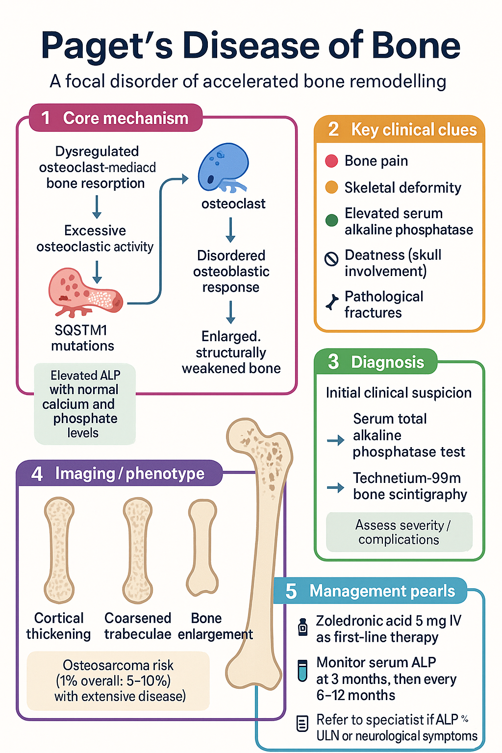
  • The hallmark biochemical finding is an elevated serum total alkaline phosphatase (ALP) with normal calcium, phosphate, and liver transaminases.
  • Plain radiographs remain the primary imaging modality — look for cortical thickening, coarsened trabeculae, and bone enlargement.
  • Technetium-99m bone scintigraphy is the best modality for assessing disease extent (polyostotic vs monostotic) and detecting occult lesions.
  • Indications for treatment: bone pain attributable to Paget's, periarticular pagetic bone causing osteoarthritis, pre-operative optimisation before orthopaedic surgery, hypercalcaemia, neurological compromise, and high-output cardiac failure.
  • First-line therapy: zoledronic acid 5 mg IV single infusion — achieves ALP normalisation in >90% of patients with durable remission >5 years.
  • Second-line/oral alternative: risedronate 30 mg PO daily for 2 months; pamidronate 60 mg IV over 2 h if zoledronic acid is contraindicated.
  • Monitor serum total ALP at 3 months post-treatment, then every 6–12 months; re-treat if ALP rises >25% above nadir with recurrence of symptoms.
  • Complications to screen for: pathological fractures, deafness (skull involvement), high-output cardiac failure (polyostotic disease), and secondary osteosarcoma (<1%).
  • All patients require adequate calcium and vitamin D supplementation before and during bisphosphonate therapy to avoid hypocalcaemia.
  • Refer to endocrinology or metabolic bone specialist if: ALP >3× ULN, skull involvement with neurological symptoms, suspected sarcomatous transformation, or refractory disease.
  • Aboriginal and Torres Strait Islander Australians may present later with more advanced disease — ensure culturally safe assessment and access to IV bisphosphonate infusion services.

🎧 Audio Brief

The Rogue Cells Warping Your Skeleton

A short clinical audio briefing generated from this article — perfect for the commute or ward round.

Introduction & Australian Epidemiology

Paget's disease of bone (PDB) is a chronic focal disorder of bone remodelling characterised by disorganised and accelerated bone turnover. This results in enlarged, structurally weakened bone that is prone to deformity, fracture, and arthritic change. Although often asymptomatic, PDB can cause significant morbidity through bone pain, skeletal deformity, deafness, and pathological fracture.

PDB is common in individuals of Anglo-Celtic descent, and Australia has one of the highest prevalences worldwide. Population-based data from the Dubbo Osteoporosis Epidemiology Study and hospital discharge records estimate prevalence of approximately 3–4% in Australians aged over 55 years, rising to 8–10% in those over 80 years. Males are affected slightly more often than females (ratio ~1.3:1). PDB is rare before age 40 years.

In Australia, the condition is more prevalent in certain regions — notably parts of New South Wales, Victoria, and Queensland — likely reflecting the predominantly British and Irish heritage of early settler populations. Prevalence appears to be declining in younger birth cohorts, consistent with trends observed in the United Kingdom and New Zealand, possibly related to changing environmental exposures and improved childhood nutrition.

The disease is characterised by three phases: an initial osteoclastic phase of excessive bone resorption, a mixed osteoclastic–osteoblastic phase, and a late osteoblastic "burned-out" sclerotic phase. Disease may affect a single bone (monostotic, ~30%) or multiple bones (polyostotic, ~70%), with the pelvis, lumbar spine, skull, femur, and tibia most commonly involved.

Paget's Disease of Bone clinical infographic — pathophysiology, clinical clues, diagnosis, imaging, and management
Tap or click image to enlarge — Paget's Disease of Bone: pathophysiology, clinical clues, diagnosis, imaging, and management.
Paget's Disease of Bone infographic, full size

Pathophysiology & Epidemiology

Pathophysiology

Paget's disease arises from dysregulated osteoclast-mediated bone resorption followed by disorganised osteoblastic bone formation. The fundamental abnormality lies within the osteoclast lineage, which produces excessive numbers of enlarged, hypernucleated osteoclasts that resorb bone at up to 20 times the normal rate. This triggers a compensatory but disordered osteoblastic response, producing woven bone with abnormal architecture.

Several genetic and environmental factors contribute:

  • Genetic: Up to 40% of patients have a positive family history. Mutations in SQSTM1 (sequestosome-1 / p62) account for ~10–50% of familial cases and ~5–10% of sporadic cases in Australian cohorts. Other implicated genes include OPTN, NFκB, and VCP.
  • Paramyxoviral hypothesis: Intracytoplasmic inclusions resembling measles virus nucleocapsids have been identified in pagetic osteoclasts, though definitive causation remains unproven.
  • RANK/RANKL/OPG axis: Pagetic osteoclasts exhibit enhanced sensitivity to RANKL (receptor activator of nuclear factor kappa-B ligand) and overexpression of RANK, driving osteoclastogenesis. Denosumab targets this pathway.
  • Environmental: Mechanical loading, low-dose radiation exposure, and possibly dietary calcium/vitamin D insufficiency in childhood may modulate risk.

Key Histological Features

Phase Histology Clinical Correlate
Osteolytic (early) Large osteoclasts with numerous nuclei (>20); excessive resorption lacunae Bone pain, "blade of grass" advancing radiolucent fronts
Mixed (active) Coupled osteoblast and osteoclast activity; disordered woven bone Bone enlargement, deformity, elevated ALP
Sclerotic (burned-out) Dense, sclerotic bone with reduced cellularity Quiescent disease; ALP may normalise

Clinical Features & Complications

The majority of patients with Paget's disease are asymptomatic at diagnosis, with the condition discovered incidentally through an elevated ALP or characteristic radiographic findings. When symptomatic, clinical features depend on the site and extent of skeletal involvement.

Presenting Features by Site

Site Symptoms Complications
Skull Headache, enlarged hat size, hot overlying skin Sensorineural or conductive deafness, cranial nerve compression (II, V, VII, VIII), platybasia
Lumbar spine Low back pain, stiffness Spinal stenosis, nerve root compression, vertebral body enlargement
Pelvis / acetabulum Hip/groin pain, waddling gait Secondary osteoarthritis of hip, pelvic deformity
Femur / tibia Anterior thigh pain, leg bowing Fissure fractures (incomplete transverse, convex lateral cortex), pathological fracture, coxa vara
Humerus Shoulder/arm pain, bowing Pathological fracture

Major Complications

  • Pathological fracture: Transverse (chalk stick) fractures of long bones, especially femoral shaft; incomplete fissure fractures on the convex (tensile) surface are a warning sign.
  • Deafness: Sensorineural (cochlear otosclerosis) or conductive (ossicular chain involvement); affects up to 50% of patients with skull Paget's.
  • Secondary osteosarcoma: Occurs in <1% of patients overall but rises to ~5–10% in those with extensive polyostotic disease with skull involvement. Presents as sudden worsening pain and rapid swelling at a pagetic site. 5-year survival is poor.
  • High-output cardiac failure: Arises when >15–20% of the skeleton is involved; the arteriovenous shunting within highly vascular pagetic bone increases cardiac output.
  • Hypercalcaemia / hypercalciuria: Uncommon; more likely if the patient is immobilised with active polyostotic disease.
  • Spinal cord / cauda equina compression: From vertebral body enlargement or epidural vascular dilatation — a neurological emergency.
  • Immobility-related complications: VTE, pressure injuries, deconditioning from pain and deformity.
⚠️
Clinical red flags: Sudden worsening of bone pain at a pagetic site, a palpable soft-tissue mass, or rapid rise in ALP should raise suspicion for sarcomatous transformation. Urgent MRI and oncology referral are indicated.

Investigations

Biochemical Investigations

Essential
Serum total alkaline phosphatase (ALP)
The single most useful biochemical marker. Elevated in >95% of patients with active disease. Levels correlate with disease extent and activity. Normal ALP does not completely exclude Paget's — monostotic disease or "burned-out" lesions may have normal or near-normal values. MBS Item 66500.
Available
Bone-specific ALP (BALP) or PINP (Procollagen type I N-terminal propeptide)
Useful when total ALP is elevated from non-skeletal causes (e.g., cholestasis). PINP is a newer marker of bone formation; available through major pathology providers in Australia (Sonic, Laverty, QML). MBS Item 66830 for PINP.
Available
Serum CTX (C-terminal telopeptide of type I collagen)
Marker of bone resorption; rises in active Paget's. Useful for monitoring response to bisphosphonate therapy. Should be collected fasting in the morning.
Essential
Serum calcium, phosphate, 25-hydroxyvitamin D, renal function (eGFR)
Baseline before bisphosphonate therapy. Hypocalcaemia may occur post-zoledronic acid infusion, especially if vitamin D is insufficient. 25-OH vitamin D target ≥75 nmol/L (RACGP position statement).
Available
Serum calcium (corrected for albumin) — post-treatment
Check at 7–10 days after IV bisphosphonate infusion. Risk of hypocalcaemia is highest in those with vitamin D deficiency, renal impairment, or high bone turnover.

Radiological Investigations

Essential
Plain radiographs (X-ray) of affected area
First-line imaging. Characteristic findings include cortical thickening, coarsened trabeculae, bone enlargement, osteolytic "blade of grass" advancing fronts (especially tibia), and mixed lytic-sclerotic pattern. Request AP and lateral views. MBS Item 57500+ (site-dependent).
Essential
Technetium-99m bone scintigraphy (bone scan)
Best modality for assessing total disease extent (polyostotic vs monostotic) and detecting clinically silent lesions. Intensely increased uptake in active pagetic bone. Also useful to evaluate response to treatment (reduced uptake = quiescent disease). MBS Item 61310. Available at most metropolitan and major regional nuclear medicine facilities in Australia.
Specialist
MRI
Indicated when sarcomatous transformation is suspected (rapidly enlarging mass at pagetic site) or to assess spinal cord/cauda equina compression. Findings: heterogeneous marrow signal, soft-tissue mass with aggressive features. Refer to tertiary centre.
Specialist
CT scan
Useful for detailed cortical assessment, pre-operative planning, and evaluating fracture risk. Also assists in biopsy guidance if malignancy suspected.
Specialist
Bone biopsy
Rarely required. Indicated if there is diagnostic uncertainty (e.g., solitary lesion mimicking metastasis) or suspected malignancy. Histology shows characteristic mosaic cement lines.

Other Assessments

  • Audiometry: If skull involvement — baseline and monitor for progressive hearing loss.
  • Echocardiography: If extensive polyostotic disease (>15% skeleton) or features of high-output cardiac failure.
  • DXA (Dual-energy X-ray Absorptiometry): Standard osteoporosis assessment; MBS Item 12322. Pagetic bone may falsely elevate DXA values at involved sites — preferentially measure non-pagetic sites.
  • Genetic testing for SQSTM1: Specialist referral. Offered if young-onset (<55 years), strong family history, or severe disease phenotype. Available through clinical genetics services in major Australian centres.

Management — Bisphosphonates & Medical Therapy

Indications for Treatment

Not all patients with Paget's disease require pharmacological treatment. Asymptomatic patients with isolated mild ALP elevation may be monitored. The following are established indications for bisphosphonate therapy:

  • Bone pain attributable to pagetic activity (not osteoarthritis)
  • Pre-operative optimisation before orthopaedic surgery on pagetic bone
  • Prevention of complications: pathological fracture, neurological compression, spinal stenosis
  • Hypercalcaemia or hypercalciuria due to active disease
  • High-output cardiac failure secondary to polyostotic Paget's
  • Involvement of weight-bearing bones with advancing osteolytic fronts
  • Asymptomatic disease with ALP >2× ULN (treat to reduce long-term complications)
🚨
Pre-treatment requirement: Ensure adequate calcium intake (≥1000 mg/day) and vitamin D repletion (25-OH vitamin D ≥75 nmol/L) before initiating any bisphosphonate. Hypocalcaemia is a recognised and potentially dangerous complication, especially with IV zoledronic acid.

First-Line: Zoledronic Acid

💊
Zoledronic Acid
Aclasta® · Generic · Nitrogen-containing bisphosphonate (IV)
Adult dose 5 mg IV infusion over ≥15 minutes (single dose)
Re-dose May repeat if ALP rises >25% above nadir with recurrence of symptoms (typically >2 years)
Route Intravenous (dilute in 100 mL NaCl 0.9%)
Renal adjustment eGFR <35 mL/min: contraindicated. eGFR 35–50: use with caution, monitor renal function. eGFR >50: no adjustment.
Hepatic adjustment Not required (renally cleared)
Key adverse effects Acute phase reaction (fever, myalgia, flu-like symptoms) in ~25%; hypocalcaemia; uveitis (rare); osteonecrosis of jaw (rare); atypical femoral fracture (rare, prolonged use)
PBS status ✔ PBS General Benefit

Zoledronic acid achieves biochemical remission (ALP normalisation) in >90% of patients and has superior efficacy and durability compared with oral bisphosphonates. A single infusion provides remission lasting >5 years in the majority. It is the preferred agent when IV access is available.

Second-Line: Risedronate

💊
Risedronate
Actonel® · Generic · Nitrogen-containing bisphosphonate (oral)
Adult dose 30 mg PO daily for 2 months (single course)
Administration On an empty stomach, with a full glass of plain water, remain upright for 30 minutes; no food, drink, or other medications for 30 minutes after dose
Re-dose May repeat 2-month course if ALP rises after initial response
Renal adjustment eGFR <30 mL/min: not recommended
Key adverse effects GI (dysphagia, oesophagitis — rare with correct administration); hypocalcaemia; atypical femoral fracture (prolonged use); osteonecrosis of jaw (rare)
PBS status ✔ PBS General Benefit

Alternative Agents

💊
Pamidronate
Aredia® · Generic · Nitrogen-containing bisphosphonate (IV)
Adult dose 60 mg IV over 2 hours, or 30 mg IV daily for 3 consecutive days. May repeat after 3–6 months based on ALP response.
Renal adjustment eGFR <30: reduce rate, use cautiously
PBS status ⚠ PBS Restricted Benefit
💊
Alendronate
Fosamax® · Generic · Nitrogen-containing bisphosphonate (oral)
Adult dose 40 mg PO daily for 6 months
Renal adjustment eGFR <30: not recommended
PBS status ✔ PBS General Benefit
💊
Calcitonin (salmon)
Miacalcic® · Calcitonin analogue (SC/IM)
Adult dose 100 IU SC/IM daily for 1–3 months, then reduce to 50 IU every other day
Role Reserved for patients intolerant of or contraindicated to bisphosphonates (e.g., severe renal impairment). Modest efficacy; tachyphylaxis limits long-term use.
PBS status ✘ Not PBS listed for Paget's

Monitoring Response to Treatment

Baseline
Serum ALP, calcium (corrected), phosphate, 25-OH vitamin D, renal function, serum CTX (if available). Bone scan for disease extent. Radiographs of affected sites.
7–10 days post-IV bisphosphonate
Check serum calcium. Treat hypocalcaemia with oral calcium supplements and calcitriol if needed.
3 months
Repeat ALP. Expect significant decline (≥25% reduction from baseline). If ALP unchanged, consider non-compliance (oral agents), wrong diagnosis, or resistant disease.
6–12 months
Repeat ALP. ALP normalisation or nadir is the therapeutic goal. Symptom reassessment.
Annually (long-term)
Serum ALP annually. Consider re-treatment if ALP rises >25% above nadir with recurrent symptoms. Bone scan if new symptoms develop.

Symptomatic & Supportive Measures

  • Analgesia: Paracetamol (first-line), NSAIDs (if no contraindication), or low-dose opioids for refractory pain.
  • Physiotherapy: Maintain mobility, gait aids if needed, falls prevention.
  • Calcium + vitamin D: Calcium 500–1000 mg/day + cholecalciferol 1000–2000 IU/day for all patients on bisphosphonates.
  • Hearing aids: If sensorineural deafness from skull involvement.
  • Orthopaedic referral: For significant deformity, pathological fracture, or planned joint replacement.
  • Cardiology input: If high-output cardiac failure suspected.
GP management pearls: Most patients with uncomplicated Paget's disease can be managed in general practice. Initiate zoledronic acid infusion (day procedure or outpatient infusion centre) after ensuring vitamin D repletion and renal function check. Refer to endocrinology if complex disease, refractory disease, or suspected malignancy.

Special Populations

👴 Elderly Patients
Zoledronic acid
Ensure adequate hydration and renal function assessment (eGFR). Monitor for post-infusion falls due to acute-phase reaction (fever, myalgia). Consider paracetamol pre-medication.
All bisphosphonates
Higher risk of atypical femoral fracture and osteonecrosis of jaw with prolonged use beyond 3–5 years. Review ongoing need periodically.
🫘 Renal Impairment
Zoledronic acid
Contraindicated if eGFR <35 mL/min. Use cautiously if eGFR 35–50. Monitor renal function post-infusion. Consider pamidronate 30 mg as alternative IV option at reduced dose.
Calcitonin
May be the safest option if eGFR <30 mL/min and bisphosphonates are contraindicated.
🤰 Pregnancy & Lactation
All bisphosphonates
Contraindicated in pregnancy and breastfeeding. Paget's disease is extremely rare in women of childbearing age. If treatment is needed, discuss with obstetric medicine.
👶 Paediatric / Young Adults
Genetic screening
If a young adult (<40 years) presents with Paget's, consider SQSTM1 mutation testing and screening first-degree relatives. Discuss with metabolic bone specialist.

Aboriginal and Torres Strait Islander Health Considerations

Aboriginal and Torres Strait Islander Health
Prevalence & presentation
PDB prevalence in Aboriginal and Torres Strait Islander Australians is not well characterised, partly due to lower rates of incidental biochemical testing and imaging in remote communities. Disease may present later with more advanced skeletal complications, including pathological fractures and deformity.
Diagnostic access
Bone scintigraphy and specialist endocrinology services are limited in remote and very remote Australia (MMM 5–7). Telehealth endocrinology consultation via MBS Item 91822 should be utilised for ongoing management.
Treatment access
IV zoledronic acid infusion requires access to a hospital or infusion centre. Outreach services and Remote Area Health Corps (RAHC) may assist in administering infusions in remote settings. Oral risedronate may be preferred where infusion services are unavailable, but adherence and correct administration technique require education.
Vitamin D & nutrition
Vitamin D deficiency is common in remote communities despite high UV exposure (due to indoor lifestyle, clothing). Screen 25-OH vitamin D levels and supplement aggressively before bisphosphonate therapy. Address calcium intake through dietary counselling using culturally appropriate resources (e.g., Aboriginal Health Worker input).
Holistic & culturally safe care
Engage Aboriginal Health Workers and Liaison Officers in care planning. Use the RACGP/NACCHO National Guide to a Preventive Health Assessment for Aboriginal and Torres Strait Islander People. Address broader social determinants: housing, nutrition, transport to specialist appointments.
Genetic considerations
No specific SQSTM1 prevalence data exist for Aboriginal and Torres Strait Islander populations. Genetic testing should be offered to all young-onset patients irrespective of ethnicity, through metropolitan genetics services with telehealth support.

📚 References

  1. 1. Singer FR, Bone HG, Hosking DJ, et al. Paget's disease of bone: an Endocrine Society clinical practice guideline. J Clin Endocrinol Metab. 2014;99(12):4408–4422.
  2. 2. Ralston SH, Corral-Gudino L, Cooper C, et al. Diagnosis and management of Paget's disease of bone in adults: a clinical guideline. J Bone Miner Res. 2019;34(4):579–604.
  3. 3. Tan A, Goodman K, Walker A, et al. Long-term randomized trial of intensive versus symptomatic management in Paget's disease of bone: the PRISM-EZ study. J Bone Miner Res. 2017;32(6):1165–1173.
  4. 4. Reid IR, Miller P, Lyles K, et al. Comparison of a single infusion of zoledronic acid with risedronate for Paget's disease. N Engl J Med. 2005;353(9):898–908.
  5. 5. Australian Institute of Health and Welfare (AIHW). Osteoporosis and Paget's disease in Australia. Canberra: AIHW; 2023. Cat. no. PHE 309.
  6. 6. The Royal Australian College of General Practitioners (RACGP). Guidelines for preventive activities in general practice. 10th ed. East Melbourne: RACGP; 2023.
  7. 7. Ebeling PR. Clinical practice: osteoporosis in men. N Engl J Med. 2008;358(14):1474–1482.
  8. 8. Cundy T, Naot D, Bava U, et al. SQSTM1 mutations in Paget's disease of bone in Australia: prevalence, genotype–phenotype correlation, and novel mutations. J Bone Miner Res. 2015;30(10):1892–1899.
  9. 9. National Health and Medical Research Council (NHMRC). Australian guidelines to reduce health risks from drinking alcohol. Canberra: NHMRC; 2020. [Relevant for bone health counselling.]
  10. 10. Ooi CG, Walsh CA, Gallagher JA, et al. Paget's disease of bone in Australia: prevalence and quality of life. Aust N Z J Med. 1999;29(3):382–388.
  11. 11. Devogelaer JP, Geusens P, Daci E, et al. Remission over 3 years in patients with Paget disease of bone treated with a single intravenous infusion of zoledronic acid. Arthritis Rheum. 2007;56(5):1563–1571.
  12. 12. Pharmaceutical Benefits Scheme (PBS). Zoledronic acid. Australian Government Department of Health. Available at: https://www.pbs.gov.au [Accessed 2025].
  13. 13. National Aboriginal Community Controlled Health Organisation (NACCHO) & RACGP. National guide to a preventive health assessment for Aboriginal and Torres Strait Islander people. 3rd ed. South Melbourne: RACGP; 2018.
co-pay for eligible patients).
Pregnancy & maternal health
Antenatal screening for thyroid disease should be integrated into Aboriginal Community Controlled Health Organisation (ACCHO) maternal health programmes. Untreated hypothyroidism in pregnancy disproportionately impacts communities with limited access to early antenatal care.
Comorbidity burden
Higher rates of diabetes, cardiovascular disease, and chronic kidney disease in Aboriginal and Torres Strait Islander communities mean hypothyroid-related dyslipidaemia and cardiovascular risk require particularly active management. Integrating thyroid function testing into chronic disease management plans (MBS Item 721) is recommended.
Iodine status
Although Australia-wide mandatory iodisation has improved status, some Aboriginal and Torres Strait Islander communities — particularly in very remote areas — may have borderline iodine adequacy. Urinary iodine monitoring in these communities should be maintained.

📚 References

  1. 1. Chaker L, Bianco AC, Jonklaas J, Peeters RP. Hypothyroidism. Lancet. 2017;390(10101):1550–1562.
  2. 2. Garber JR, Cobin RH, Gharib H, et al. Clinical practice guidelines for hypothyroidism in adults: cosponsored by the American Association of Clinical Endocrinologists and the American Thyroid Association. Endocr Pract. 2012;18(6):988–1028.
  3. 3. Pearce SH, Brabant G, Duntas LH, et al. 2013 ETA guideline: management of subclinical hypothyroidism. Eur Thyroid J. 2013;2(4):215–228.
  4. 4. Alexander EK, Pearce EN, Brent GA, et al. 2017 guidelines of the American Thyroid Association for the diagnosis and management of thyroid disease during pregnancy and the postpartum. Thyroid. 2017;27(3):315–389.
  5. 5. RACGP. Red Book: Guidelines for preventive activities in general practice. 9th ed. East Melbourne: RACGP; 2018.
  6. 6. Australian Institute of Health and Welfare (AIHW). Aboriginal and Torres Strait Islander health performance framework. Canberra: AIHW; 2023.
  7. 7. Li Y, Teng D, Shi X, et al. Prevalence of diabetes recorded in mainland China using 2018 diagnostic criteria from the American Diabetes Association: national cross sectional study. BMJ. 2020;369:m997. [TSH population reference data]
  8. 8. Ross DS. Diagnosis of and screening for hypothyroidism. In: UpToDate, Cooper DS (Ed). Wolters Kluwer; 2024. Accessed June 2024.
  9. 9. NHMRC. National evidence-based guideline: diagnosis, management and prevention of congenital hypothyroidism. Canberra: NHMRC; 2019.
  10. 10. Wiersinga WM, Duntas L, Fadeyev V, Nygaard B, Vanderpump MP. 2012 ETA guidelines: the use of L-T4 + L-T3 in the treatment of hypothyroidism. Eur Thyroid J. 2012;1(2):55–71.
  11. 11. Pharmaceuticals Benefits Scheme (PBS). Levothyroxine sodium. Australian Government Department of Health. Available at: pbs.gov.au. Accessed June 2024.
  12. 12. Australian Government Department of Health. National Newborn Bloodspot Screening — Congenital Hypothyroidism. Available at: www.newbornscreening.gov.au. Accessed June 2024.