📋 Key Information Summary
- Thyroid cancer is the most common endocrine malignancy; age-standardised incidence in Australia is approximately 14 per 100,000 and rising, partly due to increased detection of small papillary tumours.
- Differentiated thyroid cancer (DTC) — papillary (~80%) and follicular (~10%) — carries an excellent prognosis (5-year survival >95% for localised disease) and is managed primarily with surgery ± radioiodine (I-131).
- Medullary thyroid cancer (MTC) arises from parafollicular C-cells; ~25% are hereditary (RET proto-oncogene mutations). Calcitonin and CEA are essential tumour markers.
- Anaplastic thyroid cancer (ATC) is rare (<2%) but highly aggressive with median survival 3–6 months; requires urgent multidisciplinary team (MDT) discussion and molecular profiling.
- Total or near-total thyroidectomy is the standard surgical approach for tumours >4 cm, bilateral/multifocal disease, or extrathyroidal extension; lobectomy is appropriate for low-risk unifocal tumours ≤4 cm.
- Radioiodine (I-131) ablation is indicated post-operatively for high-risk and intermediate-risk DTC; low-risk patients may be observed without ablation (per 2015 ATA risk stratification).
- TSH suppression with levothyroxine targets TSH 0.1–0.5 mIU/L for intermediate risk and <0.1 mIU/L for high risk; low-risk patients aim for low-normal TSH.
- Tyrosine kinase inhibitors (lenvatinib, sorafenib) are PBS-authority options for progressive radioactive iodine-refractory differentiated thyroid cancer and advanced medullary thyroid cancer (vandetanib, cabozantinib).
- Molecular testing (BRAF V600E, RAS, TERT promoter, RET/PTC, NTRK fusions) is increasingly important for risk stratification, diagnosis of indeterminate cytology, and selection of targeted therapies.
- ATSI Australians have lower incidence but often present at later stage; culturally safe pathways and remote-access MDT support are essential.
- All patients require lifelong surveillance: thyroglobulin (Tg) monitoring post-thyroidectomy, neck ultrasound, and whole-body I-131 scan when indicated.
- All thyroid cancer management should be discussed at an endocrine / head-and-neck MDT meeting per Australian Cancer Care Standards.
🎧 Audio Brief
Introduction & Australian Epidemiology
Thyroid cancer encompasses a heterogeneous group of malignancies arising from thyroid follicular epithelial cells (papillary, follicular, anaplastic) and parafollicular C-cells (medullary). It is the most common endocrine malignancy worldwide and has shown a persistent increase in incidence over recent decades.
Australian Incidence & Trends
- Age-standardised incidence rate (ASR): ~14.0 per 100,000 (2022 AIHW data); approximately 4,500 new diagnoses per year in Australia.
- Female-to-male ratio approximately 3:1; peak incidence in women aged 40–59 years.
- Papillary thyroid cancer (PTC) accounts for ~80% of all thyroid cancers; follicular thyroid cancer (FTC) ~10%; medullary thyroid cancer (MTC) ~4%; anaplastic thyroid cancer (ATC) ~1–2%.
- Five-year relative survival for all thyroid cancers in Australia is approximately 96%, reflecting the favourable prognosis of DTC subtypes.
- Rising incidence attributed partly to incidental detection of small papillary microcarcinomas on imaging (ultrasound, CT, PET) — "overdiagnosis" debate ongoing.
- Thyroid cancer is the 9th most common cancer in Australian women and the 19th most common in men.
Risk Factors
- Ionising radiation exposure (especially childhood head/neck irradiation).
- Family history of thyroid cancer or hereditary syndromes (MEN2A/2B, FAP, Cowden syndrome, familial DTC).
- Iodine deficiency (linked to follicular subtype); Australia is generally iodine-replete but remote/regional areas may have borderline intake.
- Obesity — emerging epidemiological association with PTC.
- Hashimoto's thyroiditis — associated with PTC (controversial).
Classification & Epidemiology
Thyroid cancers are classified by cell of origin, histological subtype, and molecular profile. The 2022 WHO Classification of Endocrine and Neuroendocrine Tumours is the current standard.
| Subtype | Origin | Frequency | Prognosis | Key Molecular Features |
|---|---|---|---|---|
| Papillary (PTC) | Follicular epithelium | ~80% | Excellent (10-yr OS >95%) | BRAF V600E (~45%), RAS (~10%), RET/PTC rearrangements, TERT promoter co-mutations (worse prognosis) |
| Follicular (FTC) | Follicular epithelium | ~10% | Good (10-yr OS ~85%) | RAS (~40%), PIK3CA, PAX8-PPARγ rearrangement |
| Hürthle cell | Oncocytic follicular | ~3% | Moderate | Mitochondrial DNA mutations, complex genomic profiles |
| Medullary (MTC) | Parafollicular C-cells | ~4% | Moderate (10-yr OS ~75%) | RET proto-oncogene (germline in 25%, somatic in ~65%) |
| Anaplastic (ATC) | Dedifferentiated follicular | ~1–2% | Very poor (median OS 3–6 months) | BRAF V600E, TP53, TERT promoter — often co-occurring |
TNM Staging (AJCC/UICC 8th Edition)
Staging differs for DTC, MTC, and ATC. Age ≥55 years is a critical threshold in DTC staging, upstaging many patients.
| DTC Stage (age <55 yr) | Criteria |
|---|---|
| I | Any T, any N, M0 |
| II | Any T, any N, M1 |
| DTC Stage (age ≥55 yr) | Criteria |
|---|---|
| I | T1, N0/Nx, M0 (with ETE ≤T2 if N0) |
| II | T1–2 N1 M0, or T3a–3b any N M0 |
| IVA | T4a any N M0, or T1–3 N1a M0 |
| IVB | T4b any N M0, or any T N1b M0 |
| IVC | Any T, any N, M1 |
Papillary & Follicular (Differentiated) Thyroid Cancer
Differentiated thyroid cancer (DTC) encompasses papillary thyroid cancer (PTC) and follicular thyroid cancer (FTC). These tumours retain iodine avidity and TSH-responsive growth, which underpin the treatment paradigm of surgery + radioiodine + TSH suppression.
Papillary Thyroid Cancer
- Most common subtype (~80%); female predominance (3:1).
- Often multifocal within the thyroid; spreads via lymphatics to cervical lymph nodes (central and lateral compartments).
- Classic papillary, follicular variant (most common variant), tall cell, hobnail, and diffuse sclerosing subtypes — tall cell and hobnail variants have more aggressive behaviour.
- BRAF V600E mutation present in ~45% — associated with extrathyroidal extension, lymph node metastasis, and radioactive iodine (RAI) refractoriness when co-mutated with TERT promoter.
Follicular Thyroid Cancer
- ~10% of thyroid cancers; more common in iodine-deficient regions; higher prevalence in elderly patients.
- Haematogenous spread (lungs, bones) more common than lymphatic spread.
- Diagnosis requires capsular and/or vascular invasion on histology (distinguishes FTC from follicular adenoma).
- Hürthle cell (oncocytic) carcinoma — previously classified under FTC, now a separate entity in WHO 2022; generally less iodine-avid.
Medullary & Anaplastic Thyroid Cancer
Medullary Thyroid Cancer (MTC)
- Arises from parafollicular C-cells; produces calcitonin and CEA — essential tumour markers for diagnosis and surveillance.
- ~75% sporadic; ~25% hereditary (MEN2A, MEN2B, familial MTC) via germline RET proto-oncogene mutations.
- RET mutations: highest risk — M918T (MEN2B), high risk — C634R (MEN2A), moderate risk — other codon 634, 609, 611, 618, 620 mutations.
- Does NOT take up iodine — radioiodine therapy is not effective.
- Pre-operative workup must include calcitonin, CEA, plasma/urine metanephrines (to exclude phaeochromocytoma before thyroid surgery), serum calcium/PTH (hyperparathyroidism in MEN2A).
Anaplastic Thyroid Cancer (ATC)
- Rare (~1–2%) but the most aggressive thyroid malignancy; median survival 3–6 months.
- Most patients present with rapidly enlarging neck mass, dysphagia, dyspnoea, and hoarseness — symptoms of local invasion.
- Often arises from pre-existing DTC through dedifferentiation; molecular drivers include BRAF V600E, TP53, TERT promoter mutations.
- Emerging targeted therapies: dabrafenib + trametinib for BRAF V600E-mutant ATC (PBS available via authority); pembrolizumab may be considered.
- Requires urgent MDT discussion, supportive care goals-of-care planning, and molecular profiling of tumour tissue.
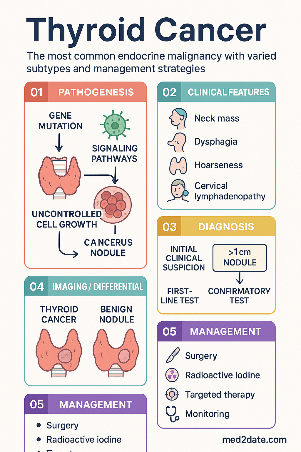
Clinical Presentation & Diagnostic Criteria
Typical Presentations
- Solitary thyroid nodule: Most common presentation — usually painless, firm, palpable; majority are benign (90–95% of nodules).
- Incidental finding: Increasingly detected on ultrasound, CT, PET-CT performed for other indications.
- Cervical lymphadenopathy: May be the presenting feature of PTC, especially in younger patients.
- Rapidly enlarging neck mass: Suggestive of ATC or aggressive DTC variant.
- Symptoms of local invasion: Hoarseness (recurrent laryngeal nerve), dysphagia (oesophageal compression), dyspnoea (tracheal compression), Horner's syndrome (sympathetic chain).
- Diarrhoea / flushing: May indicate metastatic MTC (calcitonin-mediated secretory diarrhoea).
- Bone pain / pathological fracture: Bone metastases, particularly FTC.
Diagnostic Workup of a Thyroid Nodule
- Thyroid function tests: TSH, free T4, free T3. Suppressed TSH may suggest a benign autonomous nodule; do not exclude malignancy.
- Neck ultrasound: First-line imaging; assess nodule size, composition, echogenicity, margins, calcifications, and vascularity. Use ACR TI-RADS or EU-TIRADS classification.
- Fine-needle aspiration biopsy (FNAB): Gold standard for cytological diagnosis; indicated for nodules meeting size threshold on TI-RADS score (e.g., TI-RADS 4–5 ≥10 mm; TI-RADS 3 ≥25 mm). Performed under ultrasound guidance.
- Bethesda classification of FNAB: Six categories (I–VI) from non-diagnostic to malignant; guides management.
- Molecular testing (Afirma, ThyroSeq): Consider for Bethesda III (AUS/FLUS) or Bethesda IV (follicular neoplasm) cytology to reduce unnecessary diagnostic surgery.
| Bethesda Category | Risk of Malignancy | Recommended Action |
|---|---|---|
| I — Non-diagnostic | 5–10% | Repeat FNAB (ultrasound-guided) or molecular testing |
| II — Benign | 0–3% | Surveillance ultrasound at 12–24 months |
| III — AUS/FLUS | 10–30% | Repeat FNAB, molecular testing, or diagnostic lobectomy |
| IV — Follicular neoplasm | 25–40% | Diagnostic lobectomy or molecular testing |
| V — Suspicious for malignancy | 50–75% | Total thyroidectomy or lobectomy (based on clinical factors) |
| VI — Malignant | 97–99% | Total thyroidectomy (most cases) |
Investigations
Risk Stratification
The 2015 American Thyroid Association (ATA) risk stratification system, adapted for Australian practice, guides the extent of initial therapy and intensity of follow-up.
- No local or distant metastases
- Complete macroscopic resection
- No vascular invasion
- No aggressive histology (tall cell, hobnail, columnar)
- cN0 or ≤5 pathologic micrometastases (<0.2 cm)
- Microscopic extrathyroidal extension
- Vascular invasion
- Aggressive histology variants
- BRAF V600E mutation (if other risk factors present)
- cN1 or >5 pathologic lymph node metastases
- Multifocal papillary microcarcinoma with ETE and BRAF+
- Gross extrathyroidal extension (T4)
- Incomplete tumour resection (R1 or R2)
- Distant metastases (M1)
- Post-operative Tg suggestive of distant disease
- pN1 with lymph nodes >3 cm
Management: Surgery, Radioiodine & TSH Suppression
Surgery
Surgery is the primary curative treatment for thyroid cancer. The extent of thyroidectomy and lymph node dissection is guided by tumour size, multifocality, extrathyroidal extension, and nodal disease.
| Surgical Procedure | Indications | Key Considerations |
|---|---|---|
| Thyroid lobectomy | Unifocal DTC ≤4 cm, no ETE, N0, low-risk histology; benign diagnostic uncertainty | Avoids lifelong thyroxine if contralateral lobe normal; requires completion thyroidectomy if high-risk features found on final histology |
| Total / near-total thyroidectomy | Tumour >4 cm, bilateral/multifocal, ETE, N1, M1, prior head/neck radiation, familial DTC | Allows RAI ablation and Tg surveillance; higher hypoparathyroidism and recurrent laryngeal nerve injury rates at low-volume centres |
| Central compartment dissection (VI) | Clinically positive central nodes (cN1a); prophylactic in DTC >4 cm or T3–T4 | Increased transient hypoparathyroidism risk |
| Lateral neck dissection (II–V) | Clinically positive lateral nodes (cN1b) — therapeutic only | Not recommended prophylactically; perform selective (not radical) dissection |
Radioactive Iodine (RAI) Therapy — I-131
RAI ablation exploits the iodine-uptake mechanism of differentiated thyroid cancer cells. It is used post-operatively to ablate residual thyroid tissue and treat microscopic or macroscopic residual/metastatic disease.
Indications for RAI Ablation
- High-risk DTC: Always indicated — high-activity I-131 (3.7–7.4 GBq / 100–200 mCi).
- Intermediate-risk DTC: Generally indicated — low-to-intermediate activity (1.1–3.7 GBq / 30–100 mCi).
- Low-risk DTC: May be omitted (ATA 2015 trend towards less aggressive ablation); individualised decision.
- MTC and ATC: NOT indicated — these tumours do not concentrate iodine.
Preparation for RAI
- TSH stimulation: TSH must be >30 mIU/L. Achieved by either thyroid hormone withdrawal (THW — 3–4 weeks of thyroxine cessation) or recombinant human TSH (rhTSH, Thyrogen® — preferred for quality of life).
- Low-iodine diet for 1–2 weeks prior.
- Check urine iodine if iodine contamination suspected.
- Avoid iodinated contrast for ≥2 months before RAI.
Side Effects of RAI
- Sialadenitis / xerostomia (most common — advise sour lollies/citrus post-RAI).
- Taste disturbance, nausea (acute).
- Transient radiation thyroiditis.
- Rare: second primary malignancies (leukaemia, salivary gland tumours) — risk increases with cumulative dose.
- Gonadal toxicity — counsel regarding fertility preservation before high-dose RAI.
TSH Suppression Therapy
Levothyroxine is used to suppress TSH below the normal range, as TSH stimulates DTC cell growth via the TSH receptor.
External Beam Radiation Therapy (EBRT)
- Consider for locally advanced DTC with gross residual disease, incomplete resection, or ATC.
- Not a routine part of DTC management; reserved for RAI-refractory locoregional disease.
- ATC: EBRT (intensity-modulated) often combined with radiosensitising chemotherapy (doxorubicin, paclitaxel) and/or targeted therapy.
Targeted Systemic Therapies for Advanced / Refractory Disease
Monitoring & Surveillance
Long-term follow-up is essential for all thyroid cancer patients. The intensity and tools depend on initial ATA risk stratification and response-to-therapy assessment.
Differentiated Thyroid Cancer — Post-Thyroidectomy Surveillance
| Time Point | Low Risk | Intermediate Risk | High Risk |
|---|---|---|---|
| 6–12 months | Neck US, TSH, Tg (±TgAb) | Neck US, TSH, Tg, diagnostic WBS if Tg detectable | Neck US, TSH, Tg, diagnostic WBS |
| Annually (years 1–5) | TSH, Tg; neck US if clinically indicated | TSH, Tg, neck US annually | TSH, Tg, neck US, ± diagnostic WBS, CT if indicated |
| After 5 years | Extend to 2–3 yearly TSH/Tg | Annual or 2-yearly TSH/Tg + US | Annual lifelong (may extend interval if excellent response) |
Response-to-Therapy Assessment
At 6–18 months post-initial therapy, patients are re-stratified based on biochemical and structural evidence:
- Excellent response: Stimulated Tg <1 ng/mL (or <0.2 ng/mL if using high-sensitivity assay), negative imaging → continue surveillance.
- Biochemical incomplete: Stimulated Tg ≥10 ng/mL or rising TgAb, but no structural disease → ongoing monitoring; consider repeat imaging.
- Structural incomplete: Persistent or new structural disease on imaging → further surgery, RAI, EBRT, or systemic therapy as indicated.
- Indeterminate response: Non-specific findings (e.g., mildly elevated Tg, equivocal US) → close follow-up.
Medullary Thyroid Cancer — Surveillance
- Calcitonin and CEA every 3–6 months (if elevated post-operatively, doubling time is prognostic).
- Cross-sectional imaging (CT chest/abdomen) annually or with rising markers.
- Calcitonin doubling time <2 years suggests aggressive disease — consider systemic therapy referral.
General Monitoring Parameters
- Thyroid function tests (TSH, free T4) every 6–12 weeks during dose titration, then every 6–12 months.
- Calcium, phosphate, PTH post-total thyroidectomy (monitor for hypoparathyroidism).
- Vitamin D levels — particularly in patients on TSH suppression (bone health).
- DEXA scan — postmenopausal women and men >50 years on long-term TSH suppression.
Special Populations
Pregnancy
Paediatric Thyroid Cancer
Elderly Patients
Renal Impairment
Hepatic Impairment
Immunocompromised Patients
Aboriginal and Torres Strait Islander Health Considerations
📚 References
- 1. Haugen BR, Alexander EK, Bible KC, et al. 2015 American Thyroid Association management guidelines for adult patients with thyroid nodules and differentiated thyroid cancer. Thyroid. 2016;26(1):1–133.
- 2. Wells SA Jr, Asa SL, Dralle H, et al. Revised American Thyroid Association guidelines for the management of medullary thyroid carcinoma. Thyroid. 2015;25(6):567–610.
- 3. Smallridge RC, Ain KB, Asa SL, et al. American Thyroid Association guidelines for management of patients with anaplastic thyroid cancer. Thyroid. 2012;22(11):1104–1139.
- 4. Amin MB, Edge SB, Greene FL, et al. (Eds). AJCC Cancer Staging Manual. 8th ed. New York: Springer; 2017.
- 5. Baloch ZW, Asa SL