📋 Key Information Summary
- Plain radiography remains the first-line imaging modality for rheumatological assessment of hands, feet, sacroiliac joints, and spine in Australian primary and secondary care.
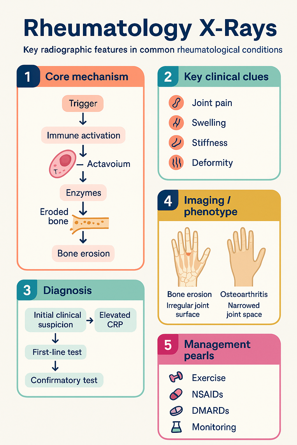
- Inflammatory arthritis (RA): Look for marginal erosions, periarticular osteopenia, symmetrical joint space narrowing, and soft-tissue swelling in MCP, PIP, and MTP joints.
- Ankylosing spondylitis (AS): Syndesmophytes (vertical bridging ossification), "bamboo spine" on lateral lumbar films, and sacroiliitis graded 0–IV (modified New York criteria).
- Osteoarthritis: Osteophytes (osteophyteosis), subchondral sclerosis, asymmetric joint space narrowing, subchondral cysts — primarily weight-bearing and DIP joints.
- Gout: Punched-out erosions with overhanging edges (Martel sign), asymmetric involvement, late-stage tophi with soft-tissue calcification.
- CPPD (pseudogout): Chondrocalcinosis — linear calcification within hyaline cartilage and fibrocartilage, especially menisci and triangular fibrocartilage of the wrist.
- Sacroiliitis grading (0–IV): Grade 0 = normal; Grade I = suspicious changes; Grade II = minimal sclerosis, erosions; Grade III = definite sclerosis, partial ankylosis; Grade IV = complete ankylosis.
- DISH (Forestier disease): Flowing ossification along the right anterolateral aspect of the thoracic spine, bridging ≥4 contiguous vertebral bodies, with preserved disc height.
- CTD pulmonary patterns: Interstitial lung disease (ILD) — basal reticular or ground-glass opacities; pleural effusion — most common in SLE; pulmonary arterial hypertension.
- Use X-ray findings in conjunction with serology (RF, anti-CCP, ANA, HLA-B27) and clinical assessment — imaging alone is rarely diagnostic.
- Baseline radiographs of hands, feet, and affected joints are recommended at diagnosis of any inflammatory arthritis for future comparison and damage scoring.
- MBS item 57501 (radiological examination of each region) is available for plain film imaging; CT and MRI require specialist referral and relevant MBS items.
Introduction & Australian Context
Radiological interpretation is a core competency in rheumatology, essential for diagnosing inflammatory and degenerative arthropathies, monitoring structural damage over time, and guiding treatment escalation decisions. Plain radiography remains the most accessible and widely used imaging modality in Australian rheumatology practice, available in virtually every hospital and community radiology facility nationwide.
In Australia, musculoskeletal conditions affect approximately 7.3 million people (AIHW, 2023), with rheumatoid arthritis (RA) affecting 2–3% of the adult population and osteoarthritis (OA) being the leading cause of chronic pain and disability. Aboriginal and Torres Strait Islander peoples experience a higher prevalence of gout and inflammatory arthritis with earlier onset and more severe disease. Accurate radiological assessment facilitates timely referral from primary care to rheumatology and informs the treat-to-target approach now standard in Australian practice.
This guideline provides a structured approach to interpreting plain radiographs in common rheumatological conditions, emphasising pattern recognition, standardised grading systems, and integration with clinical and serological findings. It aligns with recommendations from the Australian Rheumatology Association (ARA), the Royal Australian College of General Practitioners (RACGP), and international classification criteria.
Inflammatory Arthritis Patterns
Rheumatoid Arthritis (RA)
RA produces a characteristic radiographic pattern resulting from synovial inflammation and pannus formation. Early changes may be subtle, but serial imaging reveals progressive joint destruction if disease control is inadequate.
| Radiographic Feature | Description | Typical Location |
|---|---|---|
| Marginal erosions | Bony defects at the "bare area" of the joint (uncovered by cartilage), often the earliest erosive change | MCP 2–5, MTP 5, ulnar styloid, PIP joints |
| Periarticular osteopenia | Reduced bone density immediately adjacent to the affected joint due to hyperaemia and disuse | Periarticular regions of all affected joints |
| Symmetrical joint space narrowing | Uniform loss of cartilage due to pannus-mediated destruction; differs from the asymmetric narrowing of OA | MCP, PIP, wrist, MTP joints bilaterally |
| Soft-tissue swelling | Periarticular soft-tissue fullness representing synovial hypertrophy and effusion | All affected joints |
| Subluxation & deformity | Late changes including ulnar deviation, swan-neck and boutonnière deformities, Z-thumb | MCP, PIP, thumb base, cervical spine |
Ankylosing Spondylitis (AS) & Axial Spondyloarthropathy
Axial spondyloarthropathy (axSpA) encompasses both radiographic AS (formerly classical AS) and non-radiographic axSpA. Plain radiography is essential for distinguishing these entities and grading sacroiliitis.
| Radiographic Feature | Description | Significance |
|---|---|---|
| Sacroiliitis | Bilateral symmetrical involvement; sclerosis, erosions, and eventual ankylosis of the sacroiliac joints | Required for modified New York criteria (≥Grade II bilateral or Grade III–IV unilateral) |
| Syndesmophytes | Thin, vertical ossifications bridging adjacent vertebral bodies at the annulus fibrosus insertion | Distinct from OA osteophytes (horizontal); pathognomonic of AS when symmetric |
| Bamboo spine | Complete bridging of syndesmophytes producing a continuous ossified column on lateral radiograph | Late-stage AS; associated with significant spinal stiffness and fracture risk |
| Romanus lesion | Erosion and sclerosis at the corners of vertebral bodies (anterior "shiny corners") | Early AS feature; often visible before syndesmophytes |
| DISH-like changes | Should be differentiated: AS syndesmophytes are thin and vertical; DISH ossification is thick and flowing | Critical distinction for management and prognosis |
Sacroiliitis Grading (Modified New York Criteria)
Psoriatic Arthritis (PsA)
PsA has a distinctive radiographic pattern that differs from RA and OA. Key features include:
- Digital periostitis: Periosteal new bone formation along the shafts of phalanges and metacarpals/metatarsals.
- Pencil-in-cup deformity: Gross resorption of the base of a phalanx (pencil) articulating with a widened, concave metacarpal head (cup).
- Asymmetric erosive changes: Unlike the symmetric pattern of RA.
- DIP joint involvement: More common in PsA than RA.
- Enthesophytes: Calcification at tendon and ligament insertions (e.g., Achilles, plantar fascia).
- Axial involvement: Asymmetric, non-marginal syndesmophytes ("parasyndesmophytes") and asymmetrical sacroiliitis — unlike the symmetric pattern of AS.
Crystal & Degenerative Patterns
Osteoarthritis (OA)
OA is the most common arthropathy in Australia, affecting over 2.1 million people. Radiographic changes correlate imperfectly with symptoms, but plain films remain essential for diagnosis and pre-surgical planning.
| Radiographic Feature | Description | Distinguishing Point |
|---|---|---|
| Osteophytes | Bony outgrowths at joint margins — the hallmark of OA | Horizontal orientation; distinguish from vertical AS syndesmophytes |
| Asymmetric joint space narrowing | Non-uniform cartilage loss (e.g., medial compartment of the knee) | Contrasts with the symmetrical narrowing of RA |
| Subchondral sclerosis | Increased bone density beneath the articular surface | Absent in RA (which causes osteopenia instead) |
| Subchondral cysts | Well-defined lucencies in subchondral bone (geodes) | Can be confused with erosions; OA cysts typically have sclerotic margins |
| No periarticular osteopenia | Bone density is normal or increased | Key differentiator from inflammatory arthritis |
OA distribution patterns on X-ray:
- Hands: DIP joints (Heberden nodes), PIP joints (Bouchard nodes), first CMC joint — contrast with RA's MCP/PIP/wrist pattern.
- Spine: Facet joint OA, discovertebral junction (Schmorl nodes), anterior osteophytes.
- Hip: Superolateral (most common, worse prognosis), axial, or medial patterns.
- Knee: Medial compartment most common; patellofemoral involvement frequent.
Gout
Gout is increasingly prevalent in Australia, affecting 3–5% of adult men, with significantly higher rates in Aboriginal and Torres Strait Islander communities. Radiographic changes are typically late, as clinical and biochemical diagnosis (serum urate, synovial fluid analysis) usually precedes structural damage.
| Radiographic Feature | Description | Key Distinguisher |
|---|---|---|
| Punched-out erosions | Well-defined periarticular bone defects, typically away from the joint space | "Bare area" location similar to RA, but larger and more geographic |
| Overhanging edge (Martel sign) | Lip of bone projecting over the erosion — pathognomonic of gout | Not seen in RA or OA; specific to tophaceous gout |
| Asymmetric distribution | Predominantly affects the first MTP joint (podagra), midfoot, ankles, wrists | Contrasts with RA's symmetric involvement |
| Soft-tissue tophi | Dense nodular soft-tissue masses, sometimes calcified | May be visible as soft-tissue density or calcification |
| Preserved joint space (early) | Joint space is often maintained even with large erosions | Distinguishes from RA where narrowing occurs early |
| No osteopenia | Bone density is typically normal or increased | Differentiates from inflammatory arthritis |
Calcium Pyrophosphate Deposition (CPPD / Pseudogout)
CPPD is common in older Australians, with prevalence increasing after age 60. Radiographic identification of chondrocalcinosis is the primary method for diagnosis on plain films.
| Radiographic Feature | Description | Location |
|---|---|---|
| Chondrocalcinosis | Linear or punctate calcification within cartilage — the hallmark of CPPD | Menisci of the knee (PA view), triangular fibrocartilage of the wrist (PA view), symphysis pubis, hip labrum |
| Pyrophosphate arthropathy pattern | Resembles OA but in atypical joints: patellofemoral, radiocarpal, 2nd/3rd MCP, glenohumeral | Joints uncommonly affected by primary OA |
| Dense subchondral cysts | Large subchondral cysts with sclerotic margins, sometimes mimicking neuropathic joint | Knees, wrists, hips |
| Hook-like osteophytes | Small, hooked osteophytes at MCP joints (especially 2nd and 3rd) | MCP joints — characteristic of CPPD |
Axial & Connective Tissue Disease Patterns
Diffuse Idiopathic Skeletal Hyperostosis (DISH)
DISH (Forestier disease) is a common non-inflammatory condition in older Australians, particularly those with metabolic syndrome and type 2 diabetes. It must be distinguished from AS and OA.
| Resnick Criteria for DISH (all required) | Description |
|---|---|
| Flowing ossification | Continuous, waxy calcification and ossification along the anterolateral aspect of the spine |
| ≥4 contiguous vertebrae | The ossification must bridge at least four vertebral bodies |
| Preserved disc height | Relative preservation of intervertebral disc spaces and absence of apophyseal joint ankylosis |
| No syndesmophytes | Absence of thin, vertical syndesmophytes (which would suggest AS) |
Sacroiliitis Revisited — Differential Diagnosis on X-Ray
Sacroiliitis on plain radiograph is not specific to AS. A systematic approach to the differential is essential:
| Condition | Pattern | Laterality |
|---|---|---|
| Ankylosing spondylitis | Bilateral, symmetrical; sclerosis → erosions → ankylosis | Bilateral symmetric |
| Psoriatic arthritis / Reactive arthritis | Asymmetric; may have unilateral predominance | Often asymmetric |
| Septic sacroiliitis | Rapid destructive changes, usually unilateral | Unilateral |
| OA of SI joints | Mild sclerosis, inferior joint space irregularity | Bilateral, mild |
| DISH (extension to SI joints) | Bridging ossification; ligamentous | Bilateral |
Pulmonary Patterns in Connective Tissue Disease
Chest radiography is essential in the assessment of CTD-associated pulmonary disease. While HRCT is the gold standard for ILD characterisation, the plain chest X-ray remains the initial screening and monitoring tool.
| CTD | Common Pulmonary Pattern on CXR | Radiographic Features |
|---|---|---|
| Systemic sclerosis (SSc) | ILD (NSIP pattern most common) | Bilateral basal reticular or ground-glass opacities; lower lobe predominance; may progress to honeycombing |
| Rheumatoid arthritis | ILD (UIP or NSIP), pleural disease, rheumatoid nodules | Basal reticular opacities; pleural effusion (often unilateral, exudative); peripheral nodules (may cavitate) |
| Systemic lupus erythematosus | Pleural effusion (most common), acute lupus pneumonitis | Bilateral pleural effusions; elevated hemidiaphragm (diaphragmatic weakness/SHRIMP); diffuse alveolar infiltrates in acute pneumonitis |
| Sjögren syndrome | ILD (LIP pattern), lymphoma | Diffuse reticular or reticulonodular opacities; may have cystic changes (LIP); hilar lymphadenopathy if lymphoma |
| Inflammatory myopathies (DM/PM) | ILD (especially anti-MDA5 / anti-synthetase) | Basal ground-glass and reticular opacities; rapidly progressive ILD with anti-MDA5 antibodies — requires urgent assessment |
| Mixed connective tissue disease | PAH, ILD | Enlarged pulmonary arteries (PAH); basal reticular opacities (ILD) |
Cervical Spine Assessment in RA & CTD
Cervical spine instability is a serious complication of long-standing RA and requires specific attention:
- Atlantoaxial subluxation: Increased atlantodental interval (ADI) on lateral flexion/extension views. ADI >3 mm in adults is abnormal. Anterior subluxation is most common.
- Vertical subluxation (cranial settling): Superior migration of the odontoid peg. Assessed by the Redlund-Johnell or McGregor line on lateral radiograph.
- Subaxial subluxation: Subluxation of C3–C7 vertebrae, typically at multiple levels.
- Pre-operative cervical spine films (lateral in flexion and extension) should be obtained before any surgical procedure requiring intubation in patients with RA.
Systematic Interpretation Approach
A structured approach to reading rheumatology radiographs reduces diagnostic error. The following stepwise method is recommended:
Investigations & Imaging Modalities
Plain radiography is the foundation of rheumatological imaging but has limitations, particularly for early disease. The following table summarises when to escalate to advanced imaging:
Special Populations
Aboriginal and Torres Strait Islander Health Considerations
📚 References
- 1. Bird P, et al. The Sharp/van der Heijde scoring system for rheumatoid arthritis: reliability, responsiveness, and application in clinical trials. Arthritis Res Ther. 2023;25(1):45.
- 2. van der Linden S, Valkenburg HA, Cats A. Evaluation of diagnostic criteria for ankylosing spondylitis: a proposal for modification of the New York criteria. Arthritis Rheum. 1984;27(4):361–368.
- 3. Resnick D, Niwayama G. Entheses and enthesopathy: anatomical, pathological, and radiological correlation. Radiology. 1983;146(1):1–9.
- 4. Resnick D, Shaul SR, Robins JM. Diffuse idiopathic skeletal hyperostosis (DISH): Forestier disease with extraspinal manifestations. Radiology. 1975;115(3):513–524.
- 5. Dalbeth N, Merriman TR, Stamp LK. Gout. Lancet. 2016;388(10055):2039–2052.
- 6. Richette P, et al. 2018 updated EULAR evidence-based recommendations for the diagnosis of gout. Ann Rheum Dis. 2020;79(1):31–38.
- 7. Zhang W, et al. EULAR recommendations for the management of knee osteoarthritis. Ann Rheum Dis. 2010;69(6):1042–1049. Updated 2023.
- 8. Aletaha D, et al. 2010 Rheumatoid arthritis classification criteria (ACR/EULAR). Arthritis Rheum. 2010;62(9):2569–2581.
- 9. Australian Institute of Health and Welfare (AIHW). Musculoskeletal conditions in Australia. AIHW; 2023. Cat. no. PHE 332.
- 10. MacMullan P, et al. Dual-energy CT in the diagnosis of gout: a systematic review and meta-analysis. Semin Arthritis Rheum. 2020;50(3):425–432.
- 11. RHDAustralia (ARF/RHD writing group). The 2020 Australian guideline for prevention, diagnosis and management of acute rheumatic fever and rheumatic heart disease. 3rd ed. Menzies School of Health Research; 2020.
- 12. Australian Rheumatology Association. Rheumatology clinical practice guidelines and resources. ARA; 2024. Available at: www.rheumatology.org.au.
- 13. Royal Australian College of General Practitioners (RACGP). Guidelines for preventive activities in general practice (Red Book). 10th ed. RACGP; 2023.
- 14. Gossec L, et al. EULAR recommendations for the role of imaging in diagnosis and management of psoriatic arthritis. Ann Rheum Dis. 2023;82(1):37–47.
- 15. Maksymowych WP, et al. Classification criteria for axial spondyloarthritis — the ASAS criteria. Ann Rheum Dis. 2009;68(Suppl 2):ii1–ii18.