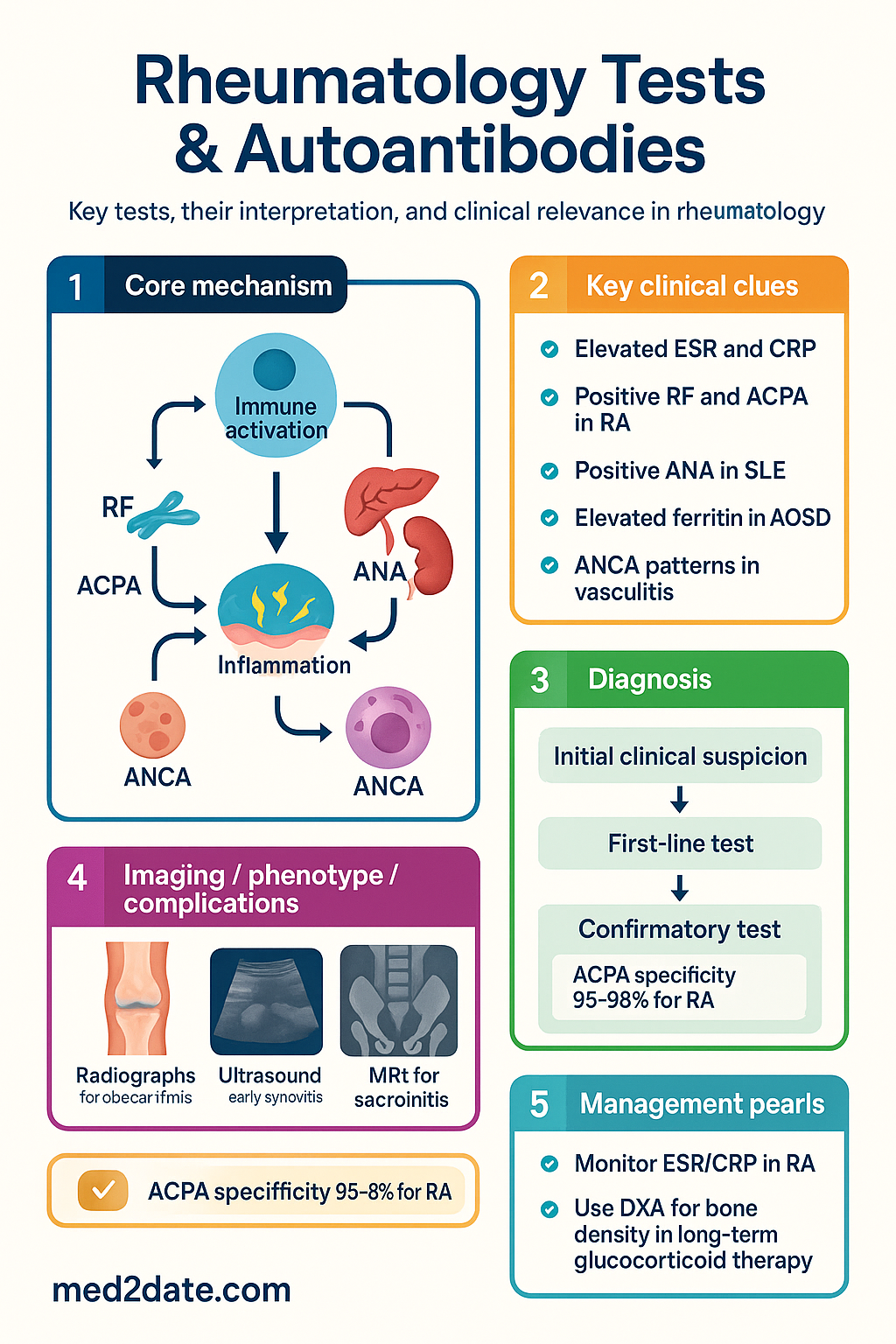
📋 Key Information Summary
- ESR and CRP are the most widely ordered inflammatory markers in rheumatology; CRP is more responsive to acute-phase changes (rising within 6–8 h, half-life ~19 h), whereas ESR reflects chronic inflammation and is influenced by age, sex, anaemia, and paraproteinaemia.
- Ferritin is an acute-phase reactant; markedly elevated levels (>1 000 µg/L) raise concern for adult-onset Still disease, haemophagocytic lymphohistiocytosis (HLH), or severe inflammatory flare.
- Rheumatoid factor (RF) has ~70–80 % sensitivity but only ~80 % specificity for RA; false positives occur in hepatitis C, Sjögren syndrome, infective endocarditis, TB, and cryoglobulinaemia.
- Anti-citrullinated peptide antibodies (ACPA/anti-CCP) have comparable sensitivity (~68–80 %) to RF but much higher specificity (~95–98 %) for RA; positive ACPA predicts more erosive disease and is included in the 2010 ACR/EULAR RA classification criteria.
- ANA is present in ~5–15 % of healthy adults; a positive ANA alone does not diagnose SLE. Specificity depends on titre, pattern (homogeneous, speckled, centromere, nucleolar), and the associated extractable nuclear antigen (ENA) profile.
- Anti-dsDNA and complement C3/C4 are essential for SLE diagnosis and disease-activity monitoring; falling C3/C4 with rising anti-dsDNA titres suggests lupus nephritis flare.
- ANCA testing uses immunofluorescence (IF) combined with ELISA; c-ANCA/PR3 is associated with granulomatosis with polyangiitis (GPA), while p-ANCA/MPO favours microscopic polyangiitis (MPA) and eosinophilic GPA.
- Antiphospholipid antibodies (lupus anticoagulant, anticardiolipin, anti-β2 glycoprotein-I) must be positive on two occasions ≥12 weeks apart to confirm antiphospholipid syndrome (APS).
- Plain radiographs remain the first-line imaging for suspected osteoarthritis, crystal arthropathy, and advanced rheumatoid arthritis; characteristic findings include periarticular erosions, joint-space narrowing, and chondrocalcinosis.
- Musculoskeletal ultrasound detects early synovitis, tenosynovitis, and erosions before radiographic changes, and is increasingly used as a point-of-care tool in Australian rheumatology clinics.
- MRI is the gold standard for sacroiliitis assessment in axial spondyloarthritis and for detecting bone marrow oedema, stress fractures, and avascular necrosis.
- DXA bone-density scanning is indicated for patients on long-term glucocorticoids (≥3 months at ≥7.5 mg/day prednisolone), postmenopausal women with RA, and anyone with fragility-fracture risk; report using T-scores with FRAX® integration.
- Test selection must be guided by the clinical pre-test probability; indiscriminate ANA and autoantibody ordering in low-risk patients leads to false positives, unnecessary anxiety, and downstream investigation costs.
Introduction & Australian Epidemiology
Rheumatology is a specialty heavily reliant on serological, immunological, and imaging investigations to establish diagnoses, prognosticate, monitor disease activity, and guide treatment decisions. In Australia, rheumatic diseases collectively affect approximately 3.6 million people, with rheumatoid arthritis (RA) alone accounting for an estimated 456 000 adults and costing the health system over AUD 3 billion annually (AIHW 2023). Musculoskeletal conditions are the leading cause of disability burden, representing 12.8 % of total disability-adjusted life years (DALYs).
Serological testing in rheumatology serves three main purposes: (1) diagnostic classification (e.g., RF and ACPA for RA, anti-dsDNA for SLE); (2) prognostic stratification (e.g., ACPA-positive RA has a higher erosive burden); and (3) disease-activity and treatment-response monitoring (e.g., ESR/CRP trends, complement levels in lupus). Imaging adds structural and inflammatory information that serology alone cannot provide.
Australian rheumatologists face unique challenges, including higher rates of axial spondyloarthritis and gout in Aboriginal and Torres Strait Islander populations, delayed referral pathways in rural and remote settings, and MBS billing considerations for ultrasound and DXA services. This guideline provides a structured overview of commonly ordered rheumatology tests, their interpretation, limitations, and appropriate clinical contexts.
Inflammatory Markers: ESR, CRP, Ferritin, CBC, LDH & Uric Acid
Inflammatory markers form the backbone of rheumatology practice, providing objective evidence of systemic or local inflammation and enabling longitudinal disease-activity monitoring. Understanding their kinetics, influencing factors, and clinical context is essential for accurate interpretation.
Erythrocyte Sedimentation Rate (ESR)
The ESR measures the rate at which red blood cells sediment in a standardised tube over one hour (Westergren method). It is influenced by plasma fibrinogen, immunoglobulins, and other acute-phase proteins that promote rouleaux formation.
| Parameter | Detail |
|---|---|
| Normal range | Males: age ÷ 2; Females: (age + 10) ÷ 2 (mm/h). General adult reference <20 mm/h (varies by lab) |
| Rise kinetics | Begins at 24–48 h; peaks at 4–5 days |
| Half-life / fall | Slow normalisation over days–weeks after inflammation resolves |
| Key use in rheumatology | Polymyalgia rheumatica (PMR) diagnosis and monitoring; temporal arteritis; RA disease activity; SLE flare tracking |
| Causes of false elevation | Anaemia, pregnancy, obesity, female sex, advancing age, hypergammaglobulinaemia, renal failure |
| Causes of false depression | Polycythaemia, sickle-cell disease, spherocytosis, CHF, hypofibrinogenaemia |
| MBS item | MBS Item 65070 — ESR |
C-Reactive Protein (CRP)
CRP is a pentraxin synthesised by hepatocytes in response to IL-6. It rises earlier and falls faster than ESR, making it superior for monitoring acute-phase changes and treatment response.
| Parameter | Detail |
|---|---|
| Normal range | <5 mg/L (high-sensitivity CRP: <3 mg/L) |
| Rise kinetics | 6–8 h after stimulus; peaks at 36–48 h |
| Half-life | ~19 h (constant); falls rapidly once stimulus removed |
| Key rheumatology uses | Active RA flares, PMR monitoring, infection vs. flare differentiation, giant cell arteritis, post-operative inflammation tracking |
| Advantages over ESR | Not affected by anaemia, age, sex, or paraproteins; more responsive to change |
| MBS item | MBS Item 65090 — CRP |
Ferritin
Ferritin is both an iron-storage protein and an acute-phase reactant. In rheumatology, it is primarily used as an inflammatory marker rather than an iron-status indicator.
| Ferritin Level | Clinical Significance |
|---|---|
| Elevated (300–1 000 µg/L) | Chronic inflammation, iron overload, liver disease, metabolic syndrome |
| Markedly elevated (>1 000 µg/L) | Adult-onset Still disease (AOSD), HLH/MAS, severe infection, haematological malignancy |
| Hyperferritinaemia in AOSD | Classic triad: quotidian fevers, evanescent salmon-coloured rash, ferritin often >3 000 µg/L. Glycosylated ferritin <20 % is highly suggestive (available at specialist reference labs) |
Complete Blood Count (CBC/FBC)
The CBC provides important contextual information in rheumatic diseases:
- Normochromic normocytic anaemia — the most common anaemia pattern in chronic inflammatory diseases (RA, SLE); ferritin is normal or elevated, serum iron and transferrin are low.
- Thrombocytosis — reactive thrombocytosis is common in active RA, vasculitis, and AOSD; platelets >600 × 10⁹/L warrants consideration of secondary causes.
- Leukocytosis — seen in active RA, AOSD (often >15 × 10⁹/L with neutrophilia), SLE on corticosteroids; must distinguish from infection.
- Leukopenia / lymphopenia — characteristic of active SLE (lymphopenia <1.0 × 10⁹/L is included in the 2019 EULAR/ACR SLE classification criteria) and may indicate disease activity or immunosuppressive drug toxicity (azathioprine, methotrexate, mycophenolate).
- Eosinophilia — consider eosinophilic GPA (Churg-Strauss), hypereosinophilic syndromes, parasitic infections, and drug reactions.
- Pancytopenia — raises concern for SLE with bone marrow involvement, HLH/MAS, methotrexate toxicity, or myelodysplasia.
Lactate Dehydrogenase (LDH)
LDH is a non-specific marker of cellular damage. In rheumatology, it is relevant for:
- HLH / MAS: Markedly elevated LDH is a hallmark; often >1 000 U/L alongside hyperferritinaemia and cytopenias.
- Vasculitis assessment: Elevated LDH in the context of constitutional symptoms may indicate end-organ damage.
- Interstitial lung disease: LDH can be elevated in ILD associated with systemic sclerosis, myositis, or RA.
Uric Acid
Serum urate is essential in the diagnosis and monitoring of gout and crystal arthropathies:
- Target serum urate for urate-lowering therapy (ULT): <0.36 mmol/L (general) or <0.30 mmol/L (tophaceous gout).
- A normal serum urate during an acute gout flare does not exclude gout (up to 40 % of patients have levels within the reference range during an attack).
- Asymptomatic hyperuricaemia (serum urate >0.42 mmol/L in men, >0.36 mmol/L in women) does not routinely require ULT unless tophi, frequent attacks, CKD stage ≥3, or urolithiasis are present.
- MBS Item 66510 — Uric acid.
Autoantibody Profiles
Autoantibody testing is central to the diagnosis, classification, and prognostication of autoimmune rheumatic diseases. Understanding the sensitivity, specificity, and clinical context of each antibody is essential for appropriate ordering and interpretation.
Rheumatoid Factor (RF)
RF is an immunoglobulin (usually IgM, occasionally IgG or IgA) that binds the Fc portion of IgG. It is detected by nephelometry, latex agglutination, or ELISA.
| Parameter | Detail |
|---|---|
| Sensitivity for RA | ~70–80 % |
| Specificity for RA | ~80 % |
| Positive in | RA, Sjögren syndrome (often high titre), hepatitis C/cryoglobulinaemia, infective endocarditis, TB, sarcoidosis, ILD, lymphoproliferative disorders |
| Normal population | ~5 % of healthy adults have low-titre RF |
| Prognostic value | High-titre RF in RA predicts more aggressive disease, extra-articular manifestations (vasculitis, nodules, ILD), and worse radiographic outcomes |
| MBS item | MBS Item 69410 — Rheumatoid factor |
Anti-Citrullinated Peptide Antibodies (ACPA / Anti-CCP)
ACPA (anti-cyclic citrullinated peptide, anti-CCP) antibodies target citrullinated proteins generated by peptidylarginine deiminase (PAD) enzymes. Second-generation anti-CCP assays (CCP2, CCP3) have largely superseded earlier versions.
| Parameter | Detail |
|---|---|
| Sensitivity for RA | ~68–80 % (comparable to RF) |
| Specificity for RA | ~95–98 % (significantly higher than RF) |
| Prognostic value | ACPA-positive RA: higher erosive burden, greater joint destruction rate, increased risk of cardiovascular events, and association with HLA-DRB1 shared epitope |
| Pre-disease detection | ACPA may be positive years before clinical RA onset; useful for screening at-risk individuals (e.g., first-degree relatives, undifferentiated arthralgia) |
| Dual positivity (RF + ACPA) | Highest risk subgroup; double-positive patients have ~4× risk of developing erosive RA compared to single-positive patients |
| MBS item | MBS Item 69410 — Anti-CCP (often bundled with RF) |
Antinuclear Antibody (ANA) Panel
ANA testing by indirect immunofluorescence (IIF) on HEp-2 cells is the gold-standard screening method. A positive ANA requires interpretation in conjunction with the staining pattern, titre, and clinical context.
| Pattern | Associated Antibodies | Associated Conditions |
|---|---|---|
| Homogeneous / diffuse | Anti-dsDNA, anti-histone | SLE, drug-induced lupus |
| Speckled | Anti-Sm, anti-Ro (SSA), anti-La (SSB), anti-U1 RNP | SLE, Sjögren, MCTD |
| Centromere | Anti-centromere (CENP-A/B) | Limited cutaneous SSc (CREST) |
| Nucleolar | Anti-Scl-70, anti-RNA polymerase III, anti-Th/To, anti-PM-Scl | Diffuse cutaneous SSc, PM/SSc overlap |
| Cytoplasmic | Anti-Jo-1, anti-PL-7, anti-MDA5 | Anti-synthetase syndrome, dermatomyositis with ILD |
Key ANA-associated extractable nuclear antigens (ENA):
- Anti-dsDNA: Highly specific for SLE (~95 %); sensitivity ~60 %. Titres correlate with disease activity, particularly lupus nephritis. Normal complement (C3/C4) with rising anti-dsDNA = serological flare (may precede clinical flare by weeks–months).
- Anti-Smith (anti-Sm): Most specific antibody for SLE (~99 %) but low sensitivity (~25–30 %). When present, it is a strong diagnostic marker.
- Anti-Ro/SSA: Associated with Sjögren syndrome (primary and secondary), subacute cutaneous lupus, neonatal lupus, and congenital heart block. Anti-Ro52 and anti-Ro60 are distinct epitopes; reporting should specify both.
- Anti-La/SSB: Usually co-occurs with anti-Ro; associated with Sjögren syndrome and neonatal lupus. Isolated anti-La is uncommon.
- Anti-Scl-70 (anti-topoisomerase I): Associated with diffuse cutaneous systemic sclerosis (dcSSc) and interstitial lung disease. Poor prognosis marker.
- Anti-centromere (CENP-A/B): Associated with limited cutaneous systemic sclerosis (lcSSc/CREST) and pulmonary arterial hypertension (PAH). Rarely seen in diffuse SSc.
- Anti-U1 RNP: High titre is diagnostic of mixed connective tissue disease (MCTD); also seen in SLE.
- Anti-Jo-1 (anti-histidyl-tRNA synthetase): Most common anti-synthetase antibody; associated with polymyositis, interstitial lung disease, mechanic's hands, and Raynaud phenomenon.
- Anti-RNA polymerase III: Associated with diffuse SSc and increased risk of scleroderma renal crisis; also linked with concurrent malignancy.
Antineutrophil Cytoplasmic Antibodies (ANCA)
ANCA testing requires a two-step approach: initial screening by indirect immunofluorescence (IF) on ethanol-fixed neutrophils, followed by antigen-specific ELISA for proteinase 3 (PR3) and myeloperoxidase (MPO).
| ANCA Type | IF Pattern | ELISA Target | Associated Vasculitis |
|---|---|---|---|
| c-ANCA | Cytoplasmic, granular | PR3 | Granulomatosis with polyangiitis (GPA, Wegener's) |
| p-ANCA | Perinuclear | MPO | Microscopic polyangiitis (MPA), eosinophilic GPA (Churg-Strauss) |
| Atypical ANCA | Perinuclear or cytoplasmic | No PR3/MPO | Inflammatory bowel disease, drug-induced (e.g., hydralazine, propylthiouracil), primary sclerosing cholangitis |
Clinical utility of ANCA titres:
- Rising PR3/MPO titres may predict relapse, but serial monitoring is not universally recommended and should be interpreted alongside clinical findings.
- ANCA negativity does not exclude ANCA-associated vasculitis — approximately 10 % of GPA and 40 % of EGPA cases are ANCA-negative.
- In Australia, ANCA-associated vasculitis incidence is estimated at 10–20 per million per year, with a median age at diagnosis of 55–65 years.
Antiphospholipid Antibodies (aPL)
Antiphospholipid syndrome (APS) is diagnosed when clinical criteria (vascular thrombosis or pregnancy morbidity) are met alongside persistent laboratory positivity. The lupus anticoagulant (LA), anticardiolipin antibodies (aCL), and anti-β2 glycoprotein-I antibodies (anti-β2GPI) form the "triple test" and must be positive on two occasions at least 12 weeks apart.
| Test | Method | Positive Threshold | Clinical Association |
|---|---|---|---|
| Lupus anticoagulant (LA) | dRVVT, SCT, or APTT-based mixing/confirmatory | Prolonged screen + mixing study + confirmatory step | Highest thrombotic risk; arterial & venous thrombosis |
| Anticardiolipin IgG/IgM | ELISA | >40 GPL or MPL, or >99th percentile | Thrombosis, pregnancy morbidity |
| Anti-β2 glycoprotein-I IgG/IgM | ELISA | >99th percentile | Thrombosis; may be sole positive marker |
Imaging in Rheumatology
Imaging is essential for confirming structural damage, detecting subclinical inflammation, guiding interventional procedures, and monitoring disease progression. The choice of modality depends on the clinical question, availability, and cost.
Plain Radiography (X-ray)
Plain X-rays remain the first-line imaging modality for many rheumatic conditions due to wide availability, low cost, and established scoring systems.
| Condition | Characteristic X-ray Findings |
|---|---|
| Rheumatoid arthritis | Periarticular erosions (MCP, MTP, ulnar styloid), joint-space narrowing (uniform), periosteal new bone, juxta-articular osteopenia, subluxation (Z-thumb, ulnar deviation), atlanto-axial subluxation (cervical spine) |
| Osteoarthritis | Asymmetric joint-space narrowing, subchondral sclerosis, osteophytes, subchondral cysts, no erosions (unless erosive OA variant) |
| Psoriatic arthritis | Asymmetric erosions, periostitis, pencil-in-cup deformity (DIP predominant), new bone formation, joint-space narrowing, sacroiliitis |
| Gout | Late: punched-out erosions with overhanging edges (rat-bite erosions), asymmetric, juxta-articular, with preserved joint space until late |
| Calcium pyrophosphate deposition (CPPD) | Chondrocalcinosis (calcification of hyaline cartilage and fibrocartilage), most commonly in the knee (menisci), wrist (triangular fibrocartilage), and symphysis pubis |
| Ankylosing spondylitis | Sacroiliitis (bilateral, erosions → sclerosis → ankylosis), syndesmophytes (bamboo spine), squaring of vertebral bodies, apophyseal joint ankylosis |
| Systemic sclerosis | Acro-osteolysis (tuft resorption), soft-tissue calcinosis, joint-space preservation |
| Charcot arthropathy | Joint destruction, subluxation, debris, sclerosis — disproportionate to symptoms |
Musculoskeletal Ultrasound (MSK US)
MSK ultrasound has transformed rheumatology practice, providing real-time, dynamic, and radiation-free assessment of soft tissues, joints, and entheses.
- Synovitis detection: Power Doppler ultrasound detects hyperaemia within inflamed synovium; more sensitive than clinical examination for subclinical synovitis.
- Erosion detection: US detects bone erosions earlier than plain radiographs, particularly at the MCP, MTP, and wrist joints.
- Tenosynovitis: Excellent for detecting flexor and extensor tenosynovitis (RA, psoriatic arthritis).
- Enthesitis: Grey-scale (thickening, hypoechogenicity) and Power Doppler assessment at common entheses (plantar fascia, Achilles, lateral epicondyle).
- Gout: "Double-contour" sign on cartilage surface (monosodium urate crystal deposition); tophus detection and measurement.
- Guided injections: US-guided intra-articular and peritendinous injections improve accuracy and efficacy compared to landmark-based techniques.
- MBS considerations: MSK ultrasound is MBS-billable when performed by a specialist (rheumatologist, radiologist) but not universally rebateable for GP-performed scans. Check current MBS item descriptors (items in the 55000 range).
Magnetic Resonance Imaging (MRI)
MRI provides superior soft-tissue contrast and is the gold standard for several rheumatological assessments:
- Axial spondyloarthritis: MRI of the sacroiliac joints (STIR and T1 sequences) detects active sacroiliitis (bone marrow oedema) before radiographic changes appear; essential for early diagnosis. The Assessment of SpondyloArthritis International Society (ASAS) defines a positive MRI as bone marrow oedema in ≥2 sites on a single slice or 1 site on ≥2 slices.
- Avascular necrosis (AVN): MRI is the most sensitive modality for early AVN detection (femoral head, humeral head); X-rays are often normal in early disease.
- Myositis: MRI of affected muscles (STIR sequences) shows oedema in active inflammation and fatty replacement in chronic disease; guides muscle biopsy site selection.
- Rheumatoid arthritis: MRI detects early erosions, bone marrow oedema (predictive of future erosion), and tenosynovitis; increasingly used in clinical trials.
- Large-vessel vasculitis: MR angiography can detect vessel-wall thickening and oedema in giant cell arteritis and Takayasu arteritis.
DXA Bone Density Scan
Dual-energy X-ray absorptiometry (DXA) is the standard method for assessing bone mineral density (BMD) and fracture risk. In rheumatology, it is particularly relevant for patients on glucocorticoids and those with inflammatory arthritides.
| Parameter | Detail |
|---|---|
| DXA T-score classification | Normal: ≥ −1.0; Osteopenia: −1.0 to −2.5; Osteoporosis: ≤ −2.5 |
| Glucocorticoid threshold | DXA indicated if prednisolone ≥7.5 mg/day for ≥3 months, or any dose if duration will be prolonged |
| FRAX® integration | FRAX® (adjusted for glucocorticoid dose) used to guide treatment thresholds; consider anti-osteoporotic therapy if 10-year major fracture risk ≥20 % or hip fracture risk ≥3 % |
| RA-specific considerations | RA is an independent risk factor for osteoporosis (inflammation, immobility, corticosteroids); include RA as a secondary risk factor in FRAX® calculation |
| Sites scanned | Lumbar spine (L1–L4) and femoral neck (bilateral); distal 1/3 radius if hip/spine not evaluable |
| MBS item | MBS Item 12306 — DXA bone density; requires clinical indication (see MBS criteria) |
Special Populations
Aboriginal and Torres Strait Islander Health Considerations
Aboriginal and Torres Strait Islander peoples experience a disproportionate burden of rheumatic disease, including higher rates of gout, rheumatic heart disease (RHD), and systemic lupus erythematosus. These disparities are compounded by barriers to accessing specialist rheumatology services, pathology testing, and imaging in remote and very remote communities.
📚 References
- 1. Australian Institute of Health and Welfare (AIHW). Musculoskeletal conditions in Australia: a snapshot, 2023. Canberra: AIHW; 2023.
- 2. Kay J, Upchurch KS. ACR/EULAR 2010 rheumatoid arthritis classification criteria. Ann Rheum Dis. 2012;71(Suppl 2):e45–e48.
- 3. Aringer M, Costenbader K, Daikh D, et al. 2019 European League Against Rheumatism/American College of Rheumatology classification criteria for systemic lupus erythematosus. Ann Rheum Dis. 2019;78(9):1151–1159.
- 4. Bossuyt X, Cohen Tervaert JW, Arimura Y, et al. Position paper: Revised 2017 international consensus on testing of ANCAs in granulomatosis with polyangiitis and microscopic polyangiitis. Nat Rev Rheumatol. 2017;13(11):683–692.
- 5. Miyakis S, Lockshin MD, Atsumi T, et al. International consensus statement on an update of the classification criteria for definite antiphospholipid syndrome (APS). J Thromb Haemost. 2006;4(2):295–306.
- 6. Rudwaleit M, van der Heijde D, Landewé R, et al. The development of Assessment of SpondyloArthritis International Society classification criteria for axial spondyloarthritis (part II): validation and final selection. Ann Rheum Dis. 2009;68(6):777–783.
- 7. Doria A, Zen M, Canova M, et al. SLE diagnosis and treatment: when early is early. Autoimmun Rev. 2010;10(1):55–60.
- 8. Piga M, Gabba A, Congia M, et al. Musculoskeletal ultrasound in rheumatology: a review. Clin Exp Rheumatol. 2020;38(4):717–726.
- 9. Ledingham J, Deighton C; British Society for Rheumatology Standards, Guidelines and Audit Working Group. Update on the British Society for Rheumatology guidelines for prescribing TNFα blockers in adults with rheumatoid arthritis (update of previous guidelines of April 2001). Rheumatology. 2005;44(2):159–163.
- 10. RHDAustralia (ARF/RHD writing group). The 2020 Australian guideline for prevention, diagnosis and management of acute rheumatic fever and rheumatic heart disease. 3rd ed. Menzies School of Health Research; 2020.
- 11. Savige J, Gillis D, Benson E, et al. International consensus statement on testing and reporting of antineutrophil cytoplasmic antibodies (ANCA). Am J Clin Pathol. 1999;111(4):507–513.
- 12. Mok CC, Kwok CL, Ho LY, et al. Life expectancy, standardized mortality ratios, and causes of death in six rheumatic diseases in Hong Kong, China. Arthritis Rheum. 2011;63(5):1182–1189.
- 13. Nikiphorou E, de Lusignan S, Mallen CD, et al. Cardiovascular risk factors and outcomes in early rheumatoid arthritis: a population-based study. Heart. 2020;106(20):1566–1572.
- 14. Richette P, Doherty M, Pascual E, et al. 2018 updated European League Against Rheumatism evidence-based recommendations for the diagnosis of gout. Ann Rheum Dis. 2020;79(1):31–38.
- 15. Australian Commission on Safety and Quality in Health Care (ACSQHC). Australian Atlas of Healthcare Variation. Sydney: ACSQHC; 2021.