Home Rheumatology Henoch-Schonlein Purpura (IgA Vasculitis)

Henoch-Schonlein Purpura (IgA Vasculitis)

📋 Key Information Summary

📋
  • IgA Vasculitis (IgAV, Henoch-Schönlein Purpura) is the most common systemic small-vessel vasculitis in children, predominantly affecting those aged 3–10 years.
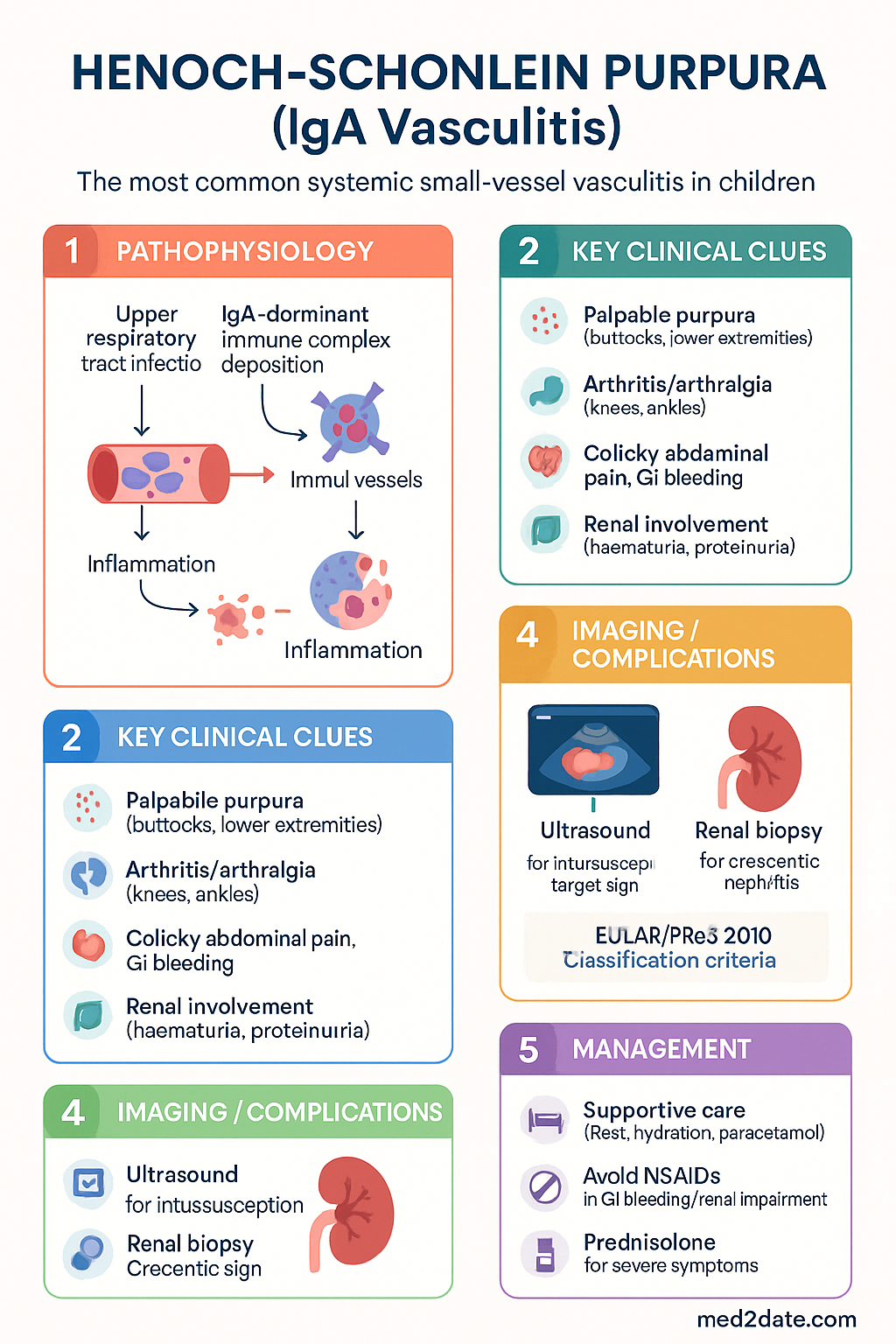
  • Pathophysiology involves IgA1-dominant immune complex deposition in vessel walls, triggered most often by upper respiratory tract infection (especially Group A Streptococcus).
  • Clinical tetrad: (1) palpable purpura — mainly buttocks and lower extremities, (2) arthritis/arthralgia — large joints (knees, ankles), (3) colicky abdominal pain with GI bleeding risk, (4) renal involvement — haematuria, proteinuria, nephritis.
  • Diagnosis is clinical using the EULAR/PReS 2010 classification criteria: palpable purpura (mandatory) plus at least one of diffuse abdominal pain, IgA deposition on biopsy, arthritis/arthralgia, or renal involvement.
  • Skin biopsy showing leucocytoclastic vasculitis with IgA deposits on immunofluorescence confirms the diagnosis; renal biopsy is reserved for persistent nephritis or nephrotic-range proteinuria.
  • The majority of children are managed supportively — disease is self-limiting in 95% of paediatric cases within 4–6 weeks.
  • NSAIDs (ibuprofen) provide analgesia for arthralgia; avoid in active GI bleeding or renal impairment.
  • Prednisolone 1 mg/kg/day (max 40 mg) for 1–2 weeks then taper is indicated for severe abdominal pain, intussusception risk, or significant renal involvement.
  • For crescentic IgA nephritis (≥50% crescents on biopsy): IV methylprednisolone pulses, mycophenolate or cyclophosphamide, and consider plasmapheresis.
  • Adult-onset IgAV carries a significantly worse renal prognosis — up to 30–40% develop chronic kidney disease versus 1–5% in children.
  • Aboriginal and Torres Strait Islander children have higher rates of post-streptococcal glomerulonephritis overlap; early renal screening and culturally safe follow-up are essential.
  • Long-term renal monitoring: urinalysis and BP at 1, 3, 6, 12 months then annually for at least 5 years; nephrology referral if persistent haematuria/proteinuria.
  • Intussusception (usually ileo-ileal, not ileo-caecal) is the most serious GI complication; ultrasound is the preferred imaging modality.
  • All patients should have throat culture/ASOT and renal function tests at diagnosis; MBS item 69309 (urinalysis) and 65070 (eGFR) are relevant for follow-up.

Introduction & Australian Epidemiology

IgA Vasculitis (IgAV), historically known as Henoch-Schönlein Purpura (HSP), is the most common systemic vasculitis of childhood and the most frequent cause of non-thrombocytopenic palpable purpura in the paediatric population. The condition is characterised by IgA1-dominant immune complex deposition within small vessel walls, leading to a triad of cutaneous, musculoskeletal, gastrointestinal, and renal manifestations.

In Australia, IgAV has an annual incidence of approximately 15–20 per 100,000 children under 16 years, with a peak incidence in autumn and winter months correlating with seasonal upper respiratory tract infections (URTIs). The disease is most common in children aged 3–10 years, with a male-to-female ratio of approximately 1.5:1. While paediatric cases dominate the epidemiological picture, adult-onset IgAV, though rarer (incidence ~1–2 per 100,000 adults), presents a more aggressive course, particularly regarding renal outcomes.

Approximately 50–75% of paediatric cases are preceded by an identifiable trigger — most commonly a Group A Streptococcal (GAS) pharyngitis or other URTI — within 1–3 weeks before symptom onset. Other documented triggers include medications (ACE inhibitors, certain antibiotics), vaccinations (influenza, hepatitis B), insect bites, and less commonly, malignancy in adults. The disease is more prevalent in Asian, Mediterranean, and Middle Eastern populations.

In the Australian context, particular attention must be paid to Aboriginal and Torres Strait Islander children, who experience higher rates of GAS infection and post-streptococcal complications in remote and regional communities. Close liaison with RHDAustralia guidelines and the Royal Darwin Hospital renal unit protocols is recommended for these populations.

Henoch-Schonlein Purpura (IgA Vasculitis) clinical infographic — pathophysiology, clinical clues, diagnosis, imaging, and management
Tap or click image to enlarge — Henoch-Schonlein Purpura (IgA Vasculitis): pathophysiology, clinical clues, diagnosis, imaging, and management.
Henoch-Schonlein Purpura (IgA Vasculitis) infographic, full size

Pathophysiology

IgA Vasculitis is a leucocytoclastic vasculitis mediated by IgA1-containing immune complexes. The pathogenesis involves three interrelated mechanisms:

  • Abnormal IgA1 glycosylation: Patients produce IgA1 with aberrantly galactosylated hinge-region O-linked glycans (galactose-deficient IgA1, or Gd-IgA1). This mirrors the defect seen in IgA nephropathy (Berger disease), suggesting a shared underlying immunological predisposition.
  • Immune complex formation: Gd-IgA1 complexes with anti-glycan IgG autoantibodies, forming pathogenic immune complexes that deposit in small vessel walls — particularly in dermal capillaries, mesenteric arterioles, and the glomerular mesangium.
  • Complement activation and inflammation: Deposited immune complexes activate the alternative complement pathway (and possibly lectin pathway), recruiting neutrophils and causing leucocytoclastic vasculitis with fibrinoid necrosis of arteriolar walls.

The inciting stimulus — commonly a respiratory tract pathogen — is believed to trigger an exaggerated mucosal IgA immune response. GAS pharyngitis is the most frequently implicated trigger in Australian practice, though Mycoplasma pneumoniae, parvovirus B19, adenovirus, and Haemophilus influenzae have also been documented. The seasonal clustering of IgAV in autumn/winter aligns with peak GAS and viral URTI transmission periods in temperate Australian climates.

⚠️
IgA nephropathy link: IgAV nephritis and IgA nephropathy (Berger disease) share the same pathogenic IgA1 abnormality. Children with IgAV nephritis who undergo renal biopsy show identical mesangial IgA deposits. Approximately 1–5% of children with IgAV develop chronic kidney disease, while adult IgAV nephritis progresses to CKD in 30–40% of cases.

Clinical Presentation & Diagnostic Criteria

Classic Clinical Tetrad

IgAV classically presents with a tetrad of clinical features, though not all are invariably present at initial assessment:

Manifestation Frequency Key Features
Palpable purpura 100% (mandatory for diagnosis) Symmetric, dependent distribution — buttocks, lower extremities, ankles. May extend to upper limbs and trunk. Non-blanching, raised, may coalesce. No thrombocytopenia.
Arthritis / Arthralgia 60–80% Migratory, non-deforming. Large joints — knees, ankles most common. Self-limiting, no permanent damage.
Gastrointestinal 50–75% Colicky periumbilical/epigastric pain. Nausea, vomiting, GI bleeding (melaena, haematochezia). Intussusception risk (usually ileo-ileal).
Renal involvement 20–60% Haematuria (micro- or macroscopic), proteinuria, nephrotic syndrome, nephritic syndrome. Crescentic GN in severe cases.

EULAR/PReS 2010 Classification Criteria

The European League Against Rheumatism (EULAR) / Paediatric Rheumatology European Society (PReS) criteria require:

Mandatory criterion: Palpable purpura (not related to thrombocytopenia) with at least one of the following:
  • Diffuse abdominal pain
  • Any biopsy showing predominant IgA deposition (skin or renal)
  • Arthritis or arthralgia
  • Renal involvement (haematuria and/or proteinuria)
These criteria have a sensitivity of 87–100% and specificity of 88–100% in paediatric populations.

Other Manifestations

  • Scrotal oedema / orchitis: 2–35% of boys; usually painless, self-limiting.
  • CNS involvement: Rare — headache, seizures, cerebral vasculitis.
  • Pulmonary haemorrhage: Rare but life-threatening; presents with haemoptysis and acute respiratory failure.
  • Testicular torsion mimicker: Important to differentiate from torsion urgently.

Investigations

IgAV is primarily a clinical diagnosis. Laboratory and histopathological investigations support the diagnosis, assess severity, and guide monitoring.

Baseline Investigations

Essential
Full blood count (FBC) with film
MBS 65070. Normal platelet count distinguishes from thrombocytopenic purpura (ITP, TTP). Mild leucocytosis common. Eosinophilia may occur.
Essential
Urinalysis ± urine microscopy
MBS 69309. Screen for haematuria (dysmorphic RBCs), proteinuria, RBC casts. Quantify with urine protein:creatinine ratio (uPCR) if abnormal.
Essential
Serum creatinine, eGFR, urea, electrolytes
MBS 65070. Baseline renal function; serial monitoring essential. In children ≥2 years, use CKiD or bedside Schwartz formula for eGFR.
Available
ESR / CRP
MBS 65070. Non-specific markers of inflammation; CRP often mildly elevated; ESR may be disproportionately high due to IgA-mediated complement activation.
Available
Serum IgA level
MBS 65097. Elevated in 30–50% of cases; not diagnostic alone. IgA:C3 ratio may have utility in distinguishing IgAV from other vasculitides.
Available
Complement levels (C3, C4)
MBS 65097. C3 normal in most IgAV; low C3 suggests alternative diagnosis (e.g., SLE, MPGN, cryoglobulinaemic vasculitis).
Available
ASOT, throat culture
MBS 69310 (ASOT). Confirm GAS trigger — important in Australian setting given high community GAS prevalence. Throat swab for culture + sensitivity.
Available
Anti-neutrophil cytoplasmic antibodies (ANCA)
MBS 65100. Usually negative in IgAV. Positive ANCA (especially pANCA/anti-MPO) suggests ANCA-associated vasculitis as alternative diagnosis.

Histopathology

  • Skin biopsy (gold standard): Leucocytoclastic vasculitis with perivascular neutrophilic infiltrate and IgA deposits on direct immunofluorescence (DIF). Preferred site: early purpuric lesion on lower extremity. MBS 13020.
  • Renal biopsy: Reserved for persistent nephrotic-range proteinuria, nephritic syndrome, declining eGFR, or suspected crescentic GN. Shows mesangial IgA deposits (IgA > C3 > IgG). Oxford classification (MEST-C) applies.

Imaging

    Available
    Abdominal ultrasound
    MBS 55030. First-line for suspected intussusception (target sign), bowel wall thickening, free fluid, scrotal oedema. No radiation exposure.
    Referral
    CT abdomen (if surgical emergency suspected)
    MBS 56001. For perforation, obstruction, or when ultrasound equivocal. Reduce radiation exposure in children — follow ALARA principles.

Risk Stratification & Severity Scoring

Risk stratification guides the intensity of treatment and frequency of monitoring. The most important prognostic determinant is the extent of renal involvement.

Mild
Cutaneous + Arthralgia Only
Palpable purpura ± mild arthralgia. Normal urinalysis. No GI symptoms. Normal renal function.
Setting: GP follow-up, urinalysis at 1, 3, 6, 12 months
Moderate
GI Involvement or Mild Nephritis
Colicky abdominal pain, nausea, GI bleeding. Microscopic haematuria ± mild proteinuria (uPCR <100 mg/mmol). Normal eGFR.
Setting: Paediatric admission. Consider prednisolone for GI symptoms. Nephrology liaison if proteinuria persists >4 weeks.
Severe
Significant Nephritis / Surgical Complication
Nephrotic-range proteinuria (uPCR ≥100 mg/mmol), nephritic syndrome, declining eGFR, crescentic GN. Intussusception, perforation, or massive GI haemorrhage.
Setting: Tertiary paediatric centre. Nephrology + surgical admission. IV pulse methylprednisolone, immunosuppression, plasmapheresis as indicated.
🚨
Poor prognostic indicators: Adult onset, nephrotic-range proteinuria at presentation, nephritic syndrome, crescentic GN (≥50% crescents), reduced eGFR at presentation, and persistent nephritis beyond 6 months are associated with progression to chronic kidney disease. These patients require aggressive immunosuppression and long-term nephrology follow-up.

Management & Prognosis

Management of IgAV is guided by the degree and severity of organ involvement. The majority of paediatric cases are self-limiting and require only supportive care. Pharmacological intervention is reserved for significant GI, renal, or other organ-threatening complications.

Supportive Care (All Patients)

  • Rest, adequate hydration, and elevation of lower limbs for oedema.
  • Paracetamol 15 mg/kg (max 1 g) Q4–6H PRN for pain.
  • Dietary modification if abdominal symptoms — small frequent meals, low-residue if GI bleeding.
  • Avoid NSAIDs if active GI bleeding or impaired renal function.
  • Reassurance — 95% of children recover fully within 4–6 weeks; relapses occur in 30–40% (usually milder) within first 4 months.

Pharmacological Management

💊
Ibuprofen
Nurofen® · Brufen® · NSAID (COX inhibitor)
Adult dose 400–600 mg PO TDS with food
Paediatric dose 5–10 mg/kg/dose PO TDS (max 400 mg/dose)
Route / Frequency Oral, TDS with food
Duration PRN for arthralgia, typically 1–2 weeks
Renal adjustment Contraindicated if eGFR <30 mL/min/1.73 m²; avoid if active nephritis
Hepatic adjustment Avoid in severe hepatic impairment
PBS status ✔ PBS General Benefit
💊
Prednisolone
Solone® · PredMix® · Panafcortelone® · Corticosteroid
Adult dose 0.5–1 mg/kg/day PO (max 60 mg), taper over 2–4 weeks
Paediatric dose 1 mg/kg/day PO (max 40 mg) for 1–2 weeks, taper over 2–4 weeks
Route / Frequency Oral, OD in the morning with food
Indications Severe colicky abdominal pain, intussusception risk, significant nephritis (nephrotic-range proteinuria)
Renal adjustment No dose adjustment; monitor for fluid retention
PBS status ✔ PBS General Benefit
💊
Methylprednisolone (IV pulse)
Solu-Medrol® · Corticosteroid (parenteral)
Adult dose 500–1000 mg IV daily × 3 days, then oral prednisolone
Paediatric dose 10–30 mg/kg/day IV (max 1000 mg) × 3 days, then oral prednisolone
Indications Crescentic IgA nephritis (≥50% crescents), rapidly progressive GN, pulmonary haemorrhage
PBS status ✔ PBS General Benefit
💊
Mycophenolate mofetil
CellCept® · Myfortic® · Immunosuppressant
Adult dose 1 g PO BD (2 g/day)
Paediatric dose 600 mg/m²/day PO divided BD
Indications Corticosteroid-resistant or dependent nephritis; crescentic GN (cyclophosphamide alternative)
Renal adjustment No dose adjustment; monitor FBC, LFTs
PBS status ⚠️ PBS Restricted Benefit — Authority Required
💊
Cyclophosphamide
Endoxan® · Alkylating agent
Adult dose IV pulse: 0.5–1 g/m² monthly × 6; OR 2 mg/kg/day PO × 3 months
Paediatric dose IV pulse: 500–1000 mg/m² monthly; adjust for renal function
Indications Severe crescentic GN not responding to corticosteroids; life-threatening organ involvement
Renal adjustment Dose reduce by 25–50% if CrCl <25 mL/min
PBS status ✔ PBS General Benefit
💊
Ramipril (ACE inhibitor)
Tritace® · ACE inhibitor (renoprotective)
Adult dose 2.5–10 mg PO OD
Paediatric dose 0.05–0.2 mg/kg/day PO OD (max 5 mg/day for children <20 kg)
Indications Persistent proteinuria; renoprotective therapy for chronic IgA nephropathy post-IgAV
Renal adjustment Start low if eGFR <30; monitor K⁺ and creatinine
PBS status ✔ PBS General Benefit

Management by Organ Involvement

Domain First-Line Second-Line / Escalation
Arthralgia Supportive: rest, paracetamol. NSAIDs (ibuprofen) if no contraindication. Corticosteroids if refractory and disabling.
GI symptoms Mild: supportive. Severe pain / bleeding: prednisolone 1 mg/kg/day. IV methylprednisolone pulse. Surgical review for perforation / intussusception.
Mild nephritis Observation; serial urinalysis + uPCR + eGFR. ACE inhibitor for proteinuria. Prednisolone if proteinuria persists >4 weeks or worsens. Nephrology referral.
Nephrotic-range / nephritic Prednisolone 1 mg/kg/day. Renal biopsy. Nephrology admission. IV methylprednisolone pulses + mycophenolate or cyclophosphamide.
Crescentic GN IV methylprednisolone pulses + cyclophosphamide (IV pulse) + plasmapheresis. Rituximab (if refractory). Consider TPE for pulmonary haemorrhage.
Pulmonary haemorrhage ICU admission. IV methylprednisolone + plasmapheresis. IVIg, cyclophosphamide. Ventilatory support as needed.
⚠️
Do NOT use NSAIDs if the patient has: active GI bleeding, declining eGFR, significant nephritis, or hypersensitivity to NSAIDs/aspirin. Use paracetamol as alternative analgesia. Monitor for signs of intussusception (sudden severe colicky pain, bloody stool, sausage-shaped mass).

Prognosis

Children: Excellent prognosis in most cases. 95% recover completely within 4–6 weeks. Disease relapses (usually milder) occur in 30–40% within the first 4 months. Long-term renal impairment develops in only 1–5% of paediatric patients — predominantly those presenting with nephrotic-range proteinuria or nephritic syndrome at onset.

Adults: Prognosis is significantly worse, particularly for renal outcomes. Up to 30–40% of adults with IgAV nephritis develop chronic kidney disease, and 10–20% progress to end-stage kidney disease over 10–15 years. Earlier nephrology involvement and aggressive immunosuppression are indicated for adult-onset IgAV.

Monitoring

All patients diagnosed with IgAV require renal surveillance for at least 5 years, as nephritis may develop weeks to months after the initial presentation. The following monitoring schedule is recommended:

Baseline
FBC, CRP, ESR, serum creatinine/eGFR, urinalysis + microscopy, urine protein:creatinine ratio (uPCR), serum IgA, C3/C4, ASOT. Blood pressure.
Weekly (first 4 weeks)
Urinalysis + BP. Monitor for new-onset haematuria, proteinuria, or rising BP.
1 month
Urinalysis, uPCR, BP, serum creatinine. If abnormal — nephrology referral.
3 months
Urinalysis, uPCR, BP, serum creatinine/eGFR.
6 months
Urinalysis, uPCR, BP. If normal — can step down to 12-monthly.
12 months
Urinalysis, BP. Review for relapse.
2–5 years
Annual urinalysis + BP. Nephrology follow-up if persistent abnormality.

When to Refer to Nephrology

  • Persistent microscopic haematuria beyond 6 months.
  • Any proteinuria (uPCR >20 mg/mmol) persisting >4 weeks.
  • Nephrotic-range proteinuria (uPCR ≥100 mg/mmol) at any time.
  • Declining eGFR or new-onset hypertension.
  • Suspected crescentic GN or nephritic syndrome.
  • Adult-onset IgAV (all cases — lower threshold for biopsy).

Long-Term Follow-Up

Patients with resolved IgAV should be counselled regarding: (1) recurrence risk with subsequent URTI triggers (30–40% relapse rate in first year), (2) importance of prompt urinalysis if rash recurs, (3) long-term renal surveillance, and (4) avoidance of nephrotoxic medications if renal impairment persists. Pregnancy counselling is recommended for women of childbearing age who had significant nephritis, as pregnancy may unmask subclinical renal disease.

Special Populations

👶 Paediatric Patients
IgAV is predominantly a childhood disease; peak incidence 3–10 years.
Prognosis is excellent — 95% complete recovery in 4–6 weeks.
Intussusception is typically ileo-ileal (not ileo-caecal as in idiopathic paediatric intussusception).
Scrotal oedema is common in boys and usually self-limiting — ultrasound to exclude torsion if acute onset.
Prednisolone: 1 mg/kg/day PO (max 40 mg). Use oral liquid (PredMix®) for younger children.
NSAIDs: ibuprofen 5–10 mg/kg/dose PO TDS. Avoid in renal impairment.
Paediatric nephrology referral threshold is lower — any persistent proteinuria beyond 4 weeks warrants referral.
🤰 Pregnancy
IgAV is rare in pregnancy but may complicate pre-existing IgA nephropathy.
NSAIDs are contraindicated in the third trimester (premature ductus arteriosus closure).
Prednisolone is Category A in Australia — generally safe in pregnancy.
Mycophenolate mofetil is Category D (teratogenic) — must be ceased ≥6 weeks pre-conception.
Cyclophosphamide is Category D — teratogenic, avoid in pregnancy.
Preferred agents in pregnancy: prednisolone, azathioprine (Category D but used under specialist supervision), cyclosporin.
Joint obstetric-nephrology management recommended for women with renal involvement.
👴 Adult / Elderly Patients
Adult-onset IgAV is rarer but has significantly worse renal prognosis (30–40% CKD risk).
Consider underlying malignancy trigger — particularly in patients >65 years with new-onset IgAV.
Lower threshold for renal biopsy in adults — nephrology referral at presentation.
More aggressive immunosuppression is generally warranted.
Prednisolone: 0.5–1 mg/kg/day PO (max 60 mg). Monitor for steroid complications in elderly (osteoporosis, glucose, infection risk).
Screen for malignancy (age-appropriate cancer screening) in adult-onset cases, especially if refractory or atypical.
🫘 Renal Impairment
Contraindicate NSAIDs if eGFR <30 mL/min/1.73 m² or active nephritis.
Dose-adjust cyclophosphamide: reduce by 25–50% if CrCl <25 mL/min.
ACE inhibitors: start low and monitor potassium + creatinine closely.
IV fluids with caution — monitor for fluid overload in nephrotic/nephritic patients.
Paracetamol is safe analgesic in renal impairment — use instead of NSAIDs.
Early nephrology involvement is essential. Consider renal biopsy if nephrotic-range proteinuria or declining eGFR.
🫁 Hepatic Impairment
Prednisolone: no dose adjustment, but monitor for fluid retention and worsening glycaemic control.
Mycophenolate: no dose adjustment, but monitor LFTs closely.
Cyclophosphamide: hepatotoxic — use with caution, monitor LFTs.
NSAIDs: avoid in severe hepatic impairment (increased bleeding risk, hepatorenal syndrome).
If hepatic involvement is from IgAV (rare), liaise with hepatology. Consider liver biopsy only if diagnostic uncertainty.
🛡️ Immunocompromised Patients
IgAV in immunocompromised hosts may present atypically or with more aggressive disease.
Corticosteroids and immunosuppressants increase infection risk further — use antimicrobial prophylaxis.
Co-trimoxazole prophylaxis (e.g., 480 mg PO on Mon/Wed/Fri) if on ≥20 mg prednisolone >2 weeks or combined immunosuppression.
Vaccination: avoid live vaccines during immunosuppression. Ensure pneumococcal and influenza vaccination are current.
Monitor for opportunistic infections — PJP, CMV, HSV, fungal infections. Low threshold for empirical antibiotics.

Aboriginal and Torres Strait Islander Health Considerations

Aboriginal and Torres Strait Islander Health

Aboriginal and Torres Strait Islander peoples experience a disproportionate burden of post-streptococcal glomerulonephritis (APSGN) and rheumatic fever (ARF), both of which can overlap clinically with or trigger IgA Vasculitis. In remote and regional communities across northern Australia, the incidence of APSGN can be 10–20 times higher than in non-Indigenous populations. It is essential to distinguish APSGN from IgAV nephritis, as management and public health responses differ significantly.

Disease burden
Higher GAS prevalence in ATSI communities (up to 30–40% throat carriage in some remote communities) increases IgAV trigger risk. APSGN outbreaks must be actively excluded and notified to the NT Centre for Disease Control or equivalent state authority.
Diagnostic access
Limited access to dermatology, rheumatology, and nephrology services in remote communities. Telehealth rheumatology/nephrology consultations via platforms like the NT Renal Services telehealth programme should be utilised early.
Renal monitoring
Higher baseline rates of CKD in ATSI populations make renal surveillance even more critical. Use point-of-care urinalysis (available in many remote clinics) for initial screening. Ensure eGFR calculation accounts for ATSI-specific reference ranges where applicable.
Cultural safety
Engage Aboriginal Health Workers and Practitioners in patient education. Use culturally appropriate resources (e.g., RHDAustralia educational materials). Explain the condition using visual aids and local language where possible. Respect family and community decision-making structures.
Treatment adherence
Medication access may be limited by Pharmacy Remote Area Allowance (PRAA) provisions. Ensure adequate PBS supply for corticosteroids and immunosuppressants before discharge. Coordinate with Remote Area Pharmacies. Consider long-acting depot formulations where available.
Follow-up
Travelling to specialist appointments is a significant barrier. Use My Health Record to document renal surveillance results across providers. Arrange local GP/AMS follow-up for urinalysis + BP at recommended intervals. Coordinate with patient travel assistance schemes (PTAS).
⚠️
APSGN notification: Acute post-streptococcal glomerulonephritis is a notifiable disease in all Australian states and territories. In ATSI communities, any child presenting with haematuria, proteinuria, oedema, and hypertension following GAS infection must have APSGN excluded (low C3, positive ASOT). Notify to the relevant state/territory communicable disease branch per jurisdictional requirements.

📚 References

  1. 1. Ozen S, Pistorio A, Iusan SM, et al. EULAR/PRINTO/PRES criteria for Henoch-Schönlein purpura, childhood polyarteritis nodosa, childhood Wegener granulomatosis and childhood Takayasu arteritis: Ankara 2008. Part II: Final classification criteria. Ann Rheum Dis. 2010;69(5):798–806.
  2. 2. Pillebout E, Thervet E, Hill G, et al. Henoch-Schönlein purpura in adults: Outcome and prognostic factors. J Am Soc Nephrol. 2002;13(5):1271–1278.
  3. 3. Davin JC, Weening JJ. Henoch-Schönlein purpura nephritis: an update. Eur J Pediatr. 2001;160(10):689–695.
  4. 4. Jauhola O, Ronkainen J, Koskimies O, et al. Clinical course of extrarenal symptoms in Henoch-Schönlein purpura: a 6-month prospective study. Arch Dis Child. 2010;95(11):871–876.
  5. 5. Dudley J, Smith G, Llewelyn-Edwards A, et al. Randomised, double-blind, placebo-controlled trial to determine whether steroids reduce the incidence and severity of nephropathy in Henoch-Schönlein purpura (HSP). Arch Dis Child. 2013;98(10):756–763.
  6. 6. Narchi H. Risk of long-term renal impairment and duration of follow up recommended for Henoch-Schönlein purpura with normal or minimal urinary findings: a systematic review. Arch Dis Child. 2005;90(9):916–920.
  7. 7. Wyatt RJ, Hogg RJ. Evidence-based assessment of management options for children with severe IgA nephropathy and Henoch-Schönlein purpura nephritis. Pediatr Nephrol. 2001;16(4):333–341.
  8. 8. Kidney Disease: Improving Global Outcomes (KDIGO). KDIGO 2021 Clinical Practice Guideline for the Management of Glomerular Diseases. Kidney Int. 2021;100(4S):S1–S276.
  9. 9. Australian Institute of Health and Welfare (AIHW). Chronic kidney disease in Aboriginal and Torres Strait Islander people. Cat. no. PHE 251. Canberra: AIHW; 2020.
  10. 10. RHDAustralia (Northern Territory Department of Health and Menzies School of Health Research). The 2020 Australian guideline for prevention, diagnosis and management of acute rheumatic fever and rheumatic heart disease (3rd edition). 2020.
  11. 11. Piram M, Mahr A. Epidemiology of IgA vasculitis (Henoch-Schönlein): current state of knowledge. Curr Opin Rheumatol. 2013;25(2):171–178.
  12. 12. Australian Commission on Safety and Quality in Health Care (ACSQHC). National Safety and Quality Health Service Standards. 2nd edition. Sydney: ACSQHC; 2017.
  13. 13. Kidney Health Australia. Chronic kidney disease (CKD) management in general practice. 3rd edition. Melbourne: Kidney Health Australia; 2015.
  14. 14. Ronkainen J, Nuutinen M, Koskimies O. The adult kidney 24 years after childhood Henoch-Schönlein purpura: a retrospective cohort study. Lancet. 2002;360(9334):666–670.
  15. 15. Pharmaceutical Benefits Scheme (PBS). Australian Government Department of Health. Available at: pbs.gov.au. Accessed 2024.