Home Rheumatology Vasculitis

Vasculitis

🎧 Vasculitis — deep-dive podcast

📋 Key Information Summary

📋
  • The Chapel Hill 2012 Consensus Conference classifies vasculitis by predominant vessel size: large vessel (GCA, TAK), medium vessel (PAN, Kawasaki), and small vessel (ANCA-associated, immune-complex mediated)
  • ANCA-associated vasculitis (AAV) comprises granulomatosis with polyangiitis (GPA), microscopic polyangiitis (MPA), and eosinophilic granulomatosis with polyangiitis (EGPA); c-ANCA/PR3 is typical of GPA; p-ANCA/MPO is typical of MPA and EGPA
  • Giant cell arteritis (GCA) is a medical emergency if vision is threatened — immediate high-dose IV methylprednisolone (500 mg–1 g daily for 3 days) is required before temporal artery biopsy
  • Rituximab is non-inferior to cyclophosphamide for remission induction in severe AAV and is preferred as first-line for relapsing GPA; PBS Authority Required
  • Tocilizumab is PBS-listed for giant cell arteritis and permits glucocorticoid-sparing in relapsing or refractory disease
  • Avacopan (complement C5a receptor inhibitor) is TGA-approved as a glucocorticoid-sparing agent in severe AAV alongside standard induction therapy
  • Polyarteritis nodosa (PAN) is associated with hepatitis B in ~30% of cases; antiviral therapy is essential alongside immunosuppression
  • IgA vasculitis (Henoch–Schönlein purpura) is the most common childhood vasculitis; adults have higher risk of renal involvement and poorer outcomes
  • All induction regimens for severe AAV require Pneumocystis jirovecii prophylaxis with trimethoprim–sulfamethoxazole (co-trimoxazole)
  • BVAS (Birmingham Vasculitis Activity Score) is the standard tool for assessing disease activity in AAV
  • Aboriginal and Torres Strait Islander peoples have higher rates of systemic inflammatory disease with later presentation and diagnostic delay
  • Relapse prevention in AAV: azathioprine (PBS-listed) or rituximab maintenance for minimum 24 months; ongoing ANCA monitoring guides risk
  • Always consider secondary causes of vasculitis — infection (HBV, HCV, HIV, endocarditis), drugs (hydralazine, propylthiouracil, allopurinol), and malignancy
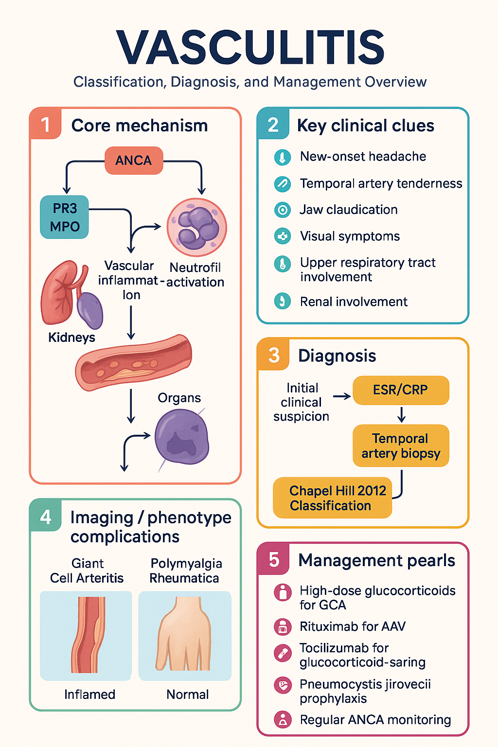
Vasculitis clinical infographic — pathophysiology, clinical clues, diagnosis, imaging, and management
Tap or click image to enlarge — Vasculitis: pathophysiology, clinical clues, diagnosis, imaging, and management.
Vasculitis infographic, full size
🎬 Vasculitis — clinical explainer

Chapel Hill 2012 Classification

The 2012 International Chapel Hill Consensus Conference (CHCC) nomenclature is the current global standard for vasculitis classification. It stratifies primary vasculitides by the calibre of predominantly affected vessel and replaces the older 1994 classification.

Vessel Size Conditions Key Features
Large vessel Giant cell arteritis (GCA)
Takayasu arteritis (TAK)
Aorta and major branches; granulomatous inflammation; ischaemia of distal organs
Medium vessel Polyarteritis nodosa (PAN)
Kawasaki disease
Mesenteric, renal, coronary arteries; necrotising inflammation without glomerulonephritis
Small vessel ANCA-associated: GPA, MPA, EGPA
Immune-complex: IgA vasculitis, cryoglobulinaemic vasculitis, anti-GBM disease
Capillaries, venules, arterioles; glomerulonephritis, alveolar haemorrhage, purpura
Variable Behçet disease, Cogan syndrome Can affect vessels of any size
⚠️
Diagnostic pitfall: The CHCC 2012 classification is a nomenclature system, not a diagnostic algorithm. Clinical diagnosis still requires integration of history, examination, histopathology, serology, and imaging.

The CHCC 2012 also introduced the category of "single-organ vasculitis" (e.g., cutaneous leukocytoclastic angiitis) and "vasculitis associated with systemic disease" or "probable aetiology" (e.g., HBV-associated PAN). These distinctions guide investigation for secondary causes.

ANCA-Associated Vasculitis (GPA, MPA, EGPA)

ANCA-associated vasculitis (AAV) encompasses three clinically overlapping but distinct entities: granulomatosis with polyangiitis (GPA, formerly Wegener's), microscopic polyangiitis (MPA), and eosinophilic granulomatosis with polyangiitis (EGPA, formerly Churg–Strauss). Australian incidence is approximately 10–20 per million per year, with a peak in the 6th–7th decade.

Granulomatosis with Polyangiitis (GPA)

GPA classically presents with a triad of upper respiratory tract (sinusitis, saddle-nose deformity, subglottic stenosis), lower respiratory tract (nodules, cavitating lesions, alveolar haemorrhage), and renal involvement (pauci-immune necrotising glomerulonephritis). c-ANCA / anti-PR3 antibodies are positive in ~70–90%.

Microscopic Polyangiitis (MPA)

MPA typically presents with rapidly progressive glomerulonephritis (RPGN) and pulmonary capillaritis without granulomatous inflammation. p-ANCA / anti-MPO antibodies are positive in ~60–80%. MPA is the most common cause of pulmonary–renal syndrome in Australia.

Eosinophilic Granulomatosis with Polyangiitis (EGPA)

EGPA is characterised by asthma, peripheral eosinophilia (≥10% or ≥1.0 × 10⁹/L), migratory pulmonary infiltrates, neuropathy (mononeuritis multiplex), and vasculitis. p-ANCA/MPO positive in ~40% (ANCA-positive subgroup has higher renal involvement). The 2022 ACR/EULAR classification criteria incorporate eosinophil count, obstructive airway disease, nasal polyps, extravascular eosinophilic predominant inflammation, cytoplasmic ANCA or anti-MPO, and maximum eosinophil count ≥1.0 × 10⁹/L.

🚨
Severe/life-threatening AAV (pulmonary haemorrhage, RPGN with creatinine >500 µmol/L, cerebral vasculitis, mononeuritis multiplex with motor deficit): requires immediate hospital admission and pulse IV methylprednisolone 500 mg–1 g daily for 3 days, followed by oral prednisolone 1 mg/kg/day (max 60 mg) plus either rituximab or cyclophosphamide.

Severity Stratification

Mild / Limited
Non-organ-threatening
Upper airway disease, arthralgia, constitutional symptoms, minor skin involvement. No renal, pulmonary, cardiac, or neurological threat.
Setting: Outpatient rheumatology/immunology
Moderate / Generalised
Active but not immediately life-threatening
Mild renal impairment (eGFR >30), pulmonary infiltrates without haemorrhage, limited neuropathy, cutaneous ulceration.
Setting: Specialist-initiated, outpatient-supervised induction
Severe / Life-threatening
Organ-threatening
Pulmonary haemorrhage, RPGN (eGFR <30 or dialysis-dependent), cerebral vasculitis, cardiac involvement, mesenteric ischaemia, motor neuropathy.
Setting: Inpatient / ICU

Induction Therapy

💊
Rituximab
MabThera®, Riximyo® · Anti-CD20 monoclonal antibody
Adult dose 375 mg/m² IV weekly × 4 doses, or 1000 mg IV × 2 doses (days 1 and 15)
Paediatric dose 375 mg/m² IV weekly × 4 doses (specialist use)
Notes Preferred for relapsing GPA, PR3-ANCA positive, and as first-line alternative to cyclophosphamide. Screen for hepatitis B before commencing.
PBS status Authority Required — Specialist
💊
Cyclophosphamide
Endoxan® · Alkylating agent
Adult dose IV pulse: 15 mg/kg every 2–3 weeks (max 1.2 g/pulse) × 6 pulses; OR oral: 2 mg/kg/day (max 200 mg) for 3–6 months
Renal adjustment Dose reduce 25–50% if eGFR <10 mL/min; IV pulse preferred in renal impairment
Key toxicity Cumulative gonadotoxicity (consider GnRH agonist), haemorrhagic cystitis (IV: use MESNA), lymphopenia, bladder cancer risk
PBS status ✔ PBS General Benefit
💊
Avacopan
Tavneos® · Complement C5a receptor inhibitor
Adult dose 30 mg PO BD with food, used alongside standard induction (rituximab or cyclophosphamide)
Role Glucocorticoid-sparing; allows accelerated steroid taper or steroid-free induction. Superior sustained remission at 52 weeks vs. prednisone taper (ADVOCATE trial).
Hepatic adjustment Contraindicated in severe hepatic impairment (Child–Pugh C)
PBS status Not PBS-listed (as of 2025) — TGA-approved, private script

Glucocorticoid Regimen

💊
Prednisolone
Solone®, Panafcortelone® · Glucocorticoid
Adult dose Induction: 1 mg/kg/day (max 60 mg) → taper to 5–7.5 mg/day by 3–5 months. Consider rapid taper (to 5 mg by week 16) with avacopan or PEXIVAS low-dose protocol
Paediatric dose 2 mg/kg/day (max 60 mg) with specialist-guided taper
PBS status ✔ PBS General Benefit

Adjunctive Therapy

💊
Trimethoprim–Sulfamethoxazole (Co-trimoxazole)
Bactrim®, Resprim® · PJP prophylaxis
Adult dose Single-strength (80/400 mg) PO daily, or double-strength (160/800 mg) PO 3 times/week; for Pneumocystis jirovecii prophylaxis during prednisolone ≥20 mg/day + cyclophosphamide/rituximab
Renal adjustment Avoid if eGFR <15; dose reduce if eGFR 15–30
PBS status ✔ PBS General Benefit

Maintenance Therapy

💊
Azathioprine
Imuran® · Purine antimetabolite
Adult dose 2 mg/kg/day (max 200 mg) PO; check TPMT/NUDT15 genotype before commencing
Duration Minimum 24 months after remission; consider longer in PR3-ANCA positive
PBS status ✔ PBS General Benefit
💊
Methotrexate
Methoblastin® · Antimetabolite
Adult dose 15–25 mg PO/SC weekly with folic acid 5 mg weekly (not on MTX day)
Role Alternative to azathioprine for maintenance in non-severe GPA/MPA; inferior to azathioprine for severe disease
PBS status ✔ PBS General Benefit
💊
Rituximab (maintenance)
MabThera® · Anti-CD20
Adult dose 500 mg IV every 6 months for minimum 24 months (MAINRISAN regimen)
Indication Superior to azathioprine for relapse prevention, especially PR3-ANCA positive GPA
PBS status Authority Required — Specialist

Refractory / Relapsing Disease

⚠️
  • Switch from cyclophosphamide to rituximab (or vice versa) if first-line induction fails
  • Consider addition of plasma exchange (PLEX) — PEXIVAS trial showed no overall benefit for routine use, but may be considered for severe AKI (creatinine >500 µmol/L) or diffuse alveolar haemorrhage
  • Mycophenolate mofetil (CellCept® 1–2 g/day BD) — PBS Restricted Benefit for lupus nephritis; off-label use in AAV with specialist approval
  • Omalizumab may be considered for EGPA with refractory asthma (not PBS-listed for EGPA)
  • Mepolizumab (Nucala®) 300 mg SC every 4 weeks — TGA-approved for EGPA; PBS Authority Required

Monitoring in AAV

Weekly × 4
FBC, LFTs, creatinine, urinalysis, CRP/ESR during induction. ANCA titres at baseline.
Monthly
FBC, LFTs, creatinine, eGFR, urinalysis, ANCA (PR3/MPO) once stable on maintenance.
3–6 monthly
ANCA titres (rising titres may predict relapse in PR3-AAV, less reliable in MPO); BVAS assessment.
Annually
Bone density (DEXA) if on ongoing corticosteroids; hepatitis B screening (if on rituximab); cervical screening (HPV risk with immunosuppression).

Giant Cell Arteritis (GCA)

Giant cell arteritis is the most common primary systemic vasculitis in Australia, predominantly affecting adults >50 years (peak 70–80 years) with a female:male ratio of 2–3:1. Australian incidence is approximately 15–25 per 100,000 per year in those aged >50. GCA commonly coexists with polymyalgia rheumatica (PMR) in 40–60% of cases.

🚨
Ischaemic optic neuropathy: Sudden irreversible vision loss occurs in up to 20% of untreated GCA. If GCA is suspected clinically, commence high-dose glucocorticoids IMMEDIATELY — do not wait for temporal artery biopsy or imaging. Biopsy within 2 weeks of treatment onset remains diagnostically valid.

Clinical Presentation

  • New-onset headache (usually temporal, may be diffuse) — most common symptom (~70%)
  • Temporal artery abnormalities: tenderness, reduced pulsation, nodularity, beading
  • Jaw claudication with chewing (~50%) — highly specific
  • Visual symptoms: amaurosis fugax, diplopia, vision loss — emergency
  • Constitutional: fever, weight loss, night sweats (may mimic sepsis or malignancy)
  • Upper limb claudication (aortic arch / large-vessel GCA)
  • PMR symptoms: bilateral shoulder and hip girdle stiffness >30 min
  • Stroke or TIA (posterior circulation)

Investigations

Essential
ESR / CRP
ESR typically >50 mm/hr (often >100); CRP elevated. Both normal in ~5% — does not exclude GCA.
Essential
Temporal artery biopsy (TAB)
Gold standard — positive in ~70–80% (skip lesions). Unilateral ≥1 cm segment; consider bilateral if first negative. MBS item: specialist surgical biopsy.
Available
Colour duplex ultrasound (CDUS) of temporal arteries
Halo sign (oedema) and compression sign — sensitivity ~75%, specificity ~95% in expert hands. Non-invasive alternative to biopsy if available.
Available
PET-CT (¹⁸F-FDG)
Large-vessel uptake — useful for diagnosis of occult large-vessel GCA and assessing extent. Not widely available acutely; may be used in refractory cases.
Available
CT angiography / MR angiography
Assess aortic dilatation and large-vessel stenosis. MRA preferred (no radiation).

Treatment

Without visual symptoms

💊
Prednisolone
Solone®
Adult dose 40–60 mg PO daily (start 40 mg for uncomplicated; 60 mg if PMR features prominent). Taper to 20 mg by 2–3 months, then slow taper 2.5 mg/month. Total duration: 1–2 years minimum.
PBS status ✔ PBS General Benefit

With visual symptoms (amaurosis fugax / vision loss)

💊
Methylprednisolone IV
Solu-Medrol®
Adult dose 500 mg–1 g IV daily for 3 days → oral prednisolone 1 mg/kg/day (max 60 mg)
PBS status ✔ PBS General Benefit (inpatient)

Glucocorticoid-Sparing Agents

💊
Tocilizumab
Actemra® · IL-6 receptor inhibitor
Adult dose 162 mg SC weekly (or fortnightly) — enables rapid steroid taper and reduces relapse rate by ~50% (GiACTA trial)
PBS status Authority Required — Specialist
💊
Methotrexate
Methoblastin®
Adult dose 15–25 mg PO/SC weekly with folic acid. Adjunct to steroids — modest relapse-reduction benefit.
PBS status ✔ PBS General Benefit

Cardiovascular Risk in GCA

GCA patients have increased cardiovascular morbidity. Address modifiable risk factors: statin therapy, antihypertensives, smoking cessation, low-dose aspirin (75 mg daily — consider in all unless contraindicated). Monitor for aortic aneurysm with imaging at baseline and periodically.

Takayasu Arteritis

Takayasu arteritis (TAK) is a granulomatous large-vessel vasculitis predominantly affecting the aorta and its major branches. It typically presents before age 40 (often 15–30 years) with a strong female predominance (8–9:1). In Australia, TAK is more frequently seen in patients of Asian and Middle Eastern descent, though it affects all ethnicities.

Clinical Presentation

  • Systemic phase: Fever, weight loss, arthralgia, myalgia — may be indolent for months
  • Occlusive phase: Limb claudication, pulse deficits, bruits (subclavian, carotid, aortic), limb blood pressure discrepancy (>10 mmHg)
  • Renovascular hypertension (renal artery stenosis)
  • Aortic regurgitation, aortic root dilatation, coronary ostial stenosis
  • Stroke/TIA (carotid/vertebral involvement)
  • Pulmonary artery involvement (uncommon but recognised)

Diagnosis

Essential
CT angiography or MR angiography
Mural thickening, stenosis, occlusion, or aneurysm of aorta and branches. MRA preferred for follow-up (no radiation).
Available
PET-CT
Early active disease — vascular wall FDG uptake before structural changes are visible on CTA/MRA.
Available
Conventional angiography
Gold standard for detail but invasive; increasingly replaced by CTA/MRA.

Treatment

💊
Prednisolone
Solone® · First-line
Adult dose 1 mg/kg/day (max 60 mg) → slow taper over 6–12 months. Relapse rate with steroid monotherapy is high (~60%).
PBS status ✔ PBS General Benefit
💊
Methotrexate
Methoblastin® · Steroid-sparing
Adult dose 15–25 mg PO/SC weekly with folic acid
PBS status ✔ PBS General Benefit
💊
Tocilizumab
Actemra® · IL-6 inhibition
Adult dose 162 mg SC weekly — effective in relapsing/refractory TAK (TAKT trial). Use with caution re: aortic graft infections.
PBS status Authority Required — Specialist (off-label for TAK; PBS-listed for GCA and RA)
⚠️
Revascularisation: Surgical bypass or endovascular angioplasty/stenting should only be performed during clinical and biochemical remission, as intervention during active disease carries high re-stenosis rates. Referral to vascular surgery is essential for significant ischaemia.

Polyarteritis Nodosa (PAN)

Polyarteritis nodosa is a necrotising medium-vessel vasculitis affecting muscular arteries. It spares the lungs and glomeruli (distinguishing it from AAV). Australian incidence is approximately 1–3 per million per year. Hepatitis B virus (HBV) is associated with ~30% of PAN cases worldwide; prevalence of HBV-PAN is lower in Australia due to vaccination programmes but remains relevant in unvaccinated populations.

Clinical Presentation

  • Constitutional: fever, weight loss, malaise (almost universal)
  • Cutaneous: livedo reticularis, nodules, purpura, digital gangrene, ulceration
  • Renal: renovascular hypertension (arterial, not glomerular), renal infarction, microaneurysms on angiography
  • Mesenteric: abdominal pain (post-prandial), bowel ischaemia/perforation, GI haemorrhage
  • Neurological: mononeuritis multiplex (common), CNS vasculitis (rare)
  • Musculoskeletal: myalgia, arthralgia (non-erosive)
  • Testicular pain (orchitis)
  • Coronary arteritis → myocardial infarction (rare)

Diagnosis

Essential
Conventional or CT angiography
Microaneurysms of renal, hepatic, and mesenteric arteries — characteristic but not pathognomonic.
Essential
Tissue biopsy
Affected organ (skin, sural nerve, muscle, testis): fibrinoid necrosis of medium-vessel walls with neutrophilic infiltrate.
Essential
Hepatitis B serology
HBsAg, anti-HBc, anti-HBs — must be performed in every PAN diagnosis.
Available
ANCA testing
Should be negative — positive ANCA suggests AAV rather than PAN.

Treatment

HBV-Negative PAN

💊
Prednisolone
Solone® · First-line
Adult dose 1 mg/kg/day (max 60 mg) for mild disease; add cyclophosphamide for severe/organ-threatening
PBS status ✔ PBS General Benefit
💊
Cyclophosphamide
Endoxan®
Adult dose IV pulse: 15 mg/kg every 2–3 weeks × 6 pulses, or oral 2 mg/kg/day for 3–6 months
PBS status ✔ PBS General Benefit

HBV-Associated PAN

⚠️
HBV-PAN management differs fundamentally:
  • Short-course corticosteroids only (1–2 weeks) for symptom control — prolonged immunosuppression impairs viral clearance
  • Antiviral therapy is the cornerstone: entecavir (Baraclude® 0.5 mg PO daily) or tenofovir (Viread® 300 mg PO daily) — PBS General Benefit
  • Consider plasma exchange (PLEX) in severe cases
  • Do NOT use rituximab or cyclophosphamide in HBV-PAN (impairs viral clearance)
  • Monitor HBV DNA and LFTs; refer to hepatologist

IgA Vasculitis (Henoch–Schönlein Purpura)

IgA vasculitis (IgAV, formerly Henoch–Schönlein purpura) is the most common systemic vasculitis in children (peak age 3–10 years; 90% of cases are paediatric). Annual incidence in children is approximately 10–20 per 100,000. Adult IgAV is rarer but associated with more severe renal involvement. Characterised by IgA1-dominant immune complex deposition in small vessels.

Clinical Features (Classic Tetrad)

  • Palpable purpura (100%): non-thrombocytopenic, gravity-dependent (buttocks, lower limbs); may be preceded by urticaria
  • Arthritis / arthralgia (~75%): large joints (knees, ankles), non-erosive, migratory
  • Abdominal pain (~65%): colicky, may precede purpura by days; risk of intussusception, bowel perforation, GI haemorrhage
  • Nephritis (~30–50% in children, higher in adults): haematuria (microscopic or macroscopic), proteinuria, nephrotic syndrome, RPGN in severe cases
  • Other: scrotal oedema, CNS vasculitis (rare), pulmonary haemorrhage (rare)
⚠️
Renal monitoring is critical: Urinalysis and BP should be checked at diagnosis, weekly for 4–6 weeks, then monthly for 6 months, then 6-monthly for 5 years. Persistent proteinuria or haematuria at 6 months warrants nephrology referral and renal biopsy. Adults are at higher risk of chronic kidney disease.

Diagnosis

Primarily clinical — EULAR/PRINTO/PRES 2010 criteria: palpable purpura (mandatory) with at least one of diffuse abdominal pain, biopsy showing predominant IgA deposition, arthritis/arthralgia, or renal involvement. No specific serological test is diagnostic; serum IgA may be elevated in 50% but is not required.

Treatment

Most cases are self-limiting in children (resolves within 4–6 weeks). Supportive care is the mainstay.

💊
Supportive care
First-line for most
Adult / Paediatric Paracetamol or NSAIDs (ibuprofen 5–10 mg/kg TDS in children) for arthralgia; adequate hydration; monitoring
PBS status ✔ PBS General Benefit
💊
Prednisolone
Solone® · For severe disease
Adult dose 1 mg/kg/day for severe abdominal pain, nephrotic syndrome, or RPGN; taper over 4–8 weeks
Paediatric dose 1–2 mg/kg/day (max 60 mg) for severe abdominal symptoms; evidence for routine use to prevent nephritis is lacking
PBS status ✔ PBS General Benefit

Severe Renal Disease

  • Nephrotic syndrome or RPGN: high-dose prednisolone ± cyclophosphamide or mycophenolate mofetil (specialist nephrology management)
  • ACE inhibitors for proteinuria control
  • Refer to paediatric or adult nephrology — renal biopsy guides therapy
  • Dapsone has been used for refractory skin disease (limited evidence)

Prognosis

Children: >95% have complete recovery. Relapse occurs in ~30% within 4 months. Adults: worse renal prognosis — up to 10–30% develop CKD or ESKD, particularly with crescentic nephritis on biopsy. Long-term nephrology follow-up is recommended for adults with any renal involvement.

Special Populations

🤰 Pregnancy
Cyclophosphamide
Contraindicated in pregnancy — teratogenic (Category D). Ensure contraception during and 3 months after therapy.
Methotrexate
Contraindicated in pregnancy — teratogenic and abortifacient. Stop 3 months before conception.
Rituximab
Avoid in pregnancy if possible; can be used if life-threatening disease and no alternatives. B-cell depletion may last 6–12 months post-dose.
Azathioprine
Safe in pregnancy (Category D — but widely used in transplant and IBD pregnancy). Preferred steroid-sparing agent during pregnancy.
Prednisolone
Can be used in pregnancy at lowest effective dose. First-pass metabolism reduces fetal exposure. Monitor for gestational diabetes.
👶 Paediatrics
AAV in children
Rare but serious. GPA most common. Cyclophosphamide dose: 2 mg/kg/day (max 150 mg). Rituximab increasingly used off-label. MPA more common than GPA in paediatric Japanese cohorts.
IgA Vasculitis
The predominant childhood vasculitis. Usually self-limiting. Long-term urinalysis monitoring essential. Referral for nephritis.
🫘 Renal Impairment
Cyclophosphamide
Reduce dose 25–50% if eGFR <10 mL/min. IV pulse preferred over oral in renal impairment to reduce cumulative dose.
Co-trimoxazole
Avoid if eGFR <15. Use dapsone or atovaquone for PJP prophylaxis as alternatives.
Methotrexate
Contraindicated if eGFR <30 mL/min — accumulates and causes toxicity.
👴 Elderly
Glucocorticoids
Higher risk of osteoporosis, diabetes, cataracts, infections. Initiate bone protection (calcium, vitamin D, bisphosphonate) with any course >3 months.
Cyclophosphamide
Prefer IV pulse over oral (reduced cumulative toxicity). Monitor FBC closely — greater myelosuppression risk.
🛡️ Immunocompromised
Infection risk
PJP prophylaxis mandatory with dual immunosuppression. Screen for latent TB (IGRA), hepatitis B/C, HIV before rituximab or cyclophosphamide. Avoid live vaccines.
COVID-19
COVID-19 vaccination recommended; time rituximab ≥4 weeks after vaccination if possible. Pre-exposure prophylaxis (tixagevimab/cilgavimab) considered during B-cell depletion.
🫁 Hepatic Impairment
Azathioprine
Metabolised hepatically — use with caution, monitor LFTs closely. Consider dose reduction.
Methotrexate
Avoid in significant hepatic impairment or heavy alcohol use. Hepatotoxic.
Avacopan
Contraindicated in Child–Pugh C. Dose adjustment not required in mild–moderate impairment.

Aboriginal and Torres Strait Islander Health Considerations

Aboriginal and Torres Strait Islander Health
Epidemiology
Aboriginal and Torres Strait Islander peoples experience higher rates of systemic inflammatory conditions and may present later with more advanced vasculitis. IgA vasculitis is commonly encountered in paediatric populations in remote communities. GCA and AAV data are limited due to under-representation in registries.
Diagnostic delay
Geographic isolation from specialist rheumatology and immunology services leads to delays in diagnosis. The median time to diagnosis for AAV in remote settings can be significantly longer. Telehealth rheumatology consultations through the Medical Specialist Outreach Assistance Programme (MSOAP) can expedite access.
HBV-associated PAN
Chronic hepatitis B prevalence is higher in Aboriginal and Torres Strait Islander communities, particularly in remote Northern Territory and Western Australia. HBV-PAN must be actively considered in any Indigenous patient presenting with medium-vessel vasculitis. Antiviral therapy and HBV vaccination are essential public health measures.
Renal comorbidity
Higher background rates of CKD mean that vasculitis-related renal involvement may compound pre-existing renal disease. Baseline eGFR and urinalysis are critical. Nephrology telehealth outreach supports management in remote settings.
Medication access
PBS medications including azathioprine, methotrexate, prednisolone, and co-trimoxazole are available through Remote Area Aboriginal Health Services (RAAHS) under Section 100 (S100) provisions. Rituximab and cyclophosphamide require infusion centre access, necessitating medical retrieval to regional centres.
Monitoring challenges
Regular blood monitoring (FBC, LFTs, creatinine, ANCA titres) is essential but logistically difficult in remote communities. Point-of-care pathology analysers (iSTAT, Abbott) and pathology collection via Remote Area Nurses (RANs) can facilitate this. Telehealth-linked pharmacist review supports medication safety.
Cultural safety
Engage Aboriginal Health Workers (AHWs) and Aboriginal Liaison Officers (ALOs) in care planning. Use interpreters where needed. Acknowledge kinship systems and sorry business in care continuity. Chronic disease management through Aboriginal Community Controlled Health Organisations (ACCHOs) improves engagement.
📊 Vasculitis — slide deck

Open slides PDF in new tab

📚 References

  1. 1. Jennette JC, Falk RJ, Bacon PA, et al. 2012 revised International Chapel Hill Consensus Conference Nomenclature of Vasculitides. Arthritis Rheum. 2013;65(1):1–11.
  2. 2. Stone JH, Merkel PA, Spiera R, et al. Rituximab versus cyclophosphamide for ANCA-associated vasculitis (RAVE trial). N Engl J Med. 2010;363(3):221–232.
  3. 3. Jones RB, Tervaert JWC, Hauser T, et al. Rituximab versus cyclophosphamide in ANCA-associated renal vasculitis (RITUXVAS trial). N Engl J Med. 2010;363(3):211–220.
  4. 4. Walsh M, Merkel PA, Peh CA, et al. Plasma exchange and glucocorticoids in severe ANCA-associated vasculitis (PEXIVAS trial). N Engl J Med. 2020;382(7):622–631.
  5. 5. Jayne DRW, Bruchfeld AN, Harper L, et al. Randomized trial of maintenance therapy in ANCA-associated vasculitis (MAINRISAN trial). N Engl J Med. 2014;371(19):1771–1780.
  6. 6. Jayne DRW, Merkel PA, Schall TJ, Bekker P. Avacopan for the treatment of ANCA-associated vasculitis (ADVOCATE trial). N Engl J Med. 2021;384(7):599–609.
  7. 7. Stone JH, Tuckwell K, Dimonaco S, et al. Trial of tocilizumab in giant-cell arteritis (GiACTA trial). N Engl J Med. 2017;377(4):317–328.
  8. 8. Nakaoka Y, Isobe M, Takei S, et al. Efficacy and safety of tocilizumab in patients with refractory Takayasu arteritis (TAKT trial). JACC. 2018;71(14):1587–1596.
  9. 9. Lightfoot RW Jr, Michel BA, Bloch DA, et al. The American College of Rheumatology 1990 criteria for the classification of polyarteritis nodosa. Arthritis Rheum. 1990;33(8):1088–1093.
  10. document.querySelectorAll('.topic-toc a[href^="#"]').forEach(o=>{o.addEventListener("click",i=>{const t=o.getAttribute("href")||"",e=document.querySelector(t);if(!e)return;i.preventDefault();const n=e.getBoundingClientRect().top+window.scrollY-70;window.scrollTo({top:n,behavior:"smooth"}),history.replaceState(null,"",t)})});const r=new Map;document.querySelectorAll(".topic-toc a").forEach(o=>{const i=o.getAttribute("href")?.slice(1);i&&r.set(i,o)});if(r.size>0&&"IntersectionObserver"in window){const o=new IntersectionObserver(i=>{let t=null;for(const e of i)if(e.isIntersecting){t=e.target.id;break}t&&r.forEach((e,n)=>e.classList.toggle("active",n===t))},{rootMargin:"-80px 0px -40% 0px",threshold:0});r.forEach((i,t)=>{const e=document.getElementById(t);e&&o.observe(e)})}window.matchMedia("(max-width: 900px)").matches&&document.querySelector(".topic-rail-collapsible")?.removeAttribute("open");const c=document.querySelector(".topic-rail-details");function s(){c&&(window.matchMedia("(max-width: 900px)").matches?c.open=!1:c.open=!0)}s();let l;window.addEventListener("resize",()=>{clearTimeout(l),l=window.setTimeout(s,120)});