Home Rheumatology Amyloidosis in Rheumatic Disease

Amyloidosis in Rheumatic Disease

📋 Key Information Summary

📋
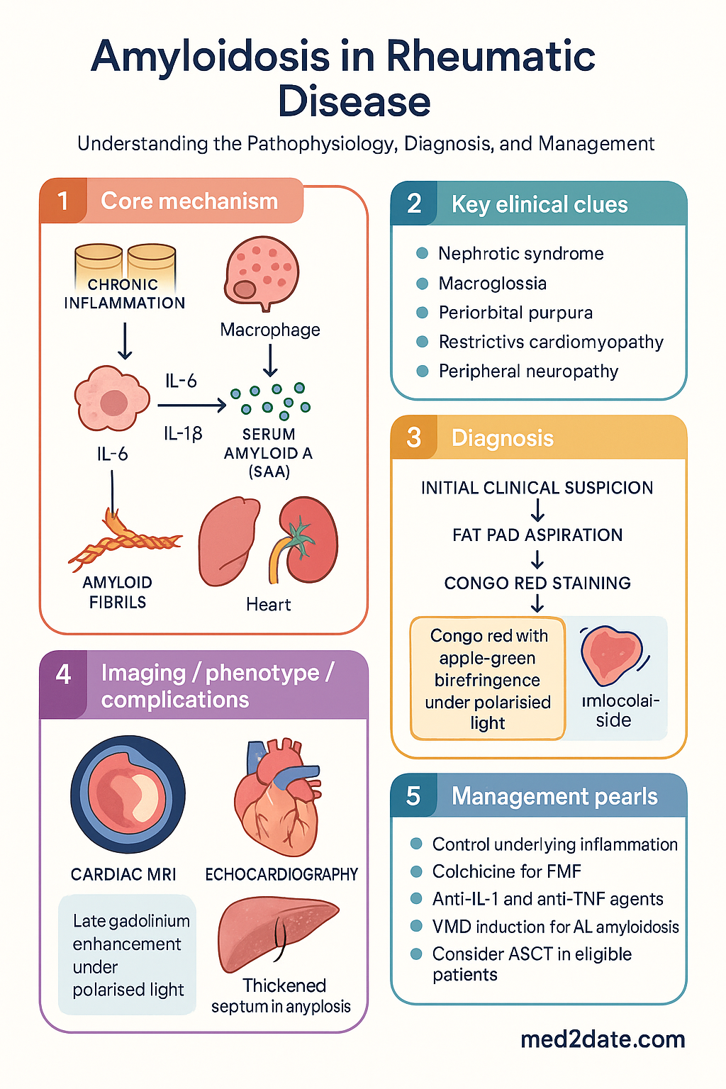
  • AA amyloidosis results from sustained elevation of serum amyloid A (SAA) protein during chronic inflammatory conditions including rheumatoid arthritis, familial Mediterranean fever, juvenile idiopathic arthritis, and ankylosing spondylitis.
  • AL amyloidosis arises from plasma cell dyscrasia (monoclonal immunoglobulin light chain deposition); associated with myeloma and monoclonal gammopathy of undetermined significance (MGUS).
  • Congo red staining with apple-green birefringence under polarised light remains the histological gold standard for amyloid confirmation.
  • AA amyloidosis primarily targets the kidney (nephrotic syndrome progressing to renal failure) and gastrointestinal tract (dysmotility, malabsorption).
  • AL amyloidosis preferentially affects the heart (restrictive cardiomyopathy), kidneys, and peripheral/autonomic nerves.
  • Macroglossia and periorbital purpura are pathognomonic clinical features of AL amyloidosis.
  • Fat pad aspiration is the safest initial biopsy technique, with sensitivity of 60–80%; rectal biopsy supplements when negative.
  • SAP scintigraphy (serum amyloid P component scan) quantifies whole-body amyloid burden but is available only at specialist centres in Australia.
  • Cardiac MRI with late gadolinium enhancement pattern is the imaging modality of choice for cardiac amyloidosis.
  • AA treatment centres on aggressive control of the underlying inflammatory disease: colchicine prevents AA in FMF; anti-IL-1 and anti-TNF agents target refractory inflammatory sources.
  • AL treatment uses bortezomib/melphalan/dexamethasone (VMD) induction with consideration of autologous stem cell transplant (ASCT) in eligible patients.
  • Prognosis in AA amyloidosis correlates with SAA levels and renal function; early suppression of inflammation halts amyloid deposition and may allow regression.

Introduction & Australian Epidemiology

Amyloidosis is a group of disorders characterised by extracellular deposition of misfolded protein in an abnormal fibrillar configuration. In the context of rheumatic disease, the two clinically relevant forms are AA (secondary) amyloidosis driven by chronic systemic inflammation, and AL (primary) amyloidosis caused by monoclonal immunoglobulin light chain deposition from plasma cell dyscrasia. Both lead to progressive organ dysfunction through disruption of normal tissue architecture and can be fatal if undiagnosed.

Secondary (AA) amyloidosis remains a serious systemic complication of inadequately controlled chronic inflammatory conditions, including rheumatoid arthritis (RA), familial Mediterranean fever (FMF), juvenile idiopathic arthritis (JIA), ankylosing spondylitis (AS), and Crohn disease. The unifying mechanism is sustained elevation of serum amyloid A (SAA), an acute-phase reactant produced by the liver under interleukin-6 (IL-6) and IL-1β stimulation.

In Australia, AA amyloidosis is rare but clinically significant. The AIHW reports that amyloidosis-related hospitalisations have a mortality rate exceeding 20% within 5 years of diagnosis. FMF is relatively prevalent among Australian communities of Middle Eastern and Mediterranean descent, particularly in Lebanese, Armenian, Turkish, and Sephardic Jewish populations. RA-related AA amyloidosis has declined markedly with early aggressive disease-modifying therapy but still occurs in patients with longstanding, poorly controlled disease. The prevalence of AL amyloidosis is estimated at 8–12 per million population per year in Australia.

⚠️
Clinical warning: Amyloidosis is frequently diagnosed late because its early manifestations (proteinuria, fatigue, peripheral oedema) overlap with common rheumatic disease complications. A high index of suspicion is essential in any patient with longstanding inflammatory disease and new-onset organ dysfunction.
Amyloidosis in Rheumatic Disease clinical infographic — pathophysiology, clinical clues, diagnosis, imaging, and management
Tap or click image to enlarge — Amyloidosis in Rheumatic Disease: pathophysiology, clinical clues, diagnosis, imaging, and management.
Amyloidosis in Rheumatic Disease infographic, full size

Pathogenesis & Types

AA Amyloidosis (Secondary)

AA amyloidosis develops when persistent elevation of serum amyloid A (SAA) — an acute-phase apolipoprotein synthesised predominantly by hepatocytes — leads to proteolytic cleavage and misfolding into β-pleated amyloid fibrils. These fibrils deposit extracellularly in parenchymal organs and progressively replace normal tissue architecture.

The key pathological events are:

  • Sustained inflammation: IL-6 and IL-1β drive hepatic SAA production. Chronic elevations (>10 mg/L persistently for months to years) are necessary but not sufficient for amyloid formation.
  • Proteolytic processing: Monocyte-derived proteases cleave SAA into the N-terminal AA fragment, which is the primary constituent of AA fibrils.
  • Fibril nucleation: AA fragments polymerise into cross-β fibrils. Glycosaminoglycans and serum amyloid P component (SAP) stabilise the fibril structure, resisting proteolysis.
  • Organ deposition: Kidney is the predominant site (glomerular mesangium, basement membrane); liver, spleen, GI tract, and adrenal glands are also affected.
Underlying Condition Estimated Risk of AA Typical Latency
Rheumatoid arthritis 2–5% (historically higher) 10–20 years of active disease
Familial Mediterranean fever Up to 75% without colchicine Childhood/adolescence
Juvenile idiopathic arthritis 1–7% 5–15 years
Ankylosing spondylitis <1% Variable, long-standing
Crohn disease 0.5–1% Variable
Other chronic infections (e.g., osteomyelitis, TB) Case reports Years of active infection

AL Amyloidosis (Primary)

AL amyloidosis results from clonal expansion of plasma cells producing monoclonal immunoglobulin light chains (typically κ or λ) that misfold and deposit as amyloid fibrils. It is classified as a plasma cell dyscrasia and may coexist with myeloma, Waldenström macroglobulinaemia, or occur in the setting of MGUS. The light chain subtype determines organ tropism: AL deposits preferentially affect the heart, kidneys, nerves, and soft tissues.

Histological Diagnosis

All amyloid subtypes share a common histological appearance:

  • Congo red staining: Amyloid deposits stain salmon-pink under conventional light microscopy.
  • Apple-green birefringence under polarised light: This is the defining diagnostic feature and must be demonstrated for definitive amyloid confirmation.
  • Subtyping: Immunohistochemistry (anti-AA antibody, anti-κ/λ antibodies) or mass spectrometry-based proteomics should be performed on all positive biopsies to determine the amyloid fibril type — critical for guiding therapy.
ℹ️
Key pathological point: Congo red with apple-green birefringence under polarised light is required for a definitive diagnosis. Staining without birefringence is non-diagnostic. If amyloid is suspected but initial Congo red is negative, immunohistochemistry for amyloid subtyping should still be performed, as some fibril variants stain weakly.

Clinical Manifestations

AA Amyloidosis

AA amyloidosis typically presents insidiously, with renal involvement as the dominant clinical feature. Patients often have long-standing inflammatory disease and may have proteinuria detected incidentally on urinalysis.

🫘
Kidney (most common)
Nephrotic-range proteinuria (≥3.5 g/24h) is the hallmark presentation
Progressive renal impairment → end-stage kidney disease
Oedema, hypoalbuminaemia, hyperlipidaemia
May present as asymptomatic proteinuria for months to years before renal function declines
🫁
Gastrointestinal & Hepatic
GI dysmotility — pseudo-obstruction, chronic diarrhoea, malabsorption
Hepatosplenomegaly with hepatomegaly more prominent than splenomegaly
Macroglossia may occur in AA but is much rarer than in AL
GI bleeding from mucosal amyloid deposition (uncommon)
💪
Other Organ Systems
Adrenal insufficiency (rare, late)
Soft tissue deposition: carpal tunnel syndrome (rare in AA)
Cardiac involvement uncommon in AA; restrictive cardiomyopathy is predominantly an AL feature

AL Amyloidosis

AL amyloidosis has multi-organ involvement and a broader clinical spectrum. It may mimic heart failure, nephrotic syndrome, neuropathy, or chronic diarrhoea — often simultaneously.

❤️
Heart
Restrictive cardiomyopathy — diastolic dysfunction with preserved ejection fraction initially
Low-voltage ECG with pseudo-infarct pattern (absent R waves in precordial leads)
Progressive heart failure; arrhythmias; sudden cardiac death
Raised NT-proBNP and troponin — prognostic staging markers
🫘
Kidney
Nephrotic syndrome with proteinuria, oedema
Renal insufficiency progressing to end-stage kidney disease
Unlike AA, may show heavier proteinuria at presentation
🧠
Neurological
Peripheral neuropathy — symmetric, length-dependent, predominantly sensory
Autonomic neuropathy — orthostatic hypotension, gastroparesis, urinary retention
Carpal tunnel syndrome (bilateral) — may precede systemic diagnosis by years
👄
Soft Tissue (Pathognomonic)
Macroglossia — pathognomonic of AL; tongue enlargement causing dysarthria, dysphagia, and sleep apnoea
Periorbital purpura ("raccoon eyes") — caused by vascular fragility; classically triggered by Valsalva or minor trauma
Skin nodules, waxy papules, nail dystrophy
Hepatosplenomegaly
⚠️
Red flags for amyloidosis in a rheumatology patient: New-onset nephrotic-range proteinuria, unexplained restrictive cardiomyopathy, bilateral carpal tunnel syndrome, unexplained neuropathy in inflammatory disease, or the combination of macroglossia with periorbital purpura should prompt urgent evaluation for amyloidosis.

Diagnosis & Investigations

Biopsy Strategy

Tissue biopsy remains the cornerstone of amyloidosis diagnosis. The biopsy strategy depends on the clinical scenario and the likelihood of organ involvement.

Essential
Fat pad aspiration (abdominal subcutaneous)
Safest initial biopsy technique. Sensitivity 60–80% for systemic amyloidosis (higher for AL than AA). Performed under local anaesthesia using a fine needle. Should be stained with Congo red and examined under polarised light. If negative, proceed to organ biopsy.
Available
Rectal biopsy
Sensitivity 75–85% for systemic amyloidosis. Complements fat pad aspiration when initial assessment is negative. Obtained at flexible sigmoidoscopy; requires Congo red staining. Widely available in Australian centres.
Specialist
Renal biopsy
Gold standard when kidney is the presenting organ. Provides Congo red confirmation, subtyping via immunohistochemistry, and prognostic information about interstitial fibrosis and tubular atrophy. Risk of bleeding; contraindicated if bilateral kidneys are small and echogenic (end-stage).
Specialist
Cardiac biopsy
Endomyocardial biopsy is the most sensitive test for cardiac amyloidosis (>95% sensitivity). Reserved for cases where cardiac involvement is suspected but extracardiac biopsy is negative. Performed at tertiary cardiac centres.
Specialist
SAP scintigraphy (serum amyloid P component scan)
Quantifies whole-body amyloid burden and distribution. Involves IV injection of radiolabelled SAP and gamma camera imaging. Available at limited specialist centres in Australia (Royal Adelaide Hospital, Royal Prince Alfred). Not essential for diagnosis but useful for monitoring response to therapy and detecting occult organ involvement.

Cardiac Assessment

Essential
Cardiac MRI with late gadolinium enhancement (LGE)
Imaging modality of choice for cardiac amyloidosis. Characteristic pattern: diffuse subendocardial or transmural LGE with abnormal gadolinium kinetics (nulling of myocardium before blood pool). T1 mapping and extracellular volume (ECV) quantification increase sensitivity. Widely available in Australian metropolitan centres.
Available
Echocardiography
Shows concentric ventricular thickening with preserved or reduced ejection fraction, diastolic dysfunction, valvular thickening, and pericardial effusion. Strain imaging may demonstrate characteristic "apical sparing" pattern. Baseline assessment in all patients.
Available
ECG
Low voltage (Sokolow–Lyon <15 mm) with pseudo-infarct pattern (absent R waves V1–V3) in the presence of thickened ventricles on echo is highly suggestive of amyloidosis.

Laboratory Investigations

Test Purpose MBS Item / Notes
SAA level (serum amyloid A) Monitor inflammatory activity in AA; target <4 mg/L to halt amyloid progression Specialist pathology (reference lab)
Serum/urine protein electrophoresis + immunofixation Detect monoclonal light chains in AL MBS 66800/66803
Serum free light chains (FLC) Quantify κ/λ ratio; essential for AL diagnosis and response monitoring MBS 66840
NT-proBNP / BNP Cardiac biomarker for AL staging and monitoring MBS 66550 (BNP); NT-proBNP limited availability
High-sensitivity troponin Prognostic staging in AL; predicts survival MBS 66514
24-hour urine protein or spot protein:creatinine ratio Quantify proteinuria and monitor renal response MBS 66506
eGFR, serum creatinine, serum albumin Renal function and nutritional status Routine MBS items
Bone marrow biopsy (for AL) Clonal plasma cell burden; Congo red for marrow amyloid Haematology referral

Risk Stratification & Severity Scoring

AL Amyloidosis — Revised Mayo Clinic Staging

AL amyloidosis staging uses four prognostic biomarkers:

  • NT-proBNP ≥1800 pg/mL
  • hs-troponin T ≥0.025 ng/mL (or hs-troponin I elevated)
  • Difference in free light chains (dFLC) ≥180 mg/L
Stage I
0 Elevated Markers
NT-proBNP <1800, troponin normal, dFLC <180
Median survival: 94 months. Consider ASCT if eligible.
Stage II
1 Elevated Marker
One of the three criteria met
Median survival: 40 months. VMD induction; ASCT if transplant-eligible.
Stage III
2 Elevated Markers
Two of the three criteria met
Median survival: 14 months. Aggressive chemotherapy. ASCT rarely suitable.
Stage IV
3 Elevated Markers
All three criteria met
Median survival: 6 months. Palliative care considerations. Daratumumab-based regimens may improve outcomes.

AA Amyloidosis — Renal Prognostic Stratification

Risk Category eGFR at Diagnosis Proteinuria 5-Year Renal Survival
Low risk ≥60 mL/min <1 g/day >85%
Intermediate risk 30–59 mL/min 1–5 g/day 50–70%
High risk <30 mL/min >5 g/day <30%

Management — AA Amyloidosis

The fundamental principle in AA amyloidosis management is aggressive suppression of the underlying inflammatory disease to reduce SAA levels. The therapeutic target is sustained SAA <4 mg/L (ideally <2 mg/L). Amyloid regression can occur if SAA levels are controlled, but this requires months to years of sustained remission.

Disease-Modifying Therapy for Underlying Condition

💊
Methotrexate
Methoblastin® · Generic · DMARD (for RA)
Adult dose 7.5–25 mg PO/SC once weekly with folic acid 5 mg weekly
Renal adjustment Avoid if eGFR <30 mL/min; dose reduce if eGFR 30–59
Hepatic adjustment Avoid in significant hepatic impairment (Child-Pugh B/C)
PBS status ✔ PBS General Benefit
💊
Sulfasalazine
Salazopyrin® · Generic · DMARD (RA, AS)
Adult dose 500 mg BD initially, titrate to 1–2 g BD
Renal adjustment Use with caution if eGFR <30; monitor closely
PBS status ✔ PBS General Benefit
💊
Leflunomide
Arava® · Generic · DMARD
Adult dose Loading: 100 mg daily × 3 days; Maintenance: 10–20 mg daily
PBS status ✔ PBS General Benefit

Biologic DMARDs — Anti-TNF and Anti-IL-1

When conventional DMARDs fail to control inflammation, biologic agents targeting TNF-α or IL-1 are critical in suppressing SAA production.

💉
Adalimumab
Humira® · Anti-TNF-α (RA, AS, Crohn)
Adult dose 40 mg SC every 2 weeks
PBS status ✔ PBS Authority Required
💉
Anakinra
Kineret® · IL-1 receptor antagonist
Adult dose 100 mg SC daily (FMF: may require dose escalation)
Indication FMF with colchicine resistance; autoinflammatory conditions
PBS status ✘ Not PBS listed (private/script)
💉
Canakinumab
Ilaris® · Anti-IL-1β monoclonal antibody
Adult dose 150 mg SC every 4–8 weeks (disease-dependent)
Indication FMF, TRAPS, HIDS/MKD; colchicine-resistant FMF
PBS status ✘ Not PBS listed for this indication

Colchicine — FMF-Specific

💊
Colchicine
Colgout® · Generic · Anti-inflammatory (FMF)
Adult dose 1–2 mg daily in divided doses (1 mg BD or 0.5 mg TDS–QID)
Paediatric dose 0.5–1 mg daily adjusted by weight; 0.03–0.05 mg/kg/day
Duration Lifelong in FMF — prevents both acute attacks and amyloidosis
Renal adjustment Dose reduce by 50% if eGFR <30; avoid in dialysis without specialist oversight; toxicity risk
PBS status ✔ PBS General Benefit
Colchicine and FMF: Regular colchicine prophylaxis prevents FMF attacks and reduces AA amyloidosis risk from ~75% to <5%. Non-adherence is the most common cause of AA amyloidosis in FMF patients. Ensure medication adherence at every consultation.

Supportive Renal Care

  • ACE inhibitors / ARBs: Recommended to reduce proteinuria and slow renal progression. Start at low dose and titrate cautiously (e.g., ramipril 2.5 mg daily → 10 mg daily).
  • Diuretics: Loop diuretics for oedema management (furosemide 20–80 mg PO daily).
  • Renal replacement therapy: Haemodialysis or peritoneal dialysis when eGFR <10 mL/min. Renal transplantation is an option but amyloid recurrence in the graft occurs in up to 30%.
  • Antiplatelet agents: Consider in patients with nephrotic syndrome (thrombotic risk).

Management — AL Amyloidosis

AL amyloidosis management requires collaboration between haematology and the relevant organ specialists (cardiology, nephrology, neurology). The goals are eradication of the clonal plasma cell population, reduction of amyloidogenic light chain production, and supportive organ management.

First-Line Chemotherapy — Bortezomib/Melphalan/Dexamethasone (VMD)

💉
Bortezomib
Velcade® · Proteasome inhibitor
Dose 1.3 mg/m² SC on days 1, 8, 15, 22 of 28-day cycle
Key toxicity Peripheral neuropathy (dose-limiting); orthostatic hypotension
PBS status ✔ PBS Authority Required
💊
Melphalan
Alkeran® · Alkylating agent
Dose 9 mg/m² PO on days 1–4 of 28-day cycle (in VMD regimen)
Renal adjustment Dose reduce if GFR <45 mL/min; use with extreme caution
PBS status ✔ PBS General Benefit
💊
Dexamethasone
Generic · Corticosteroid
Dose 20 mg PO on days 1–4, 8–11, 15–18 of each cycle (reduced in elderly)
PBS status ✔ PBS General Benefit

Daratumumab-Based Regimens

Anti-CD38 monoclonal antibody daratumumab has transformed AL treatment. The ANDROMEDA trial demonstrated that daratumumab-bortezomib-melphalan-dexamethasone (D-VMD) achieved higher rates of complete haematological response and organ response compared to VMD alone. D-VMD is increasingly used as first-line in Australia.

💉
Daratumumab
Darzalex® · Anti-CD38 monoclonal antibody
Dose 1800 mg SC weekly × 8, then fortnightly × 8, then monthly
PBS status ✔ PBS Authority Required (AL amyloidosis indication)

Autologous Stem Cell Transplant (ASCT)

ASCT may be considered in selected patients with AL amyloidosis. Eligibility criteria are strict given the high transplant-related mortality (TRM) in patients with cardiac involvement.

1
Eligibility Assessment
Age typically <70 years; ECOG 0–2; cardiac troponin T <0.06 ng/mL; NT-proBNP <5000 pg/mL; eGFR >30 mL/min; no advanced (stage IV) cardiac amyloidosis
2
Induction (if applicable)
2–4 cycles of bortezomib-based therapy to reduce light chain burden before stem cell collection
3
Stem Cell Collection & Conditioning
G-CSF mobilisation; reduced-intensity melphalan conditioning (140–200 mg/m² IV) given cardiac and renal risk
4
Post-Transplant Monitoring
Assess haematological response at 3–6 months (serum FLC); organ response at 6–12 months (cardiac biomarkers, proteinuria)
🚨
ASCT mortality: Transplant-related mortality is 5–10% in AL amyloidosis, higher than in myeloma, primarily due to cardiac complications. ASCT should only be performed at experienced centres with multidisciplinary amyloidosis programmes.

Organ Support in AL

  • Cardiac: Cautious diuretics (avoid hypotension); avoid digoxin and calcium channel blockers (bind to amyloid fibrils → toxicity); pacemaker/ICD for arrhythmias. Consider heart transplantation in selected patients with otherwise favourable prognosis.
  • Renal: ACE inhibitors/ARBs for proteinuria; dialysis if needed. Renal transplantation with concurrent systemic therapy.
  • Neuropathic: Neuropathic pain agents (gabapentin, pregabalin); physiotherapy for functional impairment; compression garments for autonomic instability.
  • Nutritional: Dietitian input for malabsorption or macroglossia-related dysphagia.

Monitoring

Baseline
Full organ assessment: serum SAA, CRP, eGFR, 24h urine protein (or UPCR), echocardiography, cardiac MRI, serum free light chains (if AL suspected), NT-proBNP, troponin, nerve conduction studies, autonomic function tests
Every 3 months (AA)
SAA level, CRP, eGFR, urine protein:creatinine ratio. Target SAA <4 mg/L. If SAA remains elevated despite therapy, escalate immunosuppression.
Every cycle (AL — during chemotherapy)
Serum free light chain ratio, NT-proBNP, troponin, blood counts (FBC), renal and liver function. Haematological response assessment at 3–6 cycles (involved FLC <40 mg/L or <10 mg/L for complete response).
6–12 monthly (stable remission)
Repeat echocardiography, cardiac biomarkers, renal function. Repeat cardiac MRI if new symptoms. SAP scintigraphy (if available) to assess amyloid burden regression.
ℹ️
Response criteria (AA): Haematological response = sustained SAA <4 mg/L. Renal response = ≥50% reduction in 24h proteinuria (if baseline >5 g/day) or ≥50% reduction if baseline 0.5–5 g/day, without worsening eGFR. Cardiac response = ≥30% reduction in NT-proBNP and ≥2 mm decrease in interventricular septal thickness on echo.

Special Populations

🤰
Pregnancy
AA amyloidosis: Pregnancy is possible if disease is well controlled (SAA normalised) and renal function preserved. Pre-conception counselling essential.
Avoid methotrexate, leflunomide, colchicine (teratogenic) — switch to pregnancy-safe DMARDs (hydroxychloroquine, sulfasalazine, low-dose prednisolone).
AL amyloidosis: Pregnancy generally contraindicated during active treatment. Chemotherapy is teratogenic. Contraception mandatory during therapy.
Nephrotic syndrome increases risk of pre-eclampsia and thromboembolism; antenatal monitoring at tertiary centre required.
👶
Paediatrics
JIA-related AA amyloidosis is the most common cause of secondary amyloidosis in children in Australia.
FMF-related amyloidosis typically manifests in childhood/adolescence if colchicine non-adherent.
Colchicine dose in FMF: 0.5–1 mg daily in children; adjusted by weight (0.03–0.05 mg/kg/day).
Biologic DMARDs (etanercept, tocilizumab) are PBS-listed for JIA and may prevent amyloid progression.
Paediatric nephrology referral essential for proteinuria assessment and management.
👴
Elderly (≥70 years)
AL amyloidosis incidence peaks in the 6th–8th decades; may be misdiagnosed as "senile" cardiac amyloidosis (wild-type ATTR).
ASCT rarely suitable; modified VMD or daratumumab-based regimens preferred.
Increased risk of chemotherapy toxicity (myelosuppression, neuropathy, cardiac events).
Falls risk from autonomic neuropathy; assess and manage orthostatic hypotension proactively.
🫘
Renal Impairment
Many amyloidosis patients present with advanced renal impairment — pharmacokinetic adjustment required.
Methotrexate: avoid if eGFR <30; dose reduce if eGFR 30–59.
Colchicine: dose reduce by 50% if eGFR <30; risk of myoneuropathy; avoid concurrent clarithromycin/cyclosporin.
Melphalan: dose reduce if eGFR <45 mL/min.
Renal transplant is viable but recurrence in graft is 20–30% (AA) and 15–20% (AL).
🫁
Hepatic Impairment
Hepatic amyloid infiltration may cause hepatomegaly and abnormal LFTs (cholestatic pattern).
Avoid hepatotoxic agents (methotrexate, leflunomide) in significant hepatic involvement.
Leflunomide has active metabolite (teriflunomide) with long half-life — cholestyramine washout if toxicity.
Liver transplantation is rarely indicated but has been performed in select AL cases.
🛡️
Immunocompromised
TNF inhibitors, anakinra, canakinumab, and chemotherapy all increase infection risk.
Screen for latent TB (QuantiFERON) and hepatitis B/C before biologic DMARDs.
Live vaccines contraindicated during biologic therapy.
Influenza and pneumococcal vaccination (Prevenar 13 then Pneumovax 23) recommended.
COVID-19 vaccination (including boosters) strongly recommended.

Aboriginal and Torres Strait Islander Health Considerations

Aboriginal and Torres Strait Islander Health
Epidemiology
Aboriginal and Torres Strait Islander Australians experience a higher burden of chronic inflammatory conditions including RA and rheumatic heart disease, both risk factors for AA amyloidosis. Delayed diagnosis of rheumatic conditions increases the window for amyloid development.
Geographic & Remote Access
Many Aboriginal and Torres Strait Islander Australians live in remote and very remote areas with limited access to rheumatology services, pathology (SAA, serum free light chains), and advanced imaging (cardiac MRI, SAP scintigraphy). Timely referral to tertiary centres is critical.
Acute Rheumatic Fever / RHD
Rheumatic heart disease (RHD) remains a significant health disparity, particularly in Northern Territory, Queensland, and Western Australia. Recurrent ARF episodes with persistent systemic inflammation may theoretically increase AA amyloidosis risk, though this association is less well-documented than RA or FMF.
Cultural Safety
Engagement with Aboriginal Health Workers and Liaison Officers is essential. Yarning-based approaches to explain kidney and heart complications improve understanding. Histological biopsy procedures require careful informed consent and cultural sensitivity. Consider female clinicians for women's health-related assessments.
Medication Access & Adherence
PBS co-payments and remote pharmacy access may affect adherence to long-term DMARDs and colchicine. Section 100 (S100) Remote Area Aboriginal Health Services supply many essential medications at no cost. Ensure colchicine supply for FMF patients via S100. Dose administration aids (DAAs) can support adherence for complex regimens.
Referral Pathways
Refer via RFDS or telehealth to tertiary rheumatology/haematology services (Royal Adelaide, Royal Darwin, Royal Prince Alfred). NT-based patients may be referred to the Top End Rheumatology Service. Ensure MBS items for telehealth (MBS 91822, 91823) are utilised for follow-up.
RHDAustralia Resources
RHDAustralia (www.rhdaustralia.org.au) provides clinical guidelines for ARF/RHD management. While amyloidosis is not a primary focus, ensuring optimal RHD management reduces chronic inflammatory burden. AIHW Closing the Gap reports emphasise the importance of early diagnosis and sustained management of chronic inflammatory conditions.

📚 References

  1. 1. Lachmann HJ, Goodman HJB, Gilbertson JA, et al. Natural history and outcome in systemic AA amyloidosis. N Engl J Med. 2007;356(23):2361–2371.
  2. 2. Merlini G, Dispenzieri A, Sanchorawala V, et al. Systemic immunoglobulin light chain amyloidosis. Nat Rev Dis Primers. 2018;4(1):38.
  3. 3. Palladini G, Dispenzieri A, Gertz MA, et al. New criteria for response to treatment in immunoglobulin light chain amyloidosis based on free light chain measurement and cardiac biomarkers: impact on survival outcomes. J Clin Oncol. 2012;30(36):4541–4549.
  4. 4. Kastritis E, Leleu X, Arnulf B, et al. Bortezomib, melphalan, and dexamethasone with or without daratumumab for newly diagnosed multiple myeloma (ANDROMEDA): a phase 3, randomized, open-label trial. Lancet Oncol. 2021;22(12):1672–1684.
  5. 5. Lachmann HJ, Kone-Paut I, Kuemmerle-Deschner JB, et al. Use of canakinumab in the cryopyrin-associated periodic syndrome. N Engl J Med. 2009;360(23):2416–2425.
  6. 6. Bély M, Apáthy Á. Clinical pathology of rheumatoid arthritis: cause of death, lethal complications and associated diseases in rheumatoid arthritis. Acta Reumatol Port. 2012;37(2):101–122.
  7. 7. Robson KJ, Anstey AV, Barwell T, et al. Guidelines for the investigation and management of amyloidosis. British Committee for Standards in Haematology. 2015.
  8. 8. Pinney JH, Lachmann HJ. Systemic AA amyloidosis. Subcell Biochem. 2012;65:541–564.
  9. 9. Australian Institute of Health and Welfare (AIHW). Amyloidosis in Australia. Cat. no. PHE 312. Canberra: AIHW; 2023.
  10. 10. RHDAustralia (ARF/RHD writing group). The 2020 Australian guideline for prevention, diagnosis and management of acute rheumatic fever and rheumatic heart disease. 3rd edn. Menzies School of Health Research, Darwin; 2020.
  11. 11. Palladini G, Milani P, Merlini G. Management of AL amyloidosis in 2020. Blood. 2020;136(23):2620–2627.
  12. 12. Lousada I, Comenzo RL, Landau H, et al. Light chain amyloidosis: patient experience survey from the Amyloidosis Research Consortium. Adv Ther. 2015;32(10):920–928.
  13. 13. Wechalekar AD, Gillmore JD, Hawkins PN. Systemic amyloidosis. Lancet. 2016;387(10038):2641–2654.
  14. 14. Fernández de Larrea C, Verga L, Morselli M, et al. The clinical value of mass spectrometry-based proteomics in amyloidosis diagnosis and prognosis. Haematologica. 2010;95(2):274–282.