Home Rheumatology Cryoglobulinaemia

Cryoglobulinaemia

📋 Key Information Summary

📋
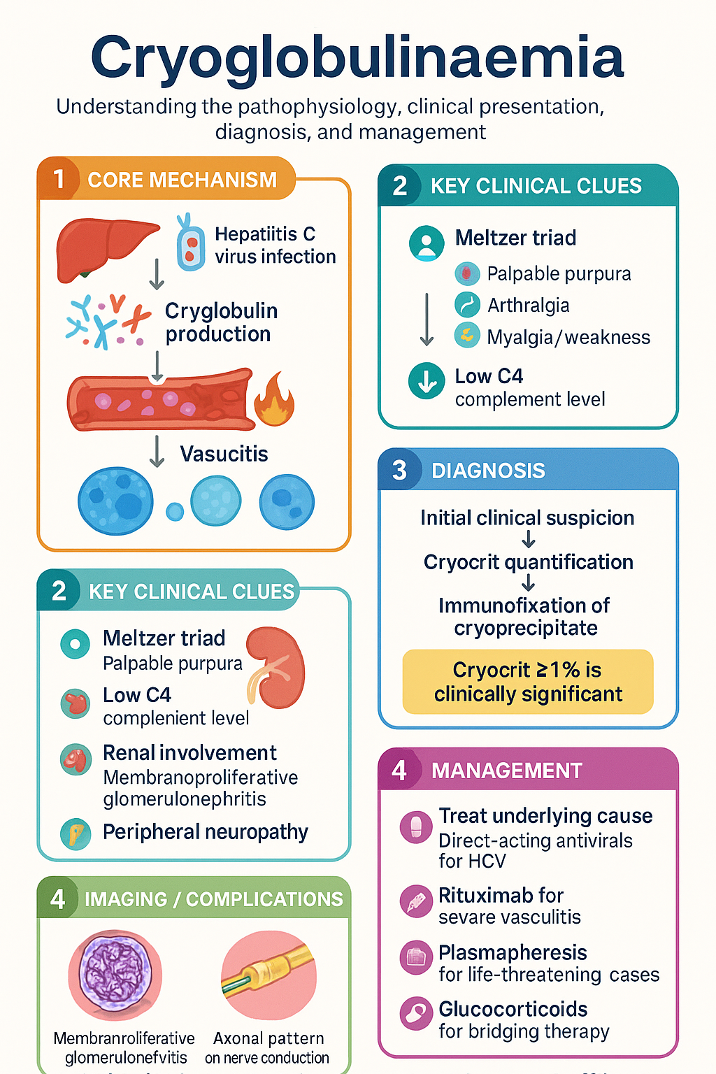
  • Cryoglobulins are immunoglobulins that reversibly precipitate at temperatures <37 °C, causing small-to-medium vessel vasculitis via immune-complex deposition.
  • Three types: Type I (monoclonal IgG or IgM; associated with haematological malignancy); Type II (mixed, monoclonal + polyclonal; majority HCV-associated); Type III (polyclonal; HCV, SLE, Sjögren).
  • Hepatitis C virus (HCV) infection is the commonest cause of mixed cryoglobulinaemia (Types II & III), accounting for ~90% of cases in endemic regions.
  • Characteristically very low C4 complement level (often the first clue); C3 may be normal or mildly reduced.
  • Meltzer triad — palpable purpura, arthralgia, myalgia/weakness — is the classic clinical presentation of mixed cryoglobulinaemic vasculitis.
  • Renal involvement (membranoproliferative glomerulonephritis) and peripheral neuropathy are important organ manifestations that determine prognosis.
  • Diagnosis requires cryocrit >1% with characterisation by immunofixation; blood must be collected and transported at 37 °C (warm-clotted tube).
  • Anti-HCV serology, HBV, HIV, ANA, RF, and serum protein electrophoresis (SPEP) with immunofixation are essential workup.
  • Treatment of the underlying cause is paramount: direct-acting antiviral (DAA) therapy for HCV-associated cryoglobulinaemia (PBS-listed).
  • Rituximab (MabThera®) is first-line immunosuppressive therapy for severe or refractory mixed cryoglobulinaemic vasculitis; PBS Authority Required for vasculitis indication.
  • Plasmapheresis (plasma exchange) is reserved for life-threatening or rapidly progressive disease (acute GN, digital necrosis, severe neuropathy).
  • Glucocorticoids (pulse IV methylprednisolone then oral prednisolone) bridge therapy while awaiting rituximab efficacy or antiviral response.
  • Type I cryoglobulinaemia management targets the underlying B-cell lymphoproliferative disorder; chemotherapy ± plasmapheresis for hyperviscosity.

Introduction & Australian Epidemiology

Cryoglobulins are serum immunoglobulins that reversibly precipitate at temperatures below 37 °C and re-dissolve upon rewarming. Their presence in the circulation can lead to immune-complex deposition in small- and medium-sized vessels, producing a systemic vasculitis known as cryoglobulinaemic vasculitis.

In Australia, mixed cryoglobulinaemia (Types II and III) is predominantly associated with chronic hepatitis C virus (HCV) infection. Despite Australia's success in reducing HCV prevalence through direct-acting antiviral (DAA) programmes since 2016, cryoglobulinaemic vasculitis persists in patients with longstanding or previously treated HCV infection. The Australian Institute of Health and Welfare (AIHW) estimates approximately 180,000 people are living with chronic HCV nationally, of whom up to 40–60% may develop low-level cryoglobulins, though clinically significant vasculitis occurs in a smaller subset (~5–10%).

Type I cryoglobulinaemia is rare and is associated with haematological malignancies, particularly Waldenström macroglobulinaemia and multiple myeloma. Non-HCV causes of mixed cryoglobulinaemia in the Australian context include systemic lupus erythematosus (SLE), Sjögren syndrome, and other autoimmune conditions. Aboriginal and Torres Strait Islander peoples have a higher prevalence of HCV and autoimmune disease, requiring specific clinical consideration.

This guideline provides an Australian-focused approach to the classification, diagnosis, and management of cryoglobulinaemia, aligned with current evidence and Australian prescribing frameworks.

Cryoglobulinaemia clinical infographic — pathophysiology, clinical clues, diagnosis, imaging, and management
Tap or click image to enlarge — Cryoglobulinaemia: pathophysiology, clinical clues, diagnosis, imaging, and management.
Cryoglobulinaemia infographic, full size

Types & Associations

Cryoglobulins are classified into three types based on the Brouet classification, which has important implications for aetiology, clinical features, and management strategy.

Feature Type I Type II (Mixed) Type III (Mixed)
Composition Monoclonal IgG or IgM (rarely IgA) Monoclonal IgM (usually IgMκ with RF activity) + polyclonal IgG Polyclonal IgM (with RF activity) + polyclonal IgG
Brouet frequency ~10–15% ~50–60% ~30–40%
Mechanism Hyperviscosity / vascular occlusion (not immune-complex) Immune-complex deposition; RF activity of monoclonal IgM Immune-complex deposition; polyclonal RF
Key associations Waldenström macroglobulinaemia, multiple myeloma, CLL, MGUS HCV (~90%), HBV, HIV, Sjögren syndrome, SLE HCV, SLE, Sjögren syndrome, other autoimmune diseases
Typical vasculitis No (occlusive rather than inflammatory) Yes — small vessel Yes — small vessel
C4 Normal or mildly low Very low (characteristic) Very low (characteristic)
RF Negative Positive (high titre) Positive (variable titre)
⚠️
Key distinction: Type I cryoglobulinaemia is a hyperviscosity/occlusive syndrome associated with underlying haematological malignancy — it does NOT cause classical immune-complex vasculitis. Types II and III are true vasculitides driven by immune-complex deposition with rheumatoid factor activity.

HCV — The Commonest Cause

Hepatitis C virus drives mixed cryoglobulinaemia through chronic antigenic stimulation of B lymphocytes, leading to clonal expansion of RF-producing B cells. In Australia, HCV genotype 1 (1a and 1b) predominates. Cryoglobulins may persist for years after HCV virological cure (SVR), and vasculitis can occasionally flare in the post-SVR period.

Other Associations

  • Sjögren syndrome: Cryoglobulins present in 5–15%; their presence predicts lymphoma risk and is a criterion for severe disease in the EULAR-SSDAI.
  • SLE: Mixed cryoglobulins may be detected; low C4 is common in SLE regardless.
  • HBV: Less common than HCV in Australia but should be excluded.
  • HIV: Polyclonal B-cell activation may produce cryoglobulins.
  • Other: Rheumatoid arthritis, malignancy (non-haematological), and rare idiopathic forms.

Clinical Manifestations & Diagnosis

Meltzer Triad

The classic presentation of mixed cryoglobulinaemic vasculitis (Types II/III) is the Meltzer triad:

  • Palpable purpura — typically lower limbs, triggered by cold exposure or prolonged standing; may be chronic and recurrent.
  • Arthralgia — usually non-erosive, affecting knees, hands, and ankles symmetrically.
  • Weakness / myalgia — constitutional symptom; may be prominent.

Organ Involvement

System Manifestation Notes
Skin Palpable purpura, livedo reticularis, digital ulceration, necrosis Most common manifestation (~90%); Raynaud phenomenon
Renal Membranoproliferative glomerulonephritis (MPGN) 20–30%; proteinuria, haematuria, declining eGFR; Type I MPGN pattern on biopsy
Peripheral nerve Peripheral neuropathy — sensory > motor; mononeuritis multiplex ~60–70%; often early and disabling; axonal pattern on NCS
GI Abdominal pain, mesenteric vasculitis (rare) Ischaemic colitis in severe cases
Pulmonary Interstitial lung disease, pulmonary haemorrhage (rare) More common in Type I
CNS Rare — stroke, transverse myelitis Consider in differential of young stroke
Type I-specific Hyperviscosity syndrome — visual disturbance, headache, mucosal bleeding Acute emergency; plasmapheresis indicated

Diagnostic Approach

The diagnosis of cryoglobulinaemic vasculitis requires both the detection of circulating cryoglobulins AND clinical evidence of vasculitis. Cryoglobulinaemia alone (without vasculitis) does not mandate immunosuppressive therapy.

1
Correct sample collection
Blood must be collected into a pre-warmed (37 °C) tube and maintained at body temperature during transport to the laboratory. A minimum of 20 mL is required. The sample must NOT be refrigerated. Processing occurs at 37 °C, then the serum is cooled to 4 °C for 7 days to detect precipitation.
2
Cryocrit quantification
Cryocrit >1% is considered clinically significant. The cryoprecipitate is quantified as a percentage of total serum volume.
3
Immunofixation of the cryoprecipitate
Characterises the cryoglobulin type (I, II, or III). Monoclonal component identification (IgMκ most common in Type II).
4
Aetiological workup
Anti-HCV (Ab + PCR if seropositive), HBV serology, HIV, ANA, anti-dsDNA, anti-Ro/SSA, anti-La/SSB, RF, SPEP + immunofixation, bone marrow biopsy if Type I.
5
Tissue biopsy (when indicated)
Skin biopsy of purpuric lesion (direct IF showing IgM, IgG, C3 in vessel walls); renal biopsy for nephritis (MPGN pattern, subendothelial deposits on EM). Nerve biopsy rarely needed.

Investigations

Essential
Cryoglobulins (cryocrit + immunofixation)
37 °C warm-clotted tube, ≥20 mL; 7-day cold precipitation protocol. Not available at all labs — refer to major tertiary or reference laboratory (e.g., Royal Melbourne, Westmead, SA Pathology). MBS Item 71137 (specialised protein studies).
Essential
Complement levels (C3, C4)
C4 is characteristically very low in mixed cryoglobulinaemia (Types II/III); often the first biochemical clue. C3 is variably reduced. MBS Item 71115.
Essential
Anti-HCV antibody + HCV RNA (PCR)
All patients with mixed cryoglobulinaemia require HCV testing. If HCV Ab positive, confirm with quantitative PCR. MBS Item 69482 (HCV Ab), 73860 (HCV RNA).
Essential
Renal function, urinalysis + urine protein-to-creatinine ratio
Screen for glomerulonephritis. ACR >30 mg/mmol warrants renal biopsy discussion.
Available
Rheumatoid factor (RF)
Positive (often high titre) in mixed cryoglobulinaemia. MBS Item 66536.
Available
SPEP + immunofixation
Essential to detect monoclonal paraprotein in Type I and some Type II cases. MBS Item 71114 (SPEP), 71137 (immunofixation).
Available
HBV serology, HIV, ANA, anti-dsDNA, ENA panel
Exclude other causes; ANA and ENA screen for SLE and Sjögren.
Specialist
Skin biopsy with direct immunofluorescence
Demonstrates IgM, IgG, C3 in vessel walls; confirms vasculitis. Dermatology or rheumatology referral.
Specialist
Renal biopsy
Indicated for unexplained proteinuria, haematuria, or declining eGFR. Shows MPGN Type I pattern with subendothelial deposits containing IgG, IgM, C3. Nephrology referral.
Specialist
Nerve conduction studies ± nerve biopsy
Axonal sensory or sensorimotor neuropathy; mononeuritis multiplex pattern. Neurology referral.
Referral
Bone marrow biopsy
For Type I cryoglobulinaemia to characterise underlying lymphoproliferative disorder (Waldenström, myeloma, MGUS). Haematology referral.

Risk Stratification & Severity Scoring

Severity assessment guides the intensity of treatment. The following classification, adapted from the Italian Group for the Study of Cryoglobulinaemia and the EULAR recommendations, stratifies mixed cryoglobulinaemic vasculitis:

Mild
Cutaneous Only
Palpable purpura (<10% body surface), arthralgia, fatigue. No organ-threatening involvement. Stable renal function.
Setting: Treat underlying cause; symptomatic Rx. Monitor 3-monthly.
Moderate
Active Vasculitis with Organ Involvement
Widespread purpura, peripheral neuropathy (sensory), mild-moderate nephritis (proteinuria <1 g/day, stable eGFR), mild CNS involvement. Cryocrit usually >5%.
Setting: Immunosuppression (rituximab ± steroids). Rheumatology + nephrology co-management.
Severe
Life- or Organ-Threatening
Rapidly progressive GN (eGFR decline, nephrotic/nephritic), severe neuropathy (motor), digital necrosis/gangrene, intestinal ischaemia, pulmonary haemorrhage, hyperviscosity (Type I).
Setting: IV methylprednisolone pulse + plasmapheresis + rituximab. Inpatient, MDT.
🚨
Emergency presentations: Hyperviscosity syndrome (Type I), rapidly progressive GN, and severe digital necrosis require urgent hospital admission, plasmapheresis, and high-dose immunosuppression. Contact rheumatology/oncology immediately.

Empirical Therapy

Empirical therapy is initiated before a complete aetiological workup is finalised, or while awaiting response to definitive treatment (e.g., antiviral therapy). The approach varies by severity.

Mild Disease — Symptomatic & Supportive

  • Cold avoidance (warm clothing, heated environments) — fundamental but often under-emphasised.
  • Compression stockings for dependent purpura.
  • NSAIDs for arthralgia (avoid if renal impairment).
  • Low-dose oral prednisolone (10–15 mg/day) may be used short-term for flares.

Moderate Disease — Glucocorticoids

💊
Prednisolone
Solone®, Panafcortelone® · Glucocorticoid
Adult dose 0.5–1 mg/kg/day PO (max 60 mg), taper over 3–6 months to ≤7.5 mg/day maintenance
Paediatric dose 1–2 mg/kg/day PO; taper as per rheumatology
Route / frequency Oral, once daily (morning)
Renal adjustment No dose adjustment; monitor fluid retention
Hepatic adjustment No adjustment
PBS status ✔ PBS General Benefit

Severe Disease — IV Pulse Methylprednisolone

💊
Methylprednisolone (IV)
Solu-Medrol® · Glucocorticoid (IV pulse)
Adult dose 500–1000 mg IV daily for 3 days (pulse), then convert to oral prednisolone 1 mg/kg/day
Route / frequency IV infusion over 30–60 min, once daily
Renal adjustment No dose adjustment
PBS status ✔ PBS General Benefit

Directed / Cause-Specific Therapy

HCV-Associated Mixed Cryoglobulinaemia

Antiviral therapy is the cornerstone of treatment for HCV-associated mixed cryoglobulinaemia. Direct-acting antivirals (DAAs) have transformed outcomes, with SVR rates >95% across most genotypes.

💊
Sofosbuvir / Velpatasvir
Epclusa® · Pan-genotypic DAA
Adult dose 1 tablet (400/100 mg) PO once daily for 12 weeks
Paediatric dose ≥6 years, ≥17 kg: weight-based dosing per PBS schedule
Renal adjustment No adjustment (sofosbuvir safe in CKD, including dialysis)
Hepatic adjustment Child-Pugh C: limited data; specialist guidance required
PBS status ✔ PBS Authority Required
💊
Glecaprevir / Pibrentasvir
Maviret® · Pan-genotypic DAA
Adult dose 3 tablets (300/120 mg total) PO once daily with food for 8 weeks (treatment-naïve, non-cirrhotic)
Renal adjustment No adjustment; safe in all stages of CKD
PBS status ✔ PBS Authority Required
⚠️
Post-SRV vasculitis: Cryoglobulinaemic vasculitis can persist or even flare after HCV SVR (virological cure). If active vasculitis persists >6 months post-SVR, immunosuppressive therapy (rituximab) is indicated. Monitor cryocrit and complement levels post-SVR.

Immunosuppressive Therapy — Rituximab

Rituximab is the preferred immunosuppressive agent for moderate-to-severe mixed cryoglobulinaemic vasculitis, based on the RITUXVAS trial and multiple observational studies demonstrating superiority over conventional immunosuppression.

💊
Rituximab
MabThera® · Anti-CD20 monoclonal antibody
Adult dose 375 mg/m² IV weekly × 4 doses (lymphoma protocol) OR 1000 mg IV on Day 1 and Day 15 (RA protocol)
Paediatric dose Not routinely used; specialist guidance only
Route / frequency IV infusion; premedicate with paracetamol, antihistamine, and corticosteroid
Renal adjustment No dose adjustment
Hepatic adjustment No dose adjustment
Key monitoring Check hepatitis B (HBsAg, anti-HBc) before first dose; screen for hypogammaglobulinaemia
PBS status ✔ PBS Authority Required (vasculitis indication)

Plasmapheresis (Plasma Exchange)

Plasmapheresis physically removes circulating cryoglobulins and is reserved for severe, life-threatening manifestations.

ℹ️
Indications for plasmapheresis:
  • Rapidly progressive glomerulonephritis with declining renal function
  • Severe digital necrosis / gangrene
  • Hyperviscosity syndrome (Type I)
  • Severe neuropathy with motor involvement
  • Pulmonary haemorrhage
Protocol: 3–5 exchanges over 7–14 days; 40–60 mL/kg per session using 4–5% albumin replacement. Must be combined with immunosuppression (rituximab or cyclophosphamide) to prevent rebound cryoglobulin production.

Type I Cryoglobulinaemia

Management targets the underlying B-cell lymphoproliferative disorder. Treatment is guided by haematology and follows the specific malignancy protocol (e.g., Bortezomib-Rituximab-Dexamethasone for Waldenström macroglobulinaemia, or R-CHOP for aggressive lymphoma). Plasmapheresis is indicated for hyperviscosity crisis. Rituximab monotherapy is appropriate for indolent disease.

HCV-Negative Mixed Cryoglobulinaemia

For cryoglobulinaemia associated with SLE, Sjögren syndrome, or other autoimmune conditions, rituximab combined with glucocorticoids is the preferred immunosuppressive strategy. Cyclophosphamide is reserved for refractory cases.

Monitoring

Ongoing monitoring assesses treatment response, disease activity, and complications of therapy.

Baseline
Full blood count, EUC, LFT, cryocrit, C3/C4, RF, HCV RNA (if applicable), urine ACR, eGFR, nerve conduction studies if symptomatic, HBV screening (prior to rituximab).
4–6 weeks
Reassess clinical symptoms (purpura, neuropathy). Repeat FBC, EUC, LFT. HCV RNA if on DAA therapy (virological response check).
12 weeks
HCV SVR12 assessment (sustained virological response). Cryocrit and C4 to assess biochemical response to immunosuppression. Urine ACR.
3–6 monthly
Ongoing monitoring: cryocrit, C4, RF, urine ACR, eGFR. Assess for rituximab-related hypogammaglobulinaemia (IgG level). Monitor for HBV reactivation if HBcAb positive.
Annually
SPEP + immunofixation (Type I/II). Bone marrow assessment if progression suspected. Vaccination review (influenza, pneumococcal — prior to immunosuppression).
⚠️
Hepatitis B reactivation: All patients requiring rituximab must be screened for HBV (HBsAg, anti-HBc). If HBcAb positive ± HBsAg positive, initiate antiviral prophylaxis (entecavir 0.5 mg PO daily) starting before rituximab and continuing for ≥12 months after the last dose, with hepatology co-management.

Special Populations

🤰 Pregnancy
Rituximab
Contraindicated in pregnancy (Category D — B-cell depletion in neonate). Avoid conception for ≥12 months after last dose. Effective contraception required during therapy.
DAA antivirals (sofosbuvir, glecaprevir/pibrentasvir)
Limited human data; generally avoid in pregnancy. Defer HCV treatment to post-partum unless severe vasculitis.
Prednisolone
Category A — safe in pregnancy at lowest effective dose. Neonatal adrenal suppression risk at high doses.
Plasmapheresis
Safe in pregnancy; may be used for severe flares. Coordinate with obstetric and rheumatology teams.
👶 Paediatrics
General
Cryoglobulinaemia is rare in children. When encountered, consider autoimmune disease (SLE) as the primary aetiology rather than HCV. Rituximab use off-label; specialist paediatric rheumatology guidance required.
DAAs
Sofosbuvir/velpatasvir PBS-listed for children ≥6 years, ≥17 kg. Glecaprevir/pibrentasvir available ≥12 years in Australia.
🧓 Elderly
Glucocorticoids
Increased risk of osteoporosis, diabetes, infection. Co-prescribe calcium, vitamin D, and consider bisphosphonate. Aim for lowest effective dose and rapid taper.
Rituximab
Higher infection risk. Monitor immunoglobulin levels. Screen for PJP risk; consider prophylaxis if prolonged immunosuppression.
🫘 Renal Impairment
Sofosbuvir/velpatasvir
Safe in all stages of CKD including dialysis — preferred DAA in renal impairment.
Rituximab
No dose adjustment; monitor for tumour lysis if high disease burden.
Plasmapheresis
Can be performed via central venous catheter in patients on haemodialysis; coordinate timing.
🫁 Hepatic Impairment
DAAs
Glecaprevir/pibrentasvir contraindicated in Child-Pugh C (hepatotoxicity risk). Sofosbuvir/velpatasvir preferred in decompensated cirrhosis with specialist hepatology input.
Rituximab
No specific hepatic dose adjustment; monitor LFTs closely in patients with cirrhosis.
🛡️ Immunocompromised
Rituximab
Profound B-cell depletion — ensure all vaccinations are up-to-date before commencing (minimum 4 weeks prior). Avoid live vaccines. Monitor immunoglobulin levels; IVIg replacement if recurrent infections with IgG <4 g/L.
HIV co-infection
DAA therapy is safe and effective in HIV/HCV co-infected patients. Drug interactions with ART must be checked (sofosbuvir/velpatasvir generally compatible).

Aboriginal and Torres Strait Islander Health Considerations

Aboriginal and Torres Strait Islander Health
HCV prevalence
Aboriginal and Torres Strait Islander peoples have a higher prevalence of chronic HCV compared with non-Indigenous Australians (estimated 3–5 times higher in some jurisdictions). DAA access through community-controlled health services has improved but gaps remain in remote and very remote areas. The PBS Authority Required pathway for DAAs is accessible through Aboriginal Community Controlled Health Organisations (ACCHOs).
Autoimmune disease burden
SLE and Sjögren syndrome are more prevalent in Aboriginal and Torres Strait Islander populations and may be more severe at presentation. Mixed cryoglobulinaemia should be considered in any Indigenous patient with unexplained purpura, neuropathy, or nephritis, even in the absence of HCV.
Remote and rural access
Specialist rheumatology, nephrology, and hepatology services are limited outside major centres. Telehealth consultations (MBS Items 91822, 91823) are essential for ongoing management. Plasmapheresis is available only at tertiary centres; aeromedical retrieval may be required for severe presentations (RFDS, CareFlight). Cryoglobulin testing requires sample transport at 37 °C, which is challenging in hot climates — coordinate with pathology services for urgent cold-chain transport arrangements.
Renal disease
Aboriginal and Torres Strait Islander peoples have a significantly higher burden of CKD and ESKD. Cryoglobulinaemic MPGN may compound pre-existing renal disease. Urine ACR monitoring should be embedded in chronic disease management programmes through primary healthcare. eGFR calculations should use CKD-EPI equations without the race coefficient.
Cultural safety
Engage Aboriginal Health Workers and Liaison Officers in care planning. Allow time for family consultation. Provide culturally appropriate patient education materials (available through NACCHO and HealthInfoNet). Respect Sorry Business and other cultural obligations that may affect treatment adherence.
Medication access
Ensure PBS co-payment support through Closing the Gap PBS co-payment measure (eligible patients pay a maximum of .70 per PBS script). Rituximab and DAA access via ACCHOs is well supported. Coordinate with local pharmacy for cold-chain requirements of certain biologics.

📚 References

  1. 1. Muchtar E, Magen H, Gertz MA. How I treat cryoglobulinemia. Blood. 2017;129(3):289-298. doi:10.1182/blood-2016-09-719773
  2. 2. Brouet JC, Clauvel JP, Danon F, Klein M, Seligmann M. Biologic and clinical significance of cryoglobulins. A report of 86 cases. Am J Med. 1974;57(5):775-788. doi:10.1016/0002-9343(74)90852-3
  3. 3. De Vita S, Quartuccio L, Isola M, et al. A randomized controlled trial of rituximab for the treatment of severe cryoglobulinemic vasculitis. Arthritis Rheum. 2012;64(3):843-853. doi:10.1002/art.34331
  4. 4. Roccatello D, Sciascia S, Baldovino S, et al. A multicentre study of the RITUXVAS trial: rituximab in cryoglobulinaemic vasculitis. RMD Open. 2018;4(2):e000662. doi:10.1136/rmdopen-2018-000662
  5. 5. Ferri C, Sebastiani M, Giuggioli D, et al. Mixed cryoglobulinemia: demographic, clinical, and serologic features and survival in 231 patients. Semin Arthritis Rheum. 2004;33(6):355-374. doi:10.1016/j.semarthrit.2003.10.001
  6. 6. Australian Institute of Health and Welfare (AIHW). Hepatitis C in Australia 2024. Canberra: AIHW; 2024. Cat. no. HIV 13.
  7. 7. The Royal Australian College of General Practitioners (RACGP). Hepatitis B and C: Testing, Management and Treatment. 4th edition. East Melbourne: RACGP; 2022.
  8. 8. Mukhtyar C, Guillevin L, Cid MC, et al. EULAR recommendations for the management of primary small and medium vessel vasculitis. Ann Rheum Dis. 2009;68(3):310-317. doi:10.1136/ard.2008.088096
  9. 9. Cacoub P, Comarmond C, Domont F, Savey L, Desbois AC, Saadoun D. Extrahepatic manifestations of chronic hepatitis C virus infection. Ther Adv Infect Dis. 2016;3(1):3-14. doi:10.1177/2049936115585942
  10. 10. Dammacco F, Sansonno D. Therapy for hepatitis C virus-related cryoglobulinemic vasculitis. N Engl J Med. 2013;369(11):1035-1045. doi:10.1056/NEJMra1208642
  11. 11. RHDAustralia (Northern Territory Department of Health and Menzies School of Health Research). Australian Guidelines for the Prevention, Diagnosis and Management of Acute Rheumatic Fever and Rheumatic Heart Disease. 3rd edition. Darwin: RHDAustralia; 2020.
  12. 12. Australasian Society for Clinical Immunology and Allergy (ASCIA). Position statement on the use of rituximab in autoimmune conditions. Sydney: ASCIA; 2023.
  13. 13. Pharmaceutical Benefits Scheme (PBS). Schedule of Pharmaceutical Benefits. Australian Government Department of Health and Aged Care. Available at: www.pbs.gov.au. Accessed 2024.
  14. 14. National Aboriginal Community Controlled Health Organisation (NACCHO). Position statement on hepatitis C elimination in Aboriginal and Torres Strait Islander communities. Canberra: NACCHO; 2023.