Home Rheumatology Necrotising Autoimmune Myopathy (NAM)

Necrotising Autoimmune Myopathy (NAM)

๐Ÿ“‹ Key Information Summary

๐Ÿ“‹
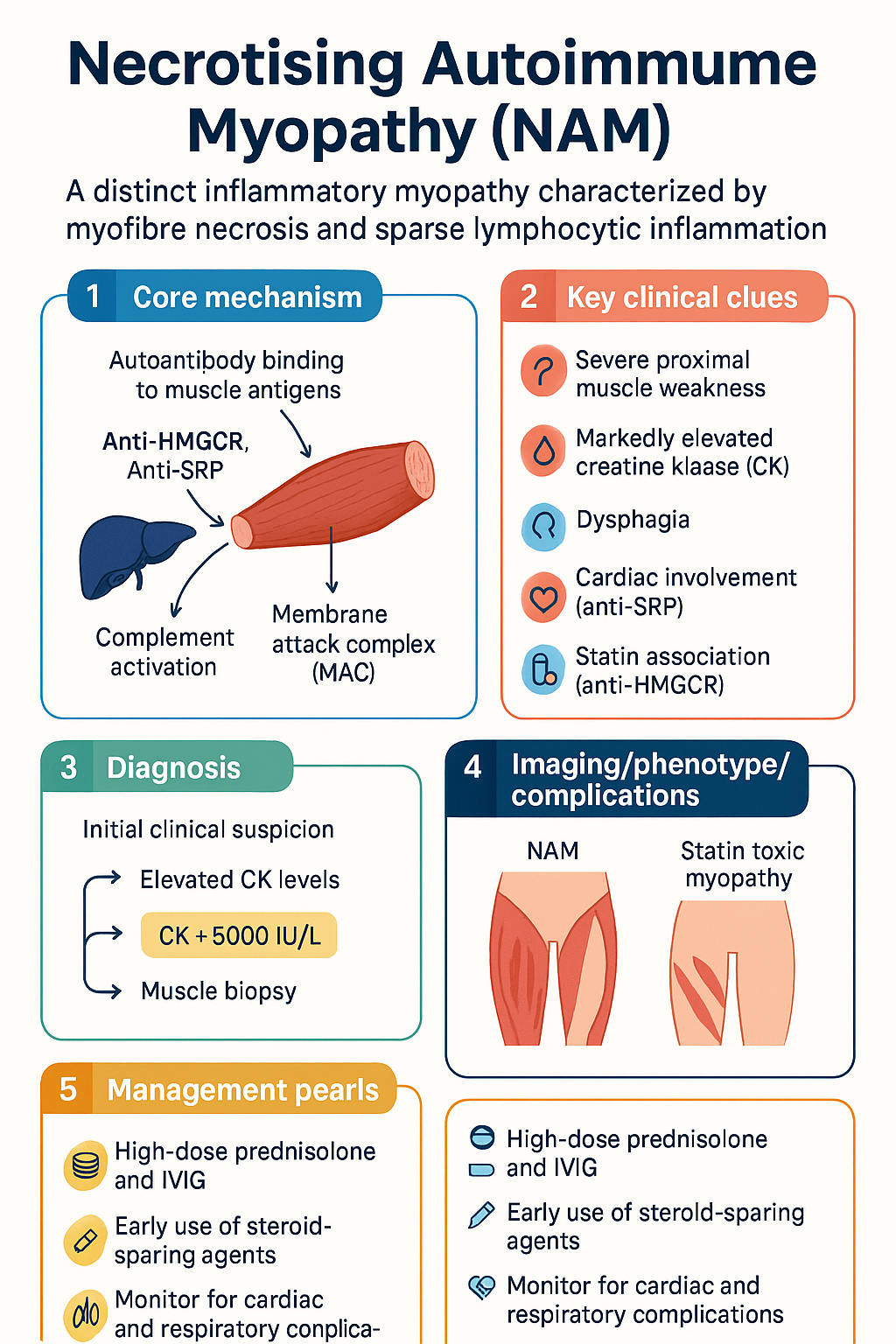
  • Necrotising autoimmune myopathy (NAM) is a distinct inflammatory myopathy defined by myofibre necrosis with sparse lymphocytic inflammation, distinguished from dermatomyositis and polymyositis by serology and histopathology.
  • Two major antibody subtypes: anti-HMGCR (3-hydroxy-3-methylglutaryl-coenzyme A reductase) โ€” classically statin-associated; anti-SRP (signal recognition particle) โ€” statin-independent and generally more severe.
  • Anti-HMGCR NAM may occur in statin-exposed patients but also in statin-naรฏve individuals, particularly children and young adults of non-Caucasian descent.
  • Anti-SRP NAM typically presents with severe, rapidly progressive proximal weakness and carries a higher risk of cardiac involvement and treatment resistance.
  • Both antibody types activate complement-mediated myocyte injury via the membrane attack complex (MAC, C5b-9), leading to necrotic and regenerating fibres.
  • Seronegative NAM exists and requires muscle biopsy for diagnosis โ€” anti-HMGCR and anti-SRP serology alone is insufficient to exclude the diagnosis.
  • Creatine kinase (CK) is typically markedly elevated, often >5000 IU/L and frequently >10,000 IU/L at presentation; CK serves as the primary treatment response marker.
  • Muscle biopsy shows necrotic and regenerating fibres with minimal inflammatory infiltrate and MAC deposition on immunohistochemistry โ€” unlike polymyositis where CD8+ T-cell infiltrates predominate.
  • Discontinuation of statin alone is insufficient; aggressive combination immunotherapy is required in nearly all cases.
  • First-line treatment: high-dose prednisolone 1 mg/kg/day (max 75 mg) PLUS intravenous immunoglobulin (IVIG) 2 g/kg over 2โ€“5 days, then 1 g/kg monthly.
  • Steroid-sparing agents: methotrexate (15โ€“25 mg/week), azathioprine (2โ€“3 mg/kg/day), or mycophenolate mofetil (2โ€“3 g/day) should be commenced early; rituximab is effective for refractory disease in both subtypes.
  • Treatment duration is prolonged โ€” typically 2โ€“5 years or longer; premature discontinuation carries a high relapse rate of 40โ€“60%.
  • Anti-SRP NAM may require more aggressive upfront therapy (e.g., early rituximab) due to higher rates of incomplete response and relapse compared with anti-HMGCR disease.
  • Distinguish NAM from statin toxic myopathy (which resolves with statin cessation and does not require immunosuppression) โ€” persistent CK elevation and weakness >2 months after statin withdrawal suggests NAM.
  • Monitor for complications: dysphagia (assess with speech pathology), respiratory muscle weakness (FVC monitoring), cardiac involvement in anti-SRP (ECG, troponin, echocardiography), and steroid side effects including osteoporosis and glycaemic deterioration.

Introduction & Australian Epidemiology

Necrotising autoimmune myopathy (NAM) โ€” also termed immune-mediated necrotising myopathy (IMNM) โ€” is a relatively recently characterised inflammatory myopathy that has been formally distinguished from dermatomyositis, polymyositis, and inclusion body myositis in the 2017 European Neuromuscular Centre (ENMC) international classification. NAM is defined by a unique clinicopathological triad: severe proximal muscle weakness, markedly elevated serum creatine kinase (CK), and a muscle biopsy demonstrating prominent myofibre necrosis and regeneration with strikingly sparse lymphocytic inflammatory infiltrate.

The identification of disease-specific autoantibodies โ€” anti-HMGCR (3-hydroxy-3-methylglutaryl-coenzyme A reductase) and anti-SRP (signal recognition particle) โ€” has refined diagnostic accuracy and improved understanding of pathogenic mechanisms. These antibodies are now considered central to the diagnosis and subclassification of NAM, with seronegative cases representing approximately 10โ€“20% of all presentations.

Epidemiology in Australia

NAM is a rare condition with an estimated annual incidence of 1โ€“2 per million population globally. Australian-specific incidence data are limited, though extrapolation from international registries suggests approximately 25โ€“50 new cases per year nationally. Key epidemiological observations in the Australian context include:

  • Anti-HMGCR NAM is the most common subtype in Australia, consistent with the widespread use of statin therapy (approximately 2.7 million Australians are prescribed statins through the PBS).
  • Anti-SRP NAM accounts for approximately 20โ€“30% of NAM cases and appears to have no significant racial predilection in Australian cohorts.
  • Statin-naรฏve anti-HMGCR NAM has been reported in children and young adults, with some evidence suggesting increased susceptibility in individuals of Southeast Asian descent โ€” relevant given Australia's multicultural population.
  • Anti-SRP NAM has been associated with cardiac involvement including myocarditis in up to 15โ€“25% of cases, a complication with significant morbidity requiring specialist cardiology input.
  • The condition affects adults across a wide age range (20โ€“80 years), with anti-HMGCR cases peaking in the 5thโ€“7th decades (correlating with statin exposure), while anti-SRP cases may present at younger ages.

Australia's universal healthcare system facilitates access to the specialist investigations required for NAM diagnosis (anti-HMGCR and anti-SRP serology available through major immunology laboratories, and PBS-subsidised access to IVIG and rituximab), though delays in diagnosis remain a significant concern โ€” the median time from symptom onset to NAM diagnosis in published series is 3โ€“6 months.

Necrotising Autoimmune Myopathy (NAM) clinical infographic โ€” pathophysiology, clinical clues, diagnosis, imaging, and management
Tap or click image to enlarge โ€” Necrotising Autoimmune Myopathy (NAM): pathophysiology, clinical clues, diagnosis, imaging, and management.
Necrotising Autoimmune Myopathy (NAM) infographic, full size

Pathogenesis & Antibodies

NAM is a humoural (antibody)-mediated myopathy in which autoantibodies directed against intracellular muscle antigens drive complement activation and myocyte necrosis. This contrasts with dermatomyositis (microangiopathy) and polymyositis (T-cell-mediated cytotoxicity). Two pathogenic antibody systems have been identified:

Anti-HMGCR Antibodies

โš ๏ธ
Statin exposure is the primary trigger: Statins upregulate HMGCR expression in regenerating muscle fibres by 3โ€“5 fold. In genetically predisposed individuals, this increased antigen expression breaks immune tolerance, leading to anti-HMGCR antibody production and complement-mediated myocyte injury. Risk correlates with statin potency (atorvastatin, rosuvastatin > pravastatin, simvastatin) rather than dose.
  • Anti-HMGCR antibodies are detected in approximately 60% of NAM cases in Australian tertiary referral series.
  • The HMGCR enzyme is the pharmacological target of statin drugs (HMG-CoA reductase inhibitors).
  • In statin-exposed patients, regenerating muscle fibres overexpress HMGCR, presenting an immunogenic target. Anti-HMGCR antibodies bind to surface-expressed HMGCR on regenerating fibres and activate the classical complement pathway, culminating in C5b-9 membrane attack complex (MAC) deposition and myofibre necrosis.
  • Statin-naรฏve anti-HMGCR NAM occurs in approximately 30โ€“40% of anti-HMGCR-positive patients, more commonly in children, young adults, and individuals of non-Caucasian ethnicity. The mechanism in this group is less well understood but may involve genetic polymorphisms in HMGCR expression regulation.
  • HLA associations: HLA-DRB1*11:01 is strongly associated with anti-HMGCR NAM (odds ratio approximately 3.6), suggesting a genetic susceptibility component.

Anti-SRP Antibodies

  • Anti-SRP antibodies target the 54 kDa subunit of the signal recognition particle, a ribonucleoprotein complex involved in co-translational protein translocation across the endoplasmic reticulum.
  • Anti-SRP NAM is statin-independent โ€” there is no established association between statin exposure and anti-SRP antibody development.
  • Anti-SRP antibodies activate complement via both the classical and alternative pathways, leading to MAC deposition on the myocyte surface and necrosis.
  • Anti-SRP NAM is generally considered more severe than anti-HMGCR disease, with higher presenting CK levels, more rapid progression, greater treatment resistance, and higher rates of extramuscular complications including cardiac involvement (myocarditis, conduction abnormalities).
  • HLA associations: HLA-DRB1*08:03 has been identified in Japanese anti-SRP cohorts; the Australian HLA association is not well characterised.

Seronegative NAM

  • Approximately 10โ€“20% of patients with clinicopathological features consistent with NAM are negative for both anti-HMGCR and anti-SRP antibodies on standard testing.
  • Seronegative NAM remains a histopathological diagnosis โ€” the biopsy must demonstrate necrotic and regenerating fibres with minimal inflammation and MAC deposition to support the diagnosis.
  • Emerging antibodies under investigation include anti-Mi2, anti-NXP2, and anti-SAE, though these are typically associated with dermatomyositis rather than pure NAM.
  • Seronegative NAM should be managed with the same aggressive immunotherapy approach as seropositive disease.

Shared Pathogenic Mechanisms

Both anti-HMGCR and anti-SRP antibodies share a final common pathway of complement-mediated myocyte injury. Key mechanistic steps include:

  1. Autoantibody binding to intracellular target antigen (HMGCR on regenerating fibres surface; SRP on myocyte surface)
  2. Classical complement pathway activation (C1q binding to antibody Fc regions)
  3. C3 convertase formation and C3b amplification loop
  4. C5 convertase generation โ†’ C5a release (pro-inflammatory) โ†’ C5b-9 MAC assembly on myocyte membrane
  5. MAC pore formation โ†’ osmotic lysis โ†’ myofibre necrosis
  6. Satellite cell activation โ†’ regenerating fibres with HMGCR upregulation (perpetuating anti-HMGCR disease)

This complement-mediated mechanism provides the rationale for IVIG (complement scavenging and anti-idiotypic antibody effects) and rituximab (B-cell depletion reducing autoantibody production) as therapeutic strategies.

Clinical Features & Diagnosis

Clinical Presentation

NAM typically presents with subacute to chronic progressive proximal muscle weakness, developing over weeks to months. The clinical phenotype differs between the two major subtypes:

Feature Anti-HMGCR NAM Anti-SRP NAM
Statin association 60โ€“70% statin-exposed; 30โ€“40% statin-naรฏve Statin-independent
Onset age Typically 50โ€“70 years (statin-associated); bimodal in children/young adults Broad range 20โ€“80 years; younger median age than anti-HMGCR
Weakness pattern Symmetric proximal; may be subacute Severe symmetric proximal; often rapidly progressive
Dysphagia 20โ€“30% 30โ€“50%
Cardiac involvement Rare (<5%) 15โ€“25% (myocarditis, conduction defects)
ILD Uncommon Reported in up to 10โ€“15%
Skin findings None (distinguishes from dermatomyositis) None
Treatment response Generally better; 70โ€“80% achieve remission More refractory; 50โ€“60% achieve complete response

Characteristic Features

  • Proximal weakness: Difficulty rising from chairs, climbing stairs, lifting arms above head, combing hair. Proximal upper and lower limbs are affected symmetrically.
  • Severity: Weakness is often severe at presentation โ€” many patients have significant functional impairment (MRC grade 3โ€“4/5 in proximal muscles).
  • Dysphagia: Occurs in 20โ€“50% of cases; may be the presenting feature; carries aspiration risk โ€” assess with speech pathology referral.
  • Fatigue: Profound and often disproportionate to the degree of weakness.
  • Myalgia: Present in approximately 30โ€“50% but is not a predominant feature; patients more commonly report weakness than pain.
  • Respiratory involvement: Respiratory muscle weakness (diaphragm, intercostals) may occur, particularly in anti-SRP NAM; monitor forced vital capacity (FVC).
  • No skin involvement: The absence of rash (heliotrope, Gottron's papules, mechanic's hands) distinguishes NAM from dermatomyositis and is a critical diagnostic feature.

Diagnostic Criteria

โœ…
The diagnosis of NAM is based on the 2017 ENMC criteria and requires: (1) Proximal muscle weakness, (2) Markedly elevated CK (typically >10ร— ULN), (3) Muscle biopsy demonstrating necrotic and regenerating fibres with minimal inflammation, (4) Positive anti-HMGCR or anti-SRP antibodies OR supportive immunohistochemistry (MAC deposition), AND (5) Exclusion of other causes of necrotising myopathy.

Differential Diagnosis

Condition Key Distinguishing Features
Statin toxic myopathy Resolves with statin cessation within 4โ€“6 weeks; CK normalises; no autoantibodies; does NOT require immunosuppression
Dermatomyositis Rash present (heliotrope, Gottron's); perifascicular atrophy on biopsy; anti-Mi2, anti-MDA5, anti-NXP2, anti-TIF1ฮณ antibodies
Polymyositis CD8+ T-cell endomysial infiltrates on biopsy; anti-Jo1 (and other antisynthetase) antibodies
Inclusion body myositis Asymmetric; finger flexor/wrist flexor/quadriceps predilection; rimmed vacuoles on biopsy; poor treatment response; older males
Muscular dystrophy Family history; slowly progressive; genetic testing diagnostic; biopsy may show dystrophic changes
Endocrine myopathy Thyroid, adrenal, parathyroid disease; CK usually mildly elevated; responds to endocrine correction
Drug/toxin myopathy Colchicine, chloroquine, alcohol, cocaine; temporal relationship; resolves with drug withdrawal
Viral myositis HIV, HCV, influenza; acute onset; viral serology positive

Investigations

Laboratory Investigations

Essential
Serum Creatine Kinase (CK)
Markedly elevated, typically >5000 IU/L (often >10,000 IU/L); serves as the primary biomarker for disease activity and treatment response. CK correlates with disease activity in most patients โ€” target CK normalisation as a treatment goal. Available through all Australian pathology services (MBS item 66514). Fasting not required.
Essential
Anti-HMGCR Antibodies
Available via major immunology laboratories (e.g., Sullivan Nicolaides, Douglass Hanly Moir, Royal Perth Hospital Immunology). ELISA-based assay; reported as positive/negative with titre. Turnaround time approximately 7โ€“14 days. Not all commercial laboratories offer this assay โ€” confirm availability with local laboratory. MBS-subsidised under autoimmune serology panel.
Essential
Anti-SRP Antibodies
ELISA and/or line blot assay; available through major immunology laboratories. Standard part of myositis antibody panels (which also include anti-Jo1, anti-Mi2, anti-MDA5, anti-NXP2, anti-TIF1ฮณ, anti-SAE). Request comprehensive myositis panel if clinical suspicion is high.
Available
Muscle Biopsy (Quadriceps or Deltoid)
The gold standard for NAM diagnosis. Findings: (1) Necrotic fibres โ€” scattered, undergoing phagocytosis; (2) Regenerating fibres โ€” basophilic, express neonatal myosin; (3) Minimal inflammatory infiltrate โ€” no CD8+ T-cell endomysial infiltrates (distinguishes from polymyositis); no perifascicular atrophy (distinguishes from dermatomyositis); (4) MAC (C5b-9) deposition on necrotic fibres and capillaries on immunohistochemistry. Refer to a neuromuscular centre for biopsy โ€” not all centres perform optimally processed muscle biopsies. MBS item 66514 (pathology) plus surgical item for biopsy procedure.
Available
Muscle MRI
Shows diffuse muscle oedema on STIR (short tau inversion recovery) sequences โ€” hyperintensity reflects active inflammation/necrosis. T1-weighted sequences show fatty replacement in chronic disease. Useful for guiding biopsy site selection and assessing disease extent. Available at all major Australian radiology practices. MBS item 63500 series.
Available
Electromyography (EMG) & Nerve Conduction Studies
Myopathic pattern: short-duration, low-amplitude, polyphasic motor unit potentials; early recruitment; fibrillation potentials and positive sharp waves in active disease. Not specific to NAM but helpful to confirm myopathy and exclude neuropathic causes. MBS item 11600 series.
Available
Additional Serology
ANA (may be positive); ESR/CRP (elevated in acute phase); LDH, aldolase (elevated, correlate with disease activity); anti-Jo1 and other myositis antibodies to exclude antisynthetase syndrome and dermatomyositis; TFTs to exclude thyroid myopathy.
Specialist
Cardiac Assessment (Anti-SRP NAM)
ECG (conduction abnormalities), troponin (myocarditis screening), echocardiography (ventricular function), cardiac MRI if troponin elevated (late gadolinium enhancement indicates myocarditis). Refer to cardiology for anti-SRP-positive patients. Investigate early if any cardiac symptoms present.
Available
Pulmonary Function Tests
FVC (forced vital capacity) to assess respiratory muscle strength โ€” particularly important in anti-SRP NAM and if dysphagia or respiratory symptoms are present. Serial FVC monitoring guides treatment response.
Referral
Genetic Testing
Consider if diagnostic uncertainty โ€” LGMD panel, limb-girdle muscular dystrophy genes; relevant in young patients or those with family history. Requires neuromuscular specialist referral.

Treatment

๐Ÿšจ
Critical: Statin discontinuation alone is insufficient to treat NAM. Unlike statin toxic myopathy, NAM is an autoimmune condition that persists after statin cessation and requires aggressive immunosuppressive therapy. Failure to initiate immunotherapy promptly results in ongoing myofibre destruction, disability, and risk of life-threatening complications including respiratory failure and dysphagia with aspiration.

Treatment Principles

  1. Combination immunotherapy is required โ€” monotherapy with corticosteroids alone has a high relapse rate.
  2. Initiate treatment early โ€” delay correlates with irreversible muscle damage and fibrosis.
  3. Anti-SRP NAM generally requires more aggressive upfront therapy than anti-HMGCR disease.
  4. CK normalisation is the primary treatment target; complete clinical remission (normal strength + normal CK) is the long-term goal.
  5. Treatment duration is prolonged โ€” minimum 2 years of sustained remission before cautious tapering; many patients require treatment for 5+ years.
  6. Premature discontinuation carries a 40โ€“60% relapse rate.

First-Line Induction Therapy

๐Ÿ’Š
Prednisolone
Generic ยท Corticosteroid
Adult dose 1 mg/kg/day PO (max 75 mg); taper after CK normalisation โ€” reduce by 5โ€“10 mg every 2โ€“4 weeks to 20 mg, then by 2.5 mg every 2โ€“4 weeks to 10 mg, then slower taper
Paediatric dose 1โ€“2 mg/kg/day PO (max 60 mg)
Route Oral; IV methylprednisolone 500โ€“1000 mg/day ร— 3 days for severe/refractory induction
Renal adjustment No dose adjustment; monitor fluid and electrolytes
Key adverse effects Osteoporosis (start bone protection โ€” alendronate 70 mg weekly if โ‰ฅ3 months prednisolone โ‰ฅ7.5 mg), hyperglycaemia, weight gain, cataracts, adrenal suppression
PBS status โœ” PBS General Benefit
๐Ÿ’‰
Intravenous Immunoglobulin (IVIG)
Intragamยฎ P / Privigenยฎ ยท Immunomodulator
Adult dose Induction: 2 g/kg IV over 2โ€“5 days; Maintenance: 1 g/kg IV every 4 weeks
Paediatric dose 2 g/kg induction; 1 g/kg maintenance every 4 weeks
Route Intravenous infusion; subcutaneous IG (Hizentraยฎ) may be used for maintenance in selected patients
Renal adjustment Use with caution if eGFR <30; avoid sucrose-containing preparations (Intragamยฎ P preferred in renal impairment); dose reduce and hydrate
Key adverse effects Headache, infusion reactions, thromboembolic risk (screen for cardiovascular risk factors), aseptic meningitis, haemolysis (anti-A/anti-B in some preparations)
PBS status PBS Authority Required โ€” Specialist Initiation ยท Approved for inflammatory myopathies under the national IVIG guidelines via Lifeblood (Australian Red Cross)

Steroid-Sparing Agents (Commence within 4โ€“8 Weeks)

A steroid-sparing agent should be started early to facilitate corticosteroid tapering and reduce cumulative steroid toxicity. Choice depends on patient factors including reproductive plans, hepatic and renal function, and comorbidities.

๐Ÿ’Š
Methotrexate
Generic ยท Antimetabolite
Adult dose Start 7.5โ€“10 mg PO/SC weekly; titrate to 15โ€“25 mg/week over 4โ€“8 weeks; co-prescribe folic acid 5 mg the day after MTX
Paediatric dose 0.3โ€“0.5 mg/kg/week PO/SC (max 25 mg/week)
Renal adjustment Contraindicated if eGFR <15; reduce dose if eGFR 15โ€“30; avoid NSAIDs
Hepatic adjustment Contraindicated in significant hepatic disease; monitor LFTs
PBS status โœ” PBS General Benefit
๐Ÿ’Š
Azathioprine
Imuranยฎ ยท Purine analogue
Adult dose Start 50 mg/day; titrate to 2โ€“3 mg/kg/day over 4โ€“6 weeks; check TPMT/NUDT15 genotype before starting
Paediatric dose 2โ€“3 mg/kg/day PO
Renal adjustment Reduce dose by 50% if eGFR <25; avoid if eGFR <15
Key monitoring TPMT/NUDT15 genotype (mandatory pre-treatment); FBC fortnightly ร— 8 weeks then monthly; LFTs monthly initially
PBS status โœ” PBS General Benefit
๐Ÿ’Š
Mycophenolate Mofetil
CellCeptยฎ ยท Mycophenolic acid inhibitor
Adult dose Start 500 mg BD; titrate to 1โ€“1.5 g BD (2โ€“3 g/day total)
Paediatric dose 300โ€“600 mg/mยฒ/day BD
Renal adjustment Dose to 1 g/day if eGFR <25
Contraception Teratogenic โ€” effective contraception required; stop 6 weeks before conception in women
PBS status โœ” PBS General Benefit

Second-Line / Refractory Disease โ€” Rituximab

โš ๏ธ
Rituximab is effective for both anti-HMGCR and anti-SRP NAM. It should be considered early in anti-SRP NAM (which is inherently more refractory) and for any patient failing first-line combination therapy. B-cell depletion reduces autoantibody production and has demonstrated efficacy in multiple published series.
๐Ÿ’‰
Rituximab
MabTheraยฎ / Generic ยท Anti-CD20 monoclonal antibody
Adult dose 1000 mg IV on day 1 and day 15 (induction); repeat cycles every 6 months based on clinical response and B-cell reconstitution (CD19+ count)
Paediatric dose 375 mg/mยฒ IV weekly ร— 4 doses (lymphoma protocol) or 750 mg/mยฒ ร— 2 doses 2 weeks apart
Route Intravenous infusion; premedicate with paracetamol, antihistamine, and corticosteroid
Renal adjustment No dose adjustment required
Key monitoring CD19/CD20 B-cell count (monthly after infusion, re-treat when CD19+ reconstitutes); immunoglobulin levels (risk of hypogammaglobulinaemia); hepatitis B screening (mandatory pre-treatment)
PBS status PBS Authority Required โ€” Specialist Initiation ยท Approved for refractory inflammatory myopathies; requires authority approval from Services Australia

Treatment Response Monitoring

Weeks 1โ€“4
Monitor CK weekly; expect 50% CK reduction by week 4 if responding. Assess swallowing function, FVC if respiratory concern. Initiate bone protection if on prednisolone โ‰ฅ7.5 mg.
Weeks 4โ€“12
CK should trend toward normalisation; begin corticosteroid taper once CK normalised and strength improving. Commence steroid-sparing agent by week 4โ€“8 if not already started. Monitor FBC, LFTs, glucose.
Months 3โ€“6
Target CK normalisation. Continue corticosteroid taper (aim for โ‰ค10 mg/day by month 6). Assess IVIG response โ€” consider switching to subcutaneous IG if venous access problematic. Repeat muscle MRI if clinical uncertainty.
Months 6โ€“12
If CK remains elevated or clinical relapse occurs: (1) increase corticosteroid, (2) escalate IVIG frequency, (3) consider rituximab. Aim for sustained CK normalisation on steroid-sparing agent ยฑ IVIG.
Years 1โ€“2+
Maintain immunotherapy for minimum 2 years of sustained remission (normal CK, normal strength). Very gradual tapering under specialist supervision. Relapse rate 40โ€“60% on premature discontinuation โ€” educate patients regarding this risk.

Special Populations

๐Ÿคฐ Pregnancy
Prednisolone
Safe in pregnancy โ€” does not cross placenta in active form (converted to prednisone then inactivated by 11ฮฒ-HSD2). Continue at lowest effective dose. Monitor for gestational diabetes.
IVIG
Safe in pregnancy โ€” Category A (ARTG). Continue maintenance infusions. No teratogenicity risk.
Methotrexate
Contraindicated โ€” teratogenic (Category X). Must stop โ‰ฅ3 months before conception. Switch to azathioprine or tacrolimus.
Mycophenolate
Contraindicated โ€” teratogenic (Category D). Stop โ‰ฅ6 weeks before planned conception.
Azathioprine
Safe in pregnancy at doses โ‰ค2 mg/kg/day (Category B2). Preferred steroid-sparing agent in pregnancy. Monitor FBC.
Rituximab
Avoid in pregnancy if possible โ€” crosses placenta, depletes fetal B cells. If essential (life-threatening refractory disease), administer in second trimester. Stop โ‰ฅ6 months before planned conception.
General
Plan pregnancies in advance with rheumatology input. Aim for stable disease on pregnancy-safe medications (prednisolone, azathioprine, IVIG) for โ‰ฅ6 months before conception. Multidisciplinary obstetric-rheumatology antenatal care recommended.
๐Ÿ‘ถ Paediatrics
Epidemiology
Anti-HMGCR NAM in children is typically statin-naรฏve. May present in adolescence. Consider muscular dystrophy and juvenile dermatomyositis in the differential.
Treatment
IVIG is first-line (1 g/kg monthly). Prednisolone 1โ€“2 mg/kg/day. Methotrexate 0.3โ€“0.5 mg/kg/week is preferred steroid-sparing agent in children. Rituximab for refractory disease โ€” experience limited but case reports support efficacy. Monitor growth, bone density, and pubertal development.
Monitoring
CK every 2โ€“4 weeks during active disease. Growth charts, bone age if on prolonged corticosteroids. School/developmental assessment. Neuropsychological monitoring for steroid-related mood disturbance.
๐Ÿ‘ด Elderly
Steroid risks
Increased risk of osteoporotic fracture, diabetes, cataracts, infection. Commence bone protection (alendronate 70 mg weekly) from outset. Aim for rapid steroid taper. Consider bone density (DEXA) at baseline.
IVIG
Higher thromboembolic risk โ€” assess venous thromboembolism risk factors; consider prophylactic anticoagulation if high risk. Ensure adequate hydration. Prefer Intragamยฎ P (lower osmolality) in renal impairment.
Steroid-sparing
Methotrexate requires careful renal dosing. Azathioprine โ€” check TPMT first. Mycophenolate may be preferred given lower infection risk than calcineurin inhibitors. Fall prevention strategies essential.
๐Ÿซ˜ Renal Impairment
IVIG
Use Intragamยฎ P (lower osmolality, no sucrose). Dose-reduce and hydrate aggressively. Monitor creatinine post-infusion. Avoid if eGFR <15 unless specialist nephrology input obtained.
Methotrexate
Contraindicated if eGFR <15. Reduce dose if eGFR 15โ€“30. Avoid concurrent NSAIDs.
Azathioprine
Reduce by 50% if eGFR <25. TPMT testing essential.
Mycophenolate
Reduce to 1 g/day if eGFR <25.
Rituximab
No dose adjustment required โ€” safe in renal impairment.
๐Ÿซ Hepatic Impairment
Methotrexate
Contraindicated in significant hepatic disease or active hepatitis. Avoid with alcohol use. Monitor LFTs closely.
Azathioprine
Hepatotoxicity risk โ€” monitor LFTs regularly. Use with caution in liver disease; may require dose reduction.
Preferred agents
Mycophenolate or rituximab may be preferred in hepatic impairment as they have less hepatic metabolism.
๐Ÿฆ  Immunocompromised
HIV
NAM may occur in HIV-positive patients. Ensure ART is optimised before immunosuppression. IVIG is preferred โ€” does not add to immunosuppression burden. Monitor CD4 count and opportunistic infection risk.
Infection screening
Mandatory pre-treatment screening: Hepatitis B (HBsAg, anti-HBc, anti-HBs), Hepatitis C, HIV, Quantiferon Gold/TST, VZV status, Strongyloides (if ATSI or tropical exposure). Pneumocystis prophylaxis (co-trimoxazole 480 mg daily) if on โ‰ฅ20 mg prednisolone for โ‰ฅ1 month plus a second immunosuppressant.

ATSI Health Considerations

Aboriginal and Torres Strait Islander Health Considerations
Epidemiology
Inflammatory myopathies, including NAM, have been reported in ATSI populations but specific prevalence data are lacking. The high burden of cardiovascular disease in ATSI communities means statin prescribing is common, potentially increasing anti-HMGCR NAM exposure. International data suggest HLA-DRB1*11:01 susceptibility to anti-HMGCR NAM โ€” the frequency of this allele in ATSI populations is not well characterised and warrants investigation.
Remote & rural access
Diagnosis of NAM requires specialist rheumatology/neurology input, anti-HMGCR/anti-SRP serology (major laboratories in metropolitan centres), and muscle biopsy โ€” all of which may be inaccessible in remote communities. Patients in remote NT, QLD, and WA may face delays of weeks to months for specialist assessment. Telehealth rheumatology consultation is available through My Health Record and MBS-rebated telehealth items โ€” use early to expedite referral.
IVIG access
IVIG administration requires hospital infusion facilities. Remote and very remote communities (MMM 6โ€“7) may lack infusion centres, necessitating patient relocation for treatment. The Retrieval Services Queensland, NT Medical Specialist Outreach Assistance Programme (MSOAP), and state patient travel assistance schemes (e.g., NT PATS, QLD IPTAAS) can help cover travel and accommodation costs for patients requiring IVIG in metropolitan centres.
Pharmacogenomics
TPMT and NUDT15 polymorphisms (relevant to azathioprine safety) have different allele frequencies in different ethnic groups. Limited data exist for ATSI populations specifically โ€” TPMT/NUDT15 genotyping before azathioprine initiation is essential and available through public hospital laboratories. Population-specific dosing recommendations are not established.
Strongyloides risk
Strongyloides stercoralis hyperinfection is a life-threatening risk in immunosuppressed patients with latent strongyloidiasis โ€” which is endemic in parts of northern Australia and disproportionately affects ATSI communities. Mandatory serological screening (Strongyloides IgG ELISA) and empirical treatment with ivermectin 200 mcg/kg PO ร— 2 doses must occur before initiating corticosteroids or other immunosuppression in any patient with remote/ATSI exposure.
Cultural safety
Engage Aboriginal and Torres Strait Islander Health Workers/Practitioners in patient education and shared decision-making. Ensure culturally appropriate communication about the chronic nature of NAM, the need for prolonged treatment, and potential medication side effects. Use the RACGP National Guide to a Preventive Health Assessment for Aboriginal and Torres Strait Islander People as a framework for comprehensive care coordination. Address social determinants โ€” housing, transport, carer support โ€” that may affect treatment adherence.
MBS item consideration
Aboriginal Health Services (AHS) can access MBS items for specialist referrals and telehealth consultations. The Closing the Gap PBS Co-payment measure reduces PBS co-payments for ATSI patients โ€” ensure patients are registered for this benefit to improve medication affordability for long-term steroid-sparing therapy.

๐Ÿ“š References

  1. 1. Allenbach Y, Mammen AL, Benveniste O, Stenzel W. 224th ENMC International Workshop: Clinico-sero-pathological classification of immune-mediated necrotizing myopathies Zandvoort, The Netherlands, 14โ€“16 October 2016. Neuromuscul Disord. 2018;28(1):87โ€“99.
  2. 2. Allenbach Y, Drouot L, Rigolet A, et al. Anti-HMGCR autoantibodies in European patients with autoimmune necrotizing myopathies: inconstant exposure to statin. Medicine (Baltimore). 2014;93(3):150โ€“157.
  3. 3. Watanabe Y, Uruha A, Suzuki S, et al. Clinical features and prognosis in anti-SRP and anti-HMGCR necrotising myopathy. J Neurol Neurosurg Psychiatry. 2016;87(10):1038โ€“1044.
  4. 4. Mammen AL, Chung T, Christopher-Stine L, et al. Autoantibodies against 3-hydroxy-3-methylglutaryl-coenzyme A reductase in patients with statin-associated autoimmune myopathy. Arthritis Rheum. 2011;63(3):713โ€“721.
  5. 5. Kassardjian CD, Lennon VA, Alfugham NB, Mahler M, Milone M. Clinical features and treatment outcomes of necrotizing autoimmune myopathy. JAMA Neurol. 2015;72(9):996โ€“1003.
  6. 6. Pinal-Fernandez I, Parks C, Werner JL, et al. Longitudinal profiling of autoantibodies in anti-HMGCR immune-mediated necrotizing myopathy. Neurology. 2017;88(6):574โ€“581.
  7. 7. Suzuki S, Nishikawa A, Kuwana M, et al. Inflammatory myopathy with anti-signal recognition particle antibodies: a nationwide survey in Japan. Arthritis Res Ther. 2020;22(1):20.
  8. 8. Australian Commission on Safety and Quality in Health Care (ACSQHC). National Safety and Quality Health Service Standards. 2nd ed. Sydney: ACSQHC; 2021.
  9. 9. Australian Institute of Health and Welfare (AIHW). Aboriginal and Torres Strait Islander Health Performance Framework 2020 summary report. Canberra: AIHW; 2020.
  10. 10. Limaye V, Lester S, Blumbergs P, Greenberg SA. Idiopathic inflammatory myopathy โ€” a review of classification, clinical features and treatment. Intern Med J. 2012;42(3):258โ€“269.
  11. 11. Rider LG, Aggarwal R, Pistorio A, et al. 2016 American College of Rheumatology/European League Against Rheumatism criteria for minimal, moderate, and major clinical response in adult dermatomyositis and polymyositis. Ann Rheum Dis. 2017;76(5):792โ€“801.
  12. 12. The Royal Australian College of General Practitioners (RACGP). National guide to a preventive health assessment for Aboriginal and Torres Strait Islander people. 3rd ed. East Melbourne: RACGP; 2018.
  13. 13. Raval NA, Bhagat K, Stenzel W, Benveniste O, Allenbach Y. Auto-immune necrotizing myopathy: updates on clinical, diagnostic and therapeutic approaches. Curr Opin Rheumatol. 2023;35(5):299โ€“308.
  14. 14. Dalakas MC. Inflammatory muscle diseases: a review of pathogenesis and treatment. Nat Rev Rheumatol. 2023;19(7):411โ€“425.
  15. 15. Services Australia. Lifeblood (Australian Red Cross Blood Service) National IVIG Guidelines โ€” clinical criteria for IVIG use in autoimmune neurological and rheumatological conditions. Canberra: Australian Government Department of Health; 2023.