Home Rheumatology Antisynthetase Syndrome

Antisynthetase Syndrome

📋 Key Information Summary

📋
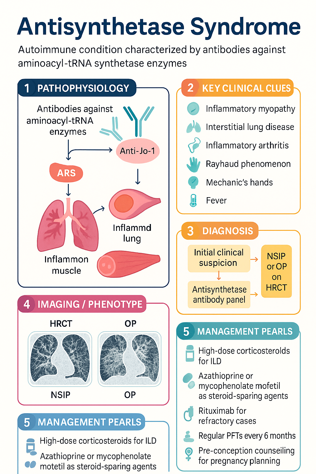
  • Antisynthetase syndrome (ASS) is an autoimmune condition defined by antibodies against aminoacyl-tRNA synthetase enzymes — most commonly anti-Jo-1 (histidyl-tRNA synthetase), followed by anti-PL-7, anti-PL-12, anti-EJ, and anti-OJ.
  • The classic six-component clinical cluster comprises inflammatory myopathy, interstitial lung disease (ILD), inflammatory arthritis, Raynaud phenomenon, mechanic's hands (hyperkeratotic fissuring), and fever.
  • ILD is often the most clinically significant and prognostically important manifestation, frequently presenting as non-specific interstitial pneumonia (NSIP) or organising pneumonia (OP) pattern on HRCT.
  • Anti-Jo-1–positive patients tend to have the most complete syndrome (myositis + ILD + arthritis); anti-PL-12– and anti-PL-7–positive patients may present with ILD-dominant disease and minimal myositis.
  • Diagnosis requires a positive antisynthetase antibody plus at least one clinical feature — confirmation relies on HRCT chest, PFTs (FVC, DLCO), CK/aldolase, muscle MRI, EMG, and/or muscle biopsy.
  • First-line ILD therapy: high-dose corticosteroids with a steroid-sparing agent — azathioprine or mycophenolate mofetil.
  • Refractory or severe ILD/myositis: rituximab (preferred biologic); cyclophosphamide for rapidly progressive ILD; IVIg for refractory myositis.
  • Pulmonary function testing (FVC, DLCO) every 6 months is mandatory to detect ILD progression; HRCT at baseline and when clinically indicated.
  • Pregnancy planning requires pre-conception counselling — mycophenolate and methotrexate are teratogenic and must be ceased ≥6 weeks and ≥3 months pre-conception respectively; azathioprine and hydroxychloroquine are preferred maintenance agents.
  • Screen for pulmonary arterial hypertension (PAH) with echocardiography; antinuclear antibody (ANA) may be negative — do not exclude ASS on ANA negativity alone.
  • Aboriginal and Torres Strait Islander peoples may have higher rates of ILD and face barriers to specialist rheumatology and respiratory access — early referral and culturally safe care pathways are essential.
  • Mortality is driven primarily by respiratory failure from progressive ILD; early aggressive treatment of lung disease improves outcomes.

Introduction & Australian Epidemiology

Antisynthetase syndrome (ASS) is a distinct autoimmune connective tissue disease characterised by the presence of autoantibodies directed against aminoacyl-tRNA synthetase enzymes, coupled with a constellation of clinical features including inflammatory myopathy, interstitial lung disease (ILD), inflammatory arthritis, Raynaud phenomenon, mechanic's hands, and fever. First described in the 1970s–1980s in patients with polymyositis and anti-Jo-1 antibodies, ASS is now recognised as a syndrome in its own right rather than a subset of dermatomyositis or polymyositis.

ASS accounts for approximately 30–40 % of all patients with idiopathic inflammatory myopathies (IIM) in Australian referral centre cohorts. The estimated prevalence in Australia is 1–2 per 100,000 population, with a female predominance (approximately 2:1 F:M). Peak incidence occurs in the 4th–6th decade of life, though ASS can present at any age including in paediatric populations.

Eight antisynthetase antibodies have been identified. Anti-Jo-1 (anti-histidyl-tRNA synthetase) is the most common, found in approximately 20–30 % of IIM patients and accounting for ~75 % of ASS cases. Other antibodies include anti-PL-7 (threonyl), anti-PL-12 (alanyl), anti-EJ (glycyl), anti-OJ (isoleucyl), anti-KS (asparaginyl), anti-Ha (tyrosyl), and anti-Zo (phenylalanyl). Each antibody defines a clinically distinct ASS phenotype — anti-PL-12 and anti-PL-7 patients more frequently present with ILD as the dominant or sole manifestation and may lack significant muscle disease.

In Australia, ILD is the leading cause of morbidity and mortality in ASS. Australian registry data indicate that approximately 70–90 % of ASS patients have evidence of ILD on HRCT at presentation, with NSIP and OP being the predominant radiological and histopathological patterns. Early recognition and treatment of ILD is therefore critical to improving long-term outcomes.

Antisynthetase Syndrome clinical infographic — pathophysiology, clinical clues, diagnosis, imaging, and management
Tap or click image to enlarge — Antisynthetase Syndrome: pathophysiology, clinical clues, diagnosis, imaging, and management.
Antisynthetase Syndrome infographic, full size

Clinical Features — The Six-Component Cluster

ASS is defined by the presence of at least one antisynthetase antibody plus at least one of the following six clinical features. Anti-Jo-1–positive patients most commonly exhibit the complete sextet, while other antibody subsets may present with fewer components.

Feature Prevalence Key Details
1. Inflammatory myopathy 60–80 % Proximal muscle weakness (shoulder/hip girdle); CK often elevated (may be normal in anti-PL-12); may be subtle or subclinical with EMG/MRI abnormalities only
2. Interstitial lung disease 70–90 % NSIP (most common), OP, or UIP pattern on HRCT; progressive dyspnoea, dry cough; may precede myositis by months–years; dominant cause of mortality
3. Inflammatory arthritis 50–70 % Symmetric, non-erosive polyarthritis; small joints of hands, wrists, knees; may mimic RA; RF and anti-CCP usually negative
4. Mechanic's hands 40–60 % Hyperkeratotic, fissuring of lateral digits; rough, cracked skin with dyschromia; pathognomonic finding in ASS
5. Raynaud phenomenon 40–60 % Episodic digital ischaemia — white → blue → red sequence; may antedate other features; less severe than in SSc
6. Fever 20–40 % Often low-grade; may be the presenting feature; exclude infection in immunosuppressed patients
⚠️
Clinical pearl: ILD may precede myositis by months to years. A patient presenting with unexplained ILD, especially NSIP pattern, should have an antisynthetase antibody panel performed — even in the absence of muscle symptoms. Anti-PL-12–positive patients frequently present with isolated ILD and clinically occult myositis.

Antibody-Specific Phenotypes

Antibody Frequency Predominant Phenotype
Anti-Jo-1 ~75 % of ASS Most complete syndrome: myositis + ILD + arthritis + mechanic's hands
Anti-PL-7 ~5–10 % ILD-dominant; lower CK; higher mortality from progressive ILD
Anti-PL-12 ~5–10 % ILD-dominant; myositis often subtle or absent; worse ILD prognosis
Anti-EJ ~3 % Myositis + ILD; overlap features
Anti-OJ ~1–2 % Rare; ILD + myositis; often incomplete syndrome
Anti-KS, anti-Ha, anti-Zo Rare Limited data; ILD reported

Pathophysiology

Aminoacyl-tRNA synthetases (ARS) are ubiquitous intracellular enzymes essential for protein synthesis, catalysing the attachment of amino acids to their cognate tRNA molecules. In ASS, autoantibodies target specific ARS enzymes. The pathogenic mechanisms linking these antibodies to tissue injury remain incompletely understood, but several mechanisms have been proposed:

  • Direct antigen-driven immune activation: ARS enzymes may be expressed on the cell surface under conditions of cellular stress (e.g., viral infection, tissue injury), becoming accessible to autoantibodies and triggering complement-mediated and antibody-dependent cellular cytotoxicity.
  • T-cell–mediated muscle injury: CD8+ cytotoxic T cells infiltrate perimysial and endomysial regions of skeletal muscle, driving perifascicular atrophy and myofibre necrosis — a pattern distinct from dermatomyositis (microangiopathy) and overlap myositis.
  • Pulmonary fibrosis driven by TGF-β and pro-fibrotic cytokines: In the lung, chronic inflammation of the alveolar epithelium and peribronchiolar interstitium leads to fibroblast activation, collagen deposition, and progressive fibrosis — particularly in NSIP and UIP patterns.
  • Molecular mimicry: Structural homology between ARS epitopes and viral proteins may initiate cross-reactive immune responses, consistent with epidemiological observations of viral triggers.
  • Genetic susceptibility: HLA-DRB1*03:01 and HLA-DQA1*05:01 are strongly associated with anti-Jo-1 ASS in Caucasian populations; HLA-B*35 and HLA-DRB1*11:01 have been linked to anti-PL-12 ILD-dominant disease.

Diagnosis

Diagnosis of ASS requires the presence of a positive antisynthetase antibody plus at least one clinical feature from the six-component cluster. No single international classification criteria has been universally adopted; however, the 2010 Connors criteria and the emerging EULAR/ACR classification for IIM (2017) are commonly applied in Australian practice.

Diagnostic Approach

1
Clinical suspicion
Unexplained ILD (especially NSIP/OP pattern), inflammatory myopathy, polyarthritis with negative RF/anti-CCP, mechanic's hands, or Raynaud phenomenon in combination.
2
Serological testing
Antisynthetase antibody panel (line immunoassay or immunoprecipitation): anti-Jo-1, anti-PL-7, anti-PL-12, anti-EJ, anti-OJ, anti-KS, anti-Ha, anti-Zo. ANA may be negative.
3
Pulmonary evaluation
HRCT chest (NSIP: bilateral ground-glass opacities, subpleural sparing; OP: migratory consolidations; UIP: honeycombing). PFTs: FVC, DLCO, TLC. 6-minute walk test.
4
Muscle assessment
CK, LDH, aldolase. Muscle MRI (STIR sequences showing oedema). EMG (irritative myopathy pattern). Muscle biopsy if diagnosis uncertain — perimysial inflammation, perifascicular atrophy.
5
Cardiac screening
Transthoracic echocardiography to screen for pulmonary arterial hypertension (PAH) and myocardial involvement.
🚨
Do not exclude ASS based on a negative ANA. Antisynthetase antibodies are anti-cytoplasmic antibodies and may not be detected on standard HEp-2 immunofluorescence. Request the specific antisynthetase antibody panel independently.

Investigations

Laboratory Investigations

Essential
Antisynthetase antibody panel (line immunoassay)
Anti-Jo-1, PL-7, PL-12, EJ, OJ, KS, Ha, Zo. Available at major reference laboratories (Royal Prince Alfred, Sullivan Nicolaides, Douglass Hanly Moir). MBS rebate available under autoimmune serology item numbers. Turnaround: 7–14 days.
Essential
Creatine kinase (CK)
Elevated in 60–80 % (typically 5–50× ULN). May be normal in anti-PL-12–positive disease. MBS Item 66500.
Available
Aldolase
More sensitive than CK for subtle myositis; elevated in ASS even when CK is normal. MBS Item 66536.
Available
LDH
Non-specific marker of muscle damage; correlates with disease activity. MBS Item 66546.
Available
ESR, CRP, FBC, LFTs, renal function
Baseline organ function and inflammatory markers. Standard MBS items.

Imaging

Essential
HRCT chest (high-resolution)
Gold standard for ILD characterisation. Patterns: NSIP (bilateral ground-glass, subpleural, basal-predominant), OP (migratory consolidation, perilobular), UIP (honeycombing, traction bronchiectasis). MBS Item 56300 (CT chest).
Essential
Pulmonary function tests (spirometry + DLCO)
FVC and DLCO for ILD detection and monitoring. Reduced DLCO may precede FVC decline. MBS Items 11503, 11505 (spirometry); 11512 (DLCO).
Available
Muscle MRI (thighs/shoulders)
STIR sequences demonstrate muscle oedema (active inflammation). T1 sequences show fatty replacement (chronic damage). Superior to EMG for defining extent of myositis. MBS Item 63000 series.
Available
Transthoracic echocardiography
Screen for PAH (estimated RVSP >36 mmHg), myocardial dysfunction. MBS Item 55124.

Electrodiagnostic & Histopathological

Available
Electromyography (EMG)
Irritative myopathy pattern: fibrillation potentials, positive sharp waves, short-duration polyphasic motor unit potentials. Useful when CK is normal. MBS Item 11600 series.
Referral
Muscle biopsy
Reserved for diagnostic uncertainty. Characteristic findings: perimysial and perivascular inflammatory infiltrates (CD8+ T cells, macrophages), perifascicular atrophy, perimysial connective tissue fragmentation. Coordinated via rheumatology/neurology referral.
Referral
Bronchoalveolar lavage (BAL) ± surgical lung biopsy
BAL lymphocytosis supports ILD diagnosis. Surgical lung biopsy (VATS) for histopathological pattern confirmation when diagnosis of ILD pattern impacts management — coordinated via respiratory medicine.

Risk Stratification & Prognosis

Prognosis in ASS is determined primarily by the severity and progression of ILD. Myositis-related mortality is low with modern immunosuppressive therapy; however, progressive pulmonary fibrosis accounts for the majority of disease-related deaths.

Favourable
Limited disease
Mild myositis (CK <5× ULN, minimal weakness), NSIP pattern ILD with stable FVC >80 % predicted, no honeycombing on HRCT, no PAH. Anti-Jo-1–positive with complete syndrome often responds well to initial therapy.
5-year survival >90 %. Monitor 6-monthly PFTs.
Intermediate
Progressive or multi-organ
Moderate myositis (CK 5–20× ULN), ILD with declining FVC (≥10 % relative decline over 12 months), OP pattern requiring treatment escalation, inflammatory arthritis requiring DMARDs, or PAH (estimated RVSP 36–50 mmHg).
Requires steroid-sparing agent ± biologic. 3-monthly PFTs during escalation.
Severe / Refractory
Rapidly progressive ILD / severe myositis
Rapidly progressive ILD (FVC decline >10 % in 3 months or >20 % in 6 months), UIP pattern with honeycombing, severe dysphagia, respiratory failure (SpO₂ <90 % on air), severe PAH (RVSP >50 mmHg), or treatment-refractory disease failing ≥2 lines of therapy.
Consider cyclophosphamide, rituximab, IVIg, or lung transplant referral. Multidisciplinary ILD MDT.
⚠️
Predictors of poor ILD outcome: UIP pattern on HRCT, anti-PL-7 or anti-PL-12 antibodies, male sex, age >60 at presentation, baseline FVC <70 % predicted, rapidly progressive decline in FVC or DLCO, and presence of PAH. These patients warrant early aggressive therapy and lung transplant assessment referral.

Management — Immunosuppressive Therapy

Treatment of ASS requires a dual approach targeting both the myositis and ILD components. ILD-directed therapy is the priority in most patients, as progressive pulmonary fibrosis drives mortality. All treatment decisions should be made in conjunction with rheumatology and respiratory medicine.

First-Line Induction Therapy

💊
Prednisolone
Solone® · Panafcortelone® · Corticosteroid
Adult dose 0.75–1 mg/kg/day (max 60 mg) PO for 4–6 weeks, then taper by 5 mg every 2–4 weeks to 10–15 mg/day maintenance
Severe myositis/ILD IV methylprednisolone 500–1000 mg/day × 3 days, then oral prednisolone 1 mg/kg/day
Paediatric dose 1–2 mg/kg/day (max 60 mg) PO; IV pulse 30 mg/kg/day (max 1 g) × 3 days
Key side effects Osteoporosis (start calcium/vitamin D + bisphosphonate if ≥3 months), diabetes, hypertension, adrenal suppression, cataracts
PBS status ✔ PBS General Benefit
💊
Mycophenolate mofetil
CellCept® · Myfortic® · Antiproliferative
Adult dose 500 mg PO BD Week 1, then 1 g PO BD (target 2–3 g/day); increase to 1.5 g BD if tolerated and required
Paediatric dose 600 mg/m²/day PO divided BD (max 2 g/day)
Renal adjustment Avoid if eGFR <25 mL/min; no dose adjustment for eGFR 25–50
Key monitoring FBC fortnightly × 3 months then monthly; LFTs; pregnancy test (teratogenic — Category D)
PBS status ⚠ PBS Authority Required
💊
Azathioprine
Imuran® · Thiopurine
Adult dose 50 mg/day PO Week 1, then 100 mg/day, target 2–2.5 mg/kg/day
Pre-treatment TPMT/NUDT15 genotype testing mandatory — reduce dose or avoid in intermediate/poor metabolisers
Paediatric dose 1–2 mg/kg/day PO (max 2.5 mg/kg/day); TPMT testing essential
Renal adjustment Reduce dose by 50 % if eGFR <25 mL/min
PBS status ✔ PBS General Benefit
ℹ️
First-line steroid-sparing agent for ASS-associated ILD: Both mycophenolate mofetil and azathioprine are accepted first-line options. Mycophenolate may have superior efficacy for ILD based on observational data and the Scleroderma Lung Study II extrapolation. Azathioprine is preferred in pregnancy planning. The choice is guided by patient factors, tolerability, and MBS/PBS considerations.

Second-Line / Refractory Disease

💊
Rituximab
MabThera® · Riximyo® · Anti-CD20 monoclonal antibody
Adult dose 1000 mg IV on Day 1 and Day 15; repeat cycle every 6 months based on clinical response and B-cell repopulation
Indication Refractory ILD or myositis failing first-line therapy (mycophenolate or azathioprine + steroids)
Pre-treatment Screen for hepatitis B (HBsAg, anti-HBc), varicella immunity, immunoglobulin levels; vaccinate ≥4 weeks prior if non-immune
PBS status ⚠ PBS Authority Required — restricted to refractory autoimmune conditions
💊
Cyclophosphamide
Endoxan® · Alkylating agent
Adult dose IV pulse: 500–750 mg/m² every 2–4 weeks × 6 pulses (Euro-Lupus or modified SLS II protocol); or oral 1–2 mg/kg/day for 3–6 months
Indication Rapidly progressive ILD; severe refractory myositis
Renal adjustment Reduce dose by 25 % if eGFR 10–25; by 50 % if eGFR <10
Key monitoring FBC (nadir Day 10–14); urinalysis for haemorrhagic cystitis; MESNA co-administration with IV dosing; cumulative lifetime dose <50 g to minimise bladder cancer risk
PBS status ✔ PBS General Benefit
💊
Intravenous immunoglobulin (IVIg)
Intragam P® · Privigen® · Immunomodulator
Adult dose 2 g/kg divided over 2–5 days every 4 weeks; maintenance 1 g/kg every 4 weeks
Indication Refractory myositis, especially dysphagia; rapidly progressive disease; adjunct to other immunosuppressants
Key side effects Headache, aseptic meningitis, thromboembolic events, haemolysis (IgA-deficient patients), renal impairment (sucrose-containing preparations)
PBS status ⚠ PBS Authority Required — restricted to specific indications

Other Agents — Limited Evidence

💊
Tacrolimus
Prograf® Calcineurin inhibitor
Adult dose 0.5–3 mg/day PO in divided doses; target trough 5–10 ng/mL for refractory ILD
Role Steroid-sparing; used in combination with low-dose prednisolone for refractory ASS-ILD; emerging evidence from Japanese studies
PBS status ⚠ PBS Authority Required (organ transplant indication; off-label for ASS may require private script)
💊
Methotrexate
Methoblastin® · Antimetabolite
Adult dose 7.5–25 mg PO/SC weekly with folic acid 5 mg weekly (not on MTX day)
Role May help myositis and arthritis; caution with ILD — avoid or use carefully in pre-existing pulmonary fibrosis; monitor PFTs closely
PBS status ✔ PBS General Benefit
🚨
Methotrexate caution: Methotrexate-induced pneumonitis may exacerbate pre-existing ILD in ASS. If methotrexate is used for the arthritis/myositis component, baseline and serial PFTs are essential. Any unexplained respiratory deterioration should prompt immediate HRCT and methotrexate cessation.

Monitoring

Structured monitoring is essential to detect ILD progression, myositis relapse, and medication toxicity. All ASS patients require long-term follow-up with rheumatology and respiratory medicine (pulmonology).

Monitoring Schedule

Baseline
HRCT chest, PFTs (FVC, DLCO, TLC), 6MWT, CK, aldolase, LFTs, FBC, renal function, ECG, echocardiography, antisynthetase antibody panel, TPMT/NUDT15 (if azathioprine planned), hepatitis B/C serology, bone density (DEXA) if starting corticosteroids.
Months 1–3
FBC, CK, LFTs every 2–4 weeks (immunosuppressant toxicity monitoring). Clinical review every 4–6 weeks. Symptom assessment: dyspnoea (MRC scale), muscle weakness, joint symptoms.
Month 6
PFTs (FVC, DLCO), CK, aldolase. Repeat HRCT if symptoms worsen or PFTs decline ≥10 %. Echocardiography if PAH suspected.
Every 6 months (ongoing)
PFTs (FVC, DLCO), CK, aldolase, FBC, LFTs. Clinical review. DEXA scan annually if on corticosteroids. Ophthalmology review annually for cataracts/glaucoma.
As clinically indicated
HRCT if ≥10 % relative decline in FVC or DLCO, or new respiratory symptoms. Repeat echocardiography for PAH screening. Muscle MRI if new weakness or rising CK.

PFT Decline Thresholds — Action Points

Change Action
FVC decline ≥10 % relative (or ≥5 % absolute) Repeat HRCT; consider treatment escalation (add/change steroid-sparing agent); multidisciplinary ILD team review
FVC decline ≥10 % in 3 months or ≥20 % in 6 months Rapidly progressive ILD — consider cyclophosphamide or rituximab; urgent respiratory/rheumatology review; lung transplant referral
DLCO <40 % predicted High mortality risk; evaluate for PAH (right heart catheterisation); lung transplant assessment

Special Populations

🤰 Pregnancy
Safe agents
Azathioprine (cease mycophenolate ≥6 weeks pre-conception), hydroxychloroquine, low-dose prednisolone (<20 mg/day), tacrolimus (with caution)
Contraindicated
Mycophenolate mofetil (Category D — teratogenic: microtia, cleft palate), methotrexate (Category X — abortifacient, neural tube defects — cease ≥3 months pre-conception), cyclophosphamide (Category D — gonadotoxic), rituximab (B-cell depleting — cease ≥6 months pre-conception ideally)
Monitoring
Monthly CK and PFTs during pregnancy; ILD may flare peripartum. Coordinate obstetric, rheumatology, and respiratory teams. Neonatal monitoring for transient neonatal myasthenia (rare).
👶 Paediatrics
Presentation
Paediatric ASS overlaps with juvenile dermatomyositis (JDM). ILD may be less common but is reported, particularly in anti-PL-12–positive adolescents. Mechanic's hands and Raynaud may be initial features.
Treatment
Prednisolone 1–2 mg/kg/day; methotrexate (first-line steroid-sparing in JDM); mycophenolate or azathioprine for ILD component; IVIg for refractory disease. Weight-based dosing required.
👴 Elderly (≥65 years)
Considerations
Higher rates of UIP pattern and progressive fibrosis. Increased corticosteroid toxicity (osteoporosis, diabetes, falls). Prefer early steroid-sparing agent introduction to enable rapid steroid taper. Monitor bone health with DEXA and initiate bisphosphonate prophylaxis early.
Polypharmacy
Review for drug interactions (azathioprine + allopurinol — TPMT shunting → severe myelosuppression). Renal dose adjustments as eGFR declines with age.
🩺 Renal Impairment
Dose adjustments
Mycophenolate: avoid if eGFR <25. Azathioprine: reduce 50 % if eGFR <25. Cyclophosphamide: reduce 25–50 % if eGFR <25. Prednisolone: no adjustment but monitor glucose closely. NSAIDs (for arthritis): avoid in CKD.
🫁 Hepatic Impairment
Dose adjustments
Azathioprine: use with caution, monitor LFTs closely. Methotrexate: avoid in significant hepatic fibrosis or active liver disease. Mycophenolate: dose adjustment not well defined — monitor LFTs. Prednisolone: no adjustment but monitor for hepatotoxicity at high doses.
🦠 Immunocompromised
Infection risk
Screen for latent TB (QuantiFERON-TB Gold), hepatitis B/C, HIV prior to immunosuppression. Varicella non-immune: vaccinate ≥4 weeks before rituximab. Pneumocystis jirovecii prophylaxis with TMP-SMX if on ≥20 mg prednisolone for ≥1 month + another immunosuppressant.
Vaccination
Annual influenza, pneumococcal (Prevenar 13 + Pneumovax 23), COVID-19 vaccination. Live vaccines contraindicated on immunosuppression. Vaccinate ≥4 weeks before rituximab or within 5 months of last dose.

ATSI Health Considerations

Aboriginal and Torres Strait Islander Health
Epidemiology
Limited population-level data on ASS prevalence in Aboriginal and Torres Strait Islander peoples. However, AIHW data demonstrate higher rates of autoimmune and connective tissue diseases, rheumatic heart disease, and chronic respiratory disease in First Nations Australians. ILD, including autoimmune-associated ILD, may be underdiagnosed due to limited specialist access.
Geographic access
Many Aboriginal and Torres Strait Islander peoples reside in regional and remote areas with limited access to rheumatology, respiratory medicine, and specialist diagnostic services (HRCT, EMG, muscle biopsy). Antisynthetase antibody testing may not be locally available. Telehealth has expanded access but may not replace the need for HRCT and PFTs.
Cultural safety
Engage Aboriginal Health Workers (AHWs) and Aboriginal Liaison Officers in care planning. Use culturally appropriate communication. Acknowledge the impact of intergenerational trauma, systemic racism, and distrust of health services. Support patient preferences for family-centred care and connection to community.
Immunosuppression risks
Higher background rates of infections (including latent TB, hepatitis B, rheumatic fever sequelae) must be screened for prior to commencing immunosuppressive therapy. Ensure pneumococcal and influenza vaccination is up to date. Consider PJP prophylaxis more readily given co-morbidity burden.
Closing the Gap
Ensure equitable access to PBS-subsidised medications (mycophenolate, azathioprine, rituximab). Engage with Closing the Gap initiatives for chronic disease management. Support access to patient-assisted travel schemes (PATS) for specialist review and HRCT/PFT services.
Referral pathways
Early referral to rheumatology and respiratory medicine via Aboriginal Community Controlled Health Organisations (ACCHOs) and regional hospital networks. Utilise RFDS and state-funded PATS for remote patients requiring HRCT, PFTs, and specialist consultation.

📚 References

  1. 1. Connors GR, Christopher-Stine L, Cosgrove O, Danoff SK. Interstitial lung disease associated with the idiopathic inflammatory myopathies: what progress has been made in the past 35 years? Chest. 2010;138(6):1464–1474.
  2. 2. Lega JC, Fabien N, Reynaud Q, et al. The clinical phenotype associated with myositis-specific and associated antibodies: a meta-analysis revisiting the so-called antisynthetase syndrome. Autoimmun Rev. 2014;13(9):883–891.
  3. 3. Lundberg IE, Tjärnlund A, Bottai M, et al. 2017 European League Against Rheumatism/American College of Rheumatology classification criteria for adult and juvenile idiopathic inflammatory myopathies and their major subgroups. Arthritis Rheumatol. 2017;69(12):2271–2282.
  4. 4. Hervier B, Devilliers H, Stanciu R, et al. Hierarchical clinical and serological classification of patients with anti-synthetase syndrome. Medicine (Baltimore). 2012;91(5):236–244.
  5. 5. Tieu J, Lundberg IE, Limaye V. Idiopathic inflammatory myositis. Best Pract Res Clin Rheumatol. 2022;36(1):101746.
  6. 6. Baccaro AC, Ceribelli A, Aoki MN, et al. Antisynthetase syndrome: clinical features and outcomes of a Brazilian cohort. Adv Rheumatol. 2020;60(1):53.
  7. 7. Tashkin DP, Elashoff R, Clements PJ, et al. Cyclophosphamide versus placebo in scleroderma lung disease. N Engl J Med. 2006;354(25):2655–2666. [Scleroderma Lung Study — extrapolated to ASS-ILD]
  8. 8. Tieu J, Giri N, Limaye V. Management of interstitial lung disease in antisynthetase syndrome: an update. Curr Treat Options Rheumatol. 2021;7:244–259.
  9. 9. Australian Institute of Health and Welfare (AIHW). Aboriginal and Torres Strait Islander health performance framework 2023 summary report. Canberra: AIHW; 2023.
  10. 10. Hallowell R, Danoff SK. Interstitial lung disease associated with the antisynthetase syndrome. J Clin Med. 2023;12(4):1391.
  11. 11. Allenbach Y, Uzunhan Y, Guenou H, et al. Different phenotypes in dermatomyositis associated with anti-Mi2 antibody. Neurology. 2020;94(1):e71–e80.
  12. 12. Royal Australian College of General Practitioners (RACGP). Guidelines for preventive activities in general practice (Red Book). 10th ed. East Melbourne: RACGP; 2023.
  13. 13. Raghu G, Remy-Jardin M, Richeldi L, et al. Idiopathic pulmonary fibrosis (an update) and progressive pulmonary fibrosis in adults: an official ATS/ERS/JRS/ALAT clinical practice guideline. Am J Respir Crit Care Med. 2022;205(9):e18–e47.
  14. 14. Australian Technical Advisory Group on Immunisation (ATAGI). Australian immunisation handbook. Canberra: Australian Government Department of Health; 2023. Available from: immunisationhandbook.health.gov.au.