📋 Key Information Summary
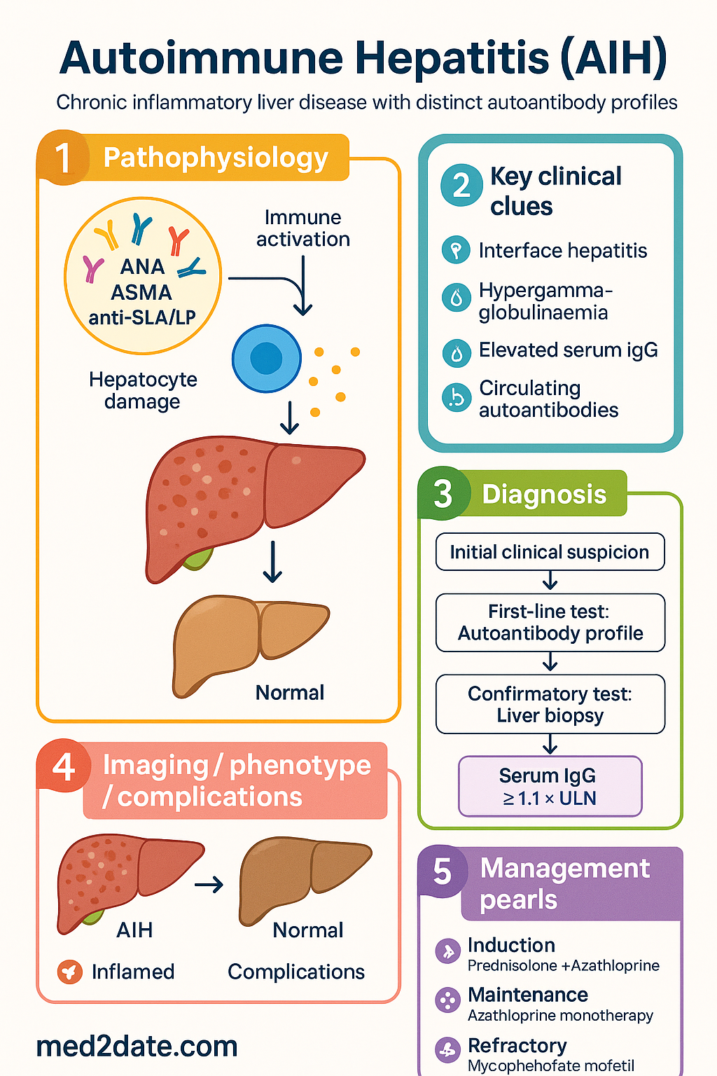
- Autoimmune hepatitis (AIH) is a chronic inflammatory liver disease characterised by interface hepatitis, hypergammaglobulinaemia, and circulating autoantibodies; without treatment it progresses to cirrhosis and liver failure.
- Two major types: Type 1 (ANA and/or ASMA positive, anti-SLA/LP — ~85% of cases) and Type 2 (anti-LKM-1 positive — rarer, typically paediatric-onset).
- Diagnosis relies on the simplified IAIHG scoring criteria incorporating autoantibodies, IgG level, histology (interface hepatitis, plasma cell infiltrate, hepatocyte rosetting), and exclusion of viral hepatitis, drug-induced liver injury, Wilson disease, and NASH.
- Elevated serum IgG (≥1.1× ULN) is present in >80% of patients and is a hallmark biochemical feature.
- Liver biopsy remains essential for diagnosis and staging; interface hepatitis with lymphoplasmacytic infiltrate is the histological hallmark.
- Induction therapy: prednisolone 1 mg/kg/day (max 60 mg) ± azathioprine 1–2 mg/kg/day; azathioprine should be started at diagnosis unless contraindicated.
- Budesonide 3 mg TDS is an alternative corticosteroid for non-cirrhotic AIH, offering reduced systemic side-effects.
- Treatment targets: biochemical remission (normal ALT and IgG) within 6–24 months; histological remission on follow-up biopsy is the gold standard.
- Long-term maintenance: azathioprine monotherapy (1–2 mg/kg/day) after steroid taper; continue for ≥3 years minimum before considering withdrawal.
- Relapse rate on treatment withdrawal is 50–87%; indefinite maintenance therapy is recommended for most patients, especially those with cirrhosis.
- Refractory disease: mycophenolate mofetil (1–2 g/day) is second-line; tacrolimus or cyclosporin reserved for third-line or refractory cases.
- AIH-PBC overlap (Paris criteria) and AIH-PSC overlap require combination therapy with immunosuppression plus ursodeoxycholic acid.
- Pregnancy: azathioprine is generally continued; mycophenolate is teratogenic and must be stopped ≥6 weeks pre-conception; postpartum flare risk is significant.
- Aboriginal and Torres Strait Islander Australians may present later with more advanced disease; access to hepatology services in remote areas requires Telehealth coordination.
Diagnosis
Antibody Profile
Autoantibody testing is central to AIH classification. Serological markers should be interpreted in conjunction with serum IgG levels and histology.
| Antibody | AIH Type | Prevalence | Notes |
|---|---|---|---|
| ANA (antinuclear antibody) | Type 1 | ~70–80% | Titre ≥1:40 (adults) or ≥1:20 (paediatric) on IIF; also seen in other autoimmune conditions |
| ASMA (anti-smooth muscle antibody) | Type 1 | ~50–70% | Anti-actin specificity increases diagnostic confidence; may occur with or without ANA |
| Anti-SLA/LP (anti-soluble liver antigen/liver-pancreas) | Type 1 | ~10–30% | Most specific antibody for AIH; associated with more severe disease and higher relapse rate |
| Anti-LKM-1 (liver-kidney microsomal type 1) | Type 2 | ~5–10% | Targets CYP2D6; typically seen in paediatric/young female patients; may be confused with anti-LKM seen in HCV |
| Anti-LC1 (liver cytosol type 1) | Type 2 | ~30% of Type 2 | May be the sole marker; useful when LKM is weakly positive |
| pANCA (perinuclear anti-neutrophil cytoplasmic) | Type 1 | ~40–50% | Non-specific but supportive when present |
Simplified IAIHG Diagnostic Criteria (2008)
The simplified IAIHG scoring system is preferred for clinical use (score ≥6 = probable AIH; ≥7 = definite AIH).
| Variable | Score 0 | Score 1 | Score 2 |
|---|---|---|---|
| ANA or ASMA | <1:40 | ≥1:40 | — |
| ANA or ASMA, or anti-LKM-1 | — | — | ≥1:80 |
| Anti-SLA/LP | — | — | Positive |
| IgG | Normal | >1.1× ULN | >1.46× ULN |
| Liver histology | Incompatible | Compatible | Typical |
| Absence of viral hepatitis | No | Yes | — |
Liver Biopsy — Histological Features
Liver biopsy is recommended in all patients with suspected AIH to confirm the diagnosis, assess severity, and establish baseline fibrosis staging. Key histological features include:
- Interface hepatitis (piecemeal necrosis): The histological hallmark — lymphoplasmacytic infiltration across the limiting plate into the hepatic lobule.
- Lobular hepatitis: Hepatocyte apoptosis, confluent necrosis, and dense portal/periportal mononuclear cell infiltrate.
- Plasma cell infiltrate: Prominent plasma cells within portal tracts — although plasma cells are not always present, their abundance increases diagnostic confidence.
- Hepatocyte rosetting: Arrangement of hepatocytes in a rosette pattern around a central bile canaliculus — a supportive feature of AIH.
- Emperipolesis: Active penetration of one cell by another (lymphocyte within hepatocyte) — recently described as relatively specific for AIH.
- Fibrosis/cirrhosis: Present in 25–50% at initial diagnosis; staged using METAVIR or Ishak scoring.
Exclusion of Other Aetiologies
AIH is a diagnosis of exclusion. The following must be actively excluded before confirming AIH:
- Viral hepatitis: Hepatitis A IgM, HBsAg / anti-HBc IgM, hepatitis C antibody with reflex PCR, hepatitis E IgM (particularly if travel history or animal exposure); CMV and EBV serologies if acute presentation.
- Drug-induced liver injury (DILI): Detailed medication history including over-the-counter, herbal (e.g., black cohosh, kava), and recreational drugs. Drug-induced autoimmune-like hepatitis (e.g., nitrofurantoin, minocycline, statins, infliximab) can mimic AIH and typically resolves on drug withdrawal.
- Wilson disease: Especially in patients <40 years; check ceruloplasmin, 24-hour urine copper, and hepatic copper on biopsy if low ceruloplasmin. Kayser-Fleischer rings on slit-lamp examination.
- Non-alcoholic steatohepatitis (NASH): Check metabolic risk factors (obesity, type 2 diabetes, dyslipidaemia), imaging for hepatic steatosis, and the NAS score on biopsy. Some patients have concurrent AIH and NASH.
- Alcoholic liver disease: Alcohol consumption history (>40 g/day in men, >20 g/day in women).
- Alpha-1 antitrypsin deficiency: Alpha-1 antitrypsin level and phenotype if suspected.
- Haemochromatosis: Transferrin saturation, ferritin, and HFE genotyping.
Investigations
Treatment
Induction Therapy
Treatment should be initiated promptly in all patients with active AIH (elevated transaminases, elevated IgG, histological activity). The goal of induction is biochemical remission.
Biochemical and Histological Remission Targets
Treatment response should be assessed at regular intervals against defined targets:
- Biochemical remission: Normalisation of serum AST/ALT (to ≤ ULN) and serum IgG (to ≤ ULN) — ideally achieved within 6–24 months of starting therapy.
- Complete biochemical response: Normal AST/ALT and IgG maintained for ≥6 months while on a stable maintenance regimen.
- Histological remission: Resolution of interface hepatitis on follow-up liver biopsy — the gold standard endpoint. Histological improvement lags behind biochemical improvement by 3–6 months.
- Relapse: Elevation of AST/ALT to >3× ULN after achieving remission; occurs in 50–87% after drug withdrawal.
A follow-up liver biopsy is recommended after 2–3 years of biochemical remission to confirm histological remission before considering any treatment withdrawal.
Long-Term Maintenance Therapy
After achieving biochemical and histological remission with induction therapy and steroid taper, the majority of patients require indefinite maintenance immunosuppression:
- Standard maintenance: Azathioprine monotherapy at 1–2 mg/kg/day after prednisolone has been weaned to 5 mg/day and then stopped.
- Duration: Minimum 3 years of remission before any consideration of withdrawal; many experts recommend lifelong therapy, especially in patients with cirrhosis, relapsing disease, or anti-SLA/LP positivity.
- Azathioprine intolerance: If azathioprine is not tolerated (myelosuppression, pancreatitis, hepatotoxicity, nausea), switch to mycophenolate mofetil.
Refractory or Relapsing Disease
Approximately 10–20% of patients do not achieve or maintain remission with standard prednisolone + azathioprine therapy.
Relapse Risk on Treatment Withdrawal
Risk factors for relapse after withdrawal include:
- Anti-SLA/LP antibody positivity
- Cirrhosis at diagnosis or during follow-up
- Persistent histological activity despite biochemical remission
- High IgG at time of withdrawal attempt
- Young age at diagnosis
- Type 2 AIH (anti-LKM-1 positive)
Monitoring Schedule
Overlap Syndromes & Pregnancy
AIH-PBC Overlap Syndrome
AIH-PBC overlap occurs in approximately 8–10% of patients with AIH and is defined by the Paris criteria (2011), which require both sets of criteria to be met simultaneously:
- ALT ≥5× ULN
- IgG ≥2× ULN or ASMA positive
- Interface hepatitis on biopsy (moderate to severe)
- ALP ≥2× ULN
- AMA positive (titre ≥1:40)
- Florid duct lesion or ductopaenia on biopsy
Management of AIH-PBC overlap: Combination therapy with ursodeoxycholic acid (UDCA) 13–15 mg/kg/day plus standard AIH immunosuppression (prednisolone ± azathioprine). UDCA alone is insufficient as it does not control the AIH component. Monitor both cholestatic and hepatitic parameters.
AIH-PSC Overlap Syndrome
AIH-PSC overlap is less common but clinically important, particularly in younger patients (paediatric and young adult). Patients present with AIH features (autoantibodies, elevated IgG, interface hepatitis) but also have cholestatic biochemistry and characteristic bile duct changes on MRCP or ERCP (beading, strictures, pruning).
- Diagnosis: MRCP is the first-line imaging investigation to evaluate for PSC-type bile duct changes. ERCP with brushings may be needed to confirm and exclude cholangiocarcinoma.
- Management: Combination of AIH immunosuppression plus UDCA 13–15 mg/kg/day. The role of UDCA in PSC remains debated, but it is commonly used in overlap syndrome to manage the cholestatic component. Immunosuppression targets the AIH component; PSC itself may not respond fully to immunosuppression.
- Surveillance: Cholangiocarcinoma risk is increased — annual MRCP or surveillance imaging; colonoscopy with biopsies every 1–3 years if concurrent IBD (which is present in ~70% of PSC patients).
Pregnancy
Pregnancy management in women with AIH requires careful pre-conception planning, multidisciplinary care (hepatologist, obstetrician, maternal-fetal medicine), and awareness of flare risk and medication teratogenicity.
- Achieve stable biochemical remission for ≥6 months before conception.
- Mycophenolate mofetil is teratogenic (Category D) — must be stopped ≥6 weeks before conception; switch to azathioprine.
- Methotrexate is contraindicated — must stop 3–6 months pre-conception.
- Tacrolimus can be continued (Category C) — benefits often outweigh risks in refractory disease.
- Optimise folic acid supplementation (5 mg/day).
- Azathioprine is generally continued throughout pregnancy at current dose — cessation risk of flare outweighs theoretical teratogenic risk.
- Prednisolone may be needed to manage flares — use lowest effective dose. Prednisolone is largely inactivated by placental 11β-HSD2 (except at high doses).
- Monitor LFTs and IgG monthly; more frequent monitoring if flaring.
- UDCA can be continued safely in pregnancy (for overlap syndromes).
- Budesonide should be avoided — limited safety data in pregnancy.
- Postpartum flare occurs in 20–40% of patients — often within the first 3 months postpartum due to immune reconstitution.
- Monitor LFTs and IgG at 2, 4, 8, and 12 weeks postpartum, then monthly for 6 months.
- Restart or escalate immunosuppression promptly if biochemical flare detected.
- Breastfeeding: azathioprine and prednisolone are generally considered compatible with breastfeeding. Low levels of 6-MMP are detected in breast milk but considered clinically insignificant.
Paediatric AIH
Paediatric AIH has distinct features: higher prevalence of Type 2 (anti-LKM-1 positive), more acute/severe presentation, higher rate of cirrhosis at diagnosis, and higher relapse rates. Treatment follows the same principles but dosing is weight-based. The Bristol criteria for evaluating treatment response in paediatric AIH may be used. Long-term follow-up with transition to adult hepatology services is essential.
Autoimmune hepatitis affects Aboriginal and Torres Strait Islander Australians, with evidence suggesting later presentation, higher rates of cirrhosis at diagnosis, and poorer outcomes related to healthcare access barriers. The following considerations apply:
📚 References
- 1. Mack CL, Adams D, Assis DN, et al. Diagnosis and Management of Autoimmune Hepatitis in Adults and Children: 2019 Practice Guidance and Guidelines From the American Association for the Study of Liver Diseases. Hepatology. 2020;72(2):671-722.
- 2. European Association for the Study of the Liver. EASL Clinical Practice Guidelines: Autoimmune hepatitis. J Hepatol. 2015;63(4):971-1004.
- 3. Hennes EM, Zeniya M, Czaja AJ, et al. Simplified criteria for the diagnosis of autoimmune hepatitis. Hepatology. 2008;48(1):169-176.
- 4. Czaja AJ. Diagnosis and Management of Autoimmune Hepatitis: Current Status and Future Directions. Gut Liver. 2016;10(2):177-203.
- 5. Kuiper EMM, Hansen BE, de Vries RA, et al. Improved prognosis of patients with primary biliary cirrhosis that overlap with autoimmune hepatitis. Hepatology. 2009;50(3):858-865. (Paris criteria for AIH-PBC overlap)
- 6. Trivedi PJ, Hirschfield GM. Review article: overlap syndromes and autoimmune liver disease. Aliment Pharmacol Ther. 2012;36(4):317-331.
- 7. Westbrook RH, Yeoman AD, Kriese S, Heneghan MA. Outcomes of pregnancy in women with autoimmune hepatitis. J Autoimmun. 2012;38(2-3):J239-J244.
- 8. Selvarajah V, Montano-Loza AJ, Czaja AJ. Systematic review: managing suboptimal treatment responses in autoimmune hepatitis with conventional and nonstandard drugs. Aliment Pharmacol Ther. 2012;36(8):691-707.
- 9. Australian Institute of Health and Welfare. Aboriginal and Torres Strait Islander Health Performance Framework 2020 summary report. Canberra: AIHW; 2020.
- 10. Yeoman AD, Westbrook RH, Al-Chalabi T, et al. Diagnostic value and utility of the simplified International Autoimmune Hepatitis Group (IAIHG) criteria in acute and chronic liver disease. Hepatology. 2009;50(2):538-545.
- 11. Libbrecht L, Van den Heuvel MC, Jansen J, Roskams T. Emperipolesis: a peculiar feature of autoimmune hepatitis. Histopathology. 2006;48(7):873-875.
- 12. Miyake Y, Iwasaki Y, Kobashi H, et al. Efficacy of azathioprine for maintenance of remission in patients with autoimmune hepatitis: a Japanese nationwide cohort study. Hepatol Res. 2016;46(8):761-768.
- 13. Than NN, Hodson J, Schmidt-Martin D, et al. Efficacy of rituximab in difficult-to-manage autoimmune hepatitis: Results from the International Autoimmune Hepatitis Group. JHEP Rep. 2019;1(5):437-445.
- 14. Australian Commission on Safety and Quality in Health Care. National Safety and Quality Health Service Standards. 2nd ed. Sydney: ACSQHC; 2017.