📋 Key Information Summary
- Abnormal LFTs are detected in up to 10% of the Australian population on routine blood work; the majority are incidental and require structured evaluation rather than reflex investigation.
- Pattern recognition is the critical first step: hepatocellular (elevated ALT/AST), cholestatic (elevated ALP/GGT ± bilirubin), or isolated hyperbilirubinaemia.
- Always exclude acetaminophen (paracetamol) toxicity and alcohol-related liver injury early — these are the two most common identifiable causes in Australian practice.
- Repeat abnormal LFTs within 2–4 weeks before initiating an extensive workup; transient elevations (exercise, medications, viral illness) are common.
- Initial serological workup should include hepatitis B surface antigen (HBsAg), hepatitis C antibody (anti-HCV), and hepatitis A IgM — all Medicare-rebatable (MBS items 69313, 69318).
- Autoimmune hepatitis screening (ANA, ASMA, anti-LKM) and primary biliary cholangitis screening (anti-mitochondrial antibody, AMA) are essential for unexplained cholestatic or mixed patterns.
- Iron studies (ferritin, transferrin saturation) and ceruloplasmin should be requested when hepatocellular pattern persists unexplained, particularly in patients <40 years.
- Right upper quadrant ultrasound is the first-line imaging modality; transient elastography (FibroScan®) is increasingly available and can non-invasively stage fibrosis.
- Refer to gastroenterology/hepatology when LFTs remain elevated >6 months without explanation, or when there is suspected advanced fibrosis, cirrhosis, or positive viral/autoimmune markers requiring treatment.
- Aboriginal and Torres Strait Islander Australians have disproportionately high rates of hepatitis B, hepatocellular carcinoma, and alcohol-related liver disease — targeted screening and culturally safe follow-up are essential.
- Pregnancy-related causes (HELLP syndrome, intrahepatic cholestasis of pregnancy) must be considered urgently in any pregnant woman with new LFT derangement.
- Medication review is mandatory — statins, antibiotics, antiepileptics, complementary medicines, and herbal preparations are frequent culprits.
Introduction & Australian Epidemiology
Liver function tests (LFTs) comprise a panel of serum biochemistry markers — including alanine aminotransferase (ALT), aspartate aminotransferase (AST), alkaline phosphatase (ALP), gamma-glutamyl transferase (GGT), total and conjugated bilirubin, albumin, and international normalised ratio (INR) — that reflect hepatocyte integrity, biliary excretion, and synthetic capacity. Abnormal LFTs are one of the most common incidental findings in Australian general practice, identified in approximately 5–10% of adults undergoing routine screening bloods.
The differential diagnosis for deranged LFTs is broad, encompassing over 100 potential aetiologies. A structured approach — beginning with pattern recognition, followed by targeted serological and imaging investigations — is essential to avoid unnecessary testing while ensuring serious pathology is not missed. The Australian burden of liver disease is significant: hepatitis B affects an estimated 220,000 Australians, hepatitis C (despite direct-acting antiviral curative therapy) remains prevalent at approximately 115,000, and alcohol-related liver disease is the leading cause of liver-related hospitalisation and mortality.
Non-alcoholic fatty liver disease (NAFLD), now increasingly termed metabolic dysfunction-associated steatotic liver disease (MASLD), is the most common liver condition globally and in Australia, estimated to affect 25–30% of adults and up to 80% of those with type 2 diabetes mellitus or obesity. It is projected to become the leading indication for liver transplantation in Australia by 2030.
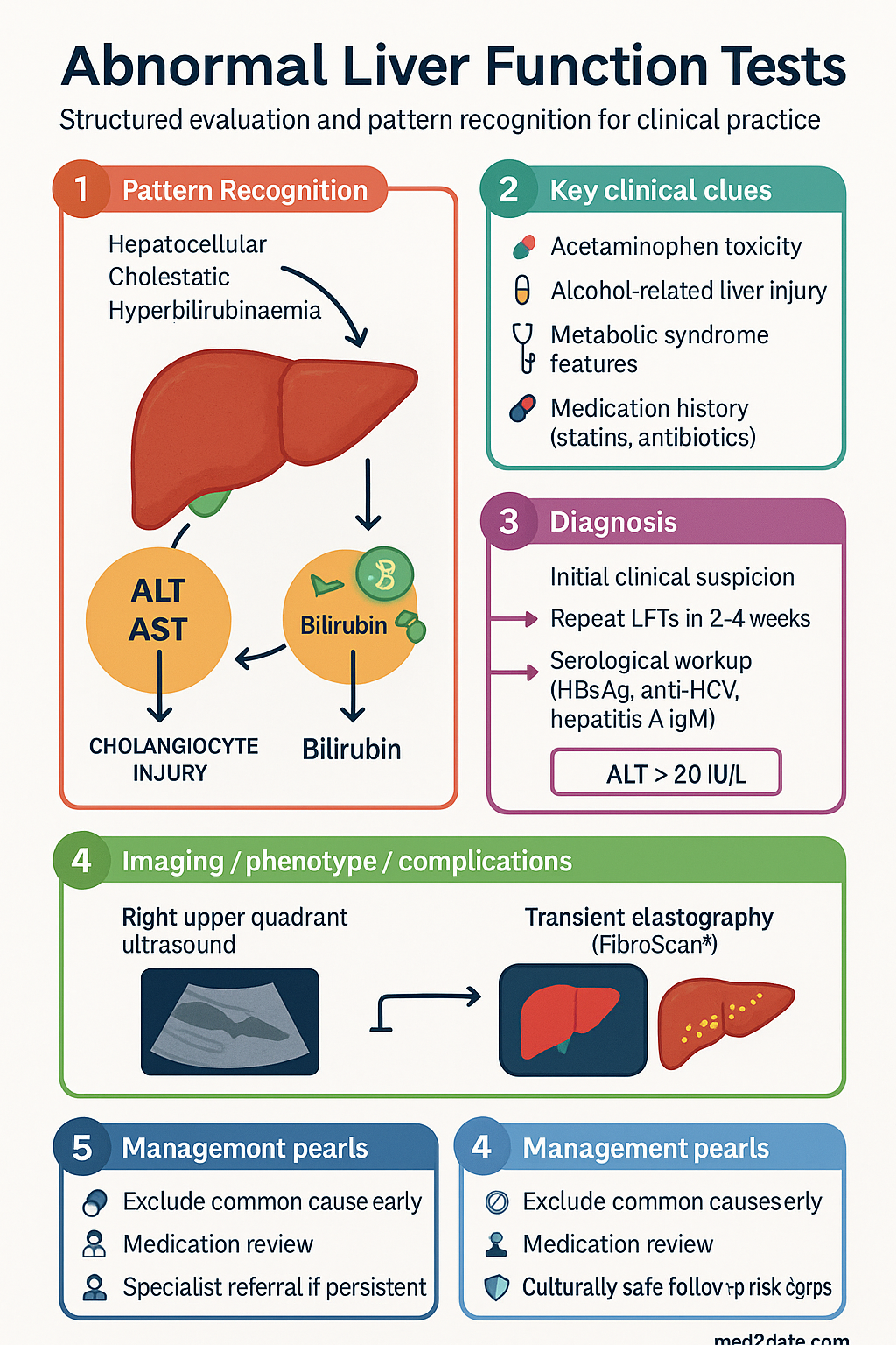
Pattern Recognition
The first step in evaluating abnormal LFTs is to determine the predominant pattern of derangement. This guides the subsequent diagnostic workup and helps prioritise the most likely aetiologies.
| Pattern | Key Markers | Common Causes | Typical AST:ALT Ratio |
|---|---|---|---|
| Hepatocellular | ALT and/or AST elevated disproportionately to ALP/GGT | MASLD/MASH, alcohol, viral hepatitis, drug-induced liver injury (DILI), autoimmune hepatitis, Wilson disease, coeliac disease | <1 (viral, MASLD) or >2 (alcoholic liver disease) |
| Cholestatic | ALP and/or GGT elevated disproportionately to transaminases | Gallstones, primary biliary cholangitis (PBC), primary sclerosing cholangitis (PSC), drug-induced cholestasis, pancreatic head mass, infiltrative liver disease | Variable; ALP typically >3× ULN |
| Isolated hyperbilirubinaemia | Conjugated or unconjugated bilirubin elevated with normal ALT, AST, ALP, GGT | Gilbert syndrome (unconjugated), haemolysis, Dubin-Johnson/Rotor syndrome (conjugated) | N/A — transaminases normal |
| Mixed | Both transaminases and ALP/GGT elevated | DILI, autoimmune hepatitis/PBC overlap, infiltrative disease, sepsis | Variable |
Hepatocellular Pattern
An ALT above the upper limit of normal (ULN, typically 35–40 IU/L for males, 25–30 IU/L for females, but varies by laboratory) with a relative preservation of ALP indicates hepatocyte injury or necrosis. Key causes to consider include:
- MASLD/MASH: The most common cause of chronically elevated ALT in Australia. Suspect in patients with metabolic syndrome features (obesity, type 2 diabetes, dyslipidaemia, hypertension). An AST:ALT ratio <1 is typical early; ratio >1 suggests progression to MASH or fibrosis.
- Alcohol-related liver disease: AST:ALT ratio >2:1, GGT markedly elevated, macrocytosis on FBE. MCV >100 fL supports chronic alcohol use.
- Viral hepatitis: Hepatitis B and C remain prevalent. Acute hepatitis A should be considered in travellers, men who have sex with men (MSM), and people who inject drugs (PWID).
- Drug-induced liver injury (DILI): A thorough medication history — including over-the-counter, herbal, and complementary preparations — is essential. Common offenders include paracetamol (dose-dependent), non-steroidal anti-inflammatory drugs (NSAIDs), statins, antiepileptics (sodium valproate, carbamazepine), antibiotics (amoxicillin-clavulanate, flucloxacillin, isoniazid), and methotrexate.
- Autoimmune hepatitis (AIH): More common in young to middle-aged women; can present acutely or insidiously. ANA, ASMA, and anti-LKM-1 antibodies are key markers.
- Wilson disease: Must be excluded in any patient <40 years with unexplained hepatocellular pattern. Serum caeruloplasmin and 24-hour urinary copper are initial tests.
Cholestatic Pattern
Predominant elevation of ALP (typically >1.5× ULN) with or without GGT elevation indicates impaired bile flow — either intrahepatic or extrahepatic. Causes include:
- Biliary obstruction: Common bile duct stones, pancreatic head tumours, cholangiocarcinoma. Ultrasound with Doppler is the first-line investigation.
- Primary biliary cholangitis (PBC): Chronic autoimmune destruction of intrahepatic bile ducts; predominantly affects middle-aged women. Anti-mitochondrial antibody (AMA) is positive in 90–95%.
- Primary sclerosing cholangitis (PSC): Chronic fibrosing inflammation of intra- and extrahepatic bile ducts; strongly associated with inflammatory bowel disease (IBD), especially ulcerative colitis. p-ANCA may be positive; MRCP is diagnostic.
- Drug-induced cholestasis: Oral contraceptive pill, anabolic steroids, flucloxacillin, co-amoxiclav, azathioprine.
- Infiltrative diseases: Sarcoidosis, amyloidosis, hepatic metastases, lymphoma.
Isolated Hyperbilirubinaemia
If bilirubin is the sole abnormality (ALT, AST, ALP, GGT all normal), determine whether the hyperbilirubinaemia is conjugated or unconjugated:
- Unconjugated (indirect): Gilbert syndrome (affects 5–10% of the Australian population; benign, no treatment needed), haemolysis (check FBE, reticulocyte count, LDH, haptoglobin), or ineffective erythropoiesis.
- Conjugated (direct): Rare — Dubin-Johnson syndrome, Rotor syndrome. More commonly indicates early biliary obstruction or hepatocellular dysfunction not yet reflected in other LFTs.
Medication and Substance Review
A comprehensive drug history is mandatory at the first encounter. Ask specifically about:
- Prescription medications — especially statins, methotrexate, antiepileptics, antituberculous therapy, antiretrovirals
- Over-the-counter analgesics — paracetamol (including combination preparations such as Panadeine Forte®), NSAIDs
- Herbal and complementary medicines — green tea extract, kava, comfrey, black cohosh, multi-ingredient formulations
- Recreational substances — alcohol (use AUDIT-C tool), anabolic steroids, cannabis, methamphetamine
Initial Laboratory Workup
After confirming persistent abnormality on repeat testing (ideally 2–4 weeks after the initial finding), a systematic laboratory workup should be undertaken based on the pattern of LFT derangement. All investigations listed below are Medicare-rebatable under the relevant MBS items when clinically indicated.
Tier 1 — First-Line Investigations (All Patterns)
| Investigation | Indication | MBS Item | Notes |
|---|---|---|---|
| Repeat LFTs (ALT, AST, ALP, GGT, bilirubin, albumin) | All patterns | 66500 | Confirms persistence; documents trend |
| Full blood examination (FBE) | All patterns | 65070 | Macrocytosis (alcohol), thrombocytopenia (cirrhosis/portal HTN), haemolysis screen |
| Coagulation studies (INR, APTT) | Elevated transaminases, suspected synthetic dysfunction | 65140 | Rising INR with normal vitamin K suggests advanced hepatocellular failure |
| Hepatitis B surface antigen (HBsAg) | All hepatocellular patterns, ATSI patients, migrants from endemic regions | 69313 | If positive → HBV DNA, HBeAg, anti-HBe, hepatitis B core antibody |
| Hepatitis C antibody (anti-HCV) | All hepatocellular patterns, PWID, birth cohort 1950–1975, HIV-positive patients | 69318 | If positive → HCV RNA (qualitative PCR) to confirm active infection |
| Hepatitis A IgM | Acute hepatitis, travellers, MSM, PWID | 69312 | Self-limiting; IgG indicates past infection or vaccination |
Tier 2 — Hepatocellular Pattern (Persistent Elevation)
| Investigation | Condition | Notes |
|---|---|---|
| ANA, anti-smooth muscle antibody (ASMA), anti-LKM-1 | Autoimmune hepatitis | ANA titre ≥1:40 and ASMA ≥1:40 are significant; IgG level often elevated |
| Serum ferritin, transferrin saturation | Haemochromatosis | Ferritin >300 µg/L (males) or >200 µg/L (females) + transferrin saturation >45% → HFE gene testing |
| Serum caeruloplasmin, 24-hour urinary copper | Wilson disease | Mandatory if patient <40 years with unexplained hepatocellular pattern. Low caeruloplasmin (<0.2 g/L) is suggestive |
| Alpha-1 antitrypsin (A1AT) level and phenotype | Alpha-1 antitrypsin deficiency | Consider in young patients with liver disease and/or early-onset emphysema |
| Tissue transglutaminase (tTG) IgA + total IgA | Coeliac disease | Unexplained isolated ALT elevation may be the sole manifestation of coeliac disease |
| Thyroid function tests (TSH) | Thyroid dysfunction | Hypothyroidism associated with MASLD; hyperthyroidism can cause isolated ALT elevation |
Tier 2 — Cholestatic Pattern
| Investigation | Condition | Notes |
|---|---|---|
| Anti-mitochondrial antibody (AMA) | Primary biliary cholangitis | Positive in 90–95% of PBC. If positive → refer for liver biopsy consideration |
| p-ANCA, IgG4 level | PSC, IgG4-related sclerosing cholangitis | MRCP is definitive for PSC; IgG4-related disease is steroid-responsive |
| CA 19-9, CEA | Pancreaticobiliary malignancy | Not diagnostic alone; useful in context of obstructive jaundice on imaging |
Tier 2 — Isolated Hyperbilirubinaemia
| Investigation | Condition | Notes |
|---|---|---|
| Split bilirubin (conjugated vs unconjugated) | Differentiates pre-hepatic from hepatobiliary | Unconjugated >80% = Gilbert syndrome or haemolysis |
| FBE, reticulocyte count, LDH, haptoglobin, blood film | Haemolysis | Low haptoglobin + high LDH + high reticulocytes = haemolysis |
| Direct Coombs test | Autoimmune haemolytic anaemia | If haemolysis confirmed |
Imaging
Right Upper Quadrant Ultrasound
Ultrasound is the first-line imaging modality for all patterns of LFT derangement. It is non-invasive, widely available across metropolitan and regional Australia, and does not involve ionising radiation. Key information obtained includes:
- Biliary duct dilatation (intrahepatic and extrahepatic) — suggests obstructive aetiology
- Gallstones and common bile duct stones
- Liver parenchymal echogenicity — increased echogenicity suggests steatosis (MASLD); coarse echotexture suggests chronic liver disease/fibrosis
- Hepatic focal lesions — cysts, haemangiomas, hepatocellular carcinoma, metastases
- Splenomegaly — suggests portal hypertension
- Ascites — suggests decompensated liver disease
- Pancreatic head mass — in cholestatic pattern with obstructive features
Transient Elastography (FibroScan®)
Transient elastography measures liver stiffness as a surrogate for fibrosis stage. It is increasingly available in Australian gastroenterology/hepatology practices, specialist liver clinics, and some larger regional centres. Key considerations:
- Availability: Mainly tertiary hospitals and specialist liver clinics in capital cities; expanding to regional centres (e.g., through Royal Flying Doctor Service outreach). Not universally available in rural and remote Australia.
- Interpretation: Liver stiffness measurement (LSM) <7 kPa generally excludes significant fibrosis (≥F2); LSM >12 kPa is highly suggestive of cirrhosis (F4). Controlled attenuation parameter (CAP) score concurrently measures steatosis.
- Limitations: Unreliable in patients with obesity (BMI >40), ascites, acute hepatitis, or elevated ALT >5× ULN. The XL probe may improve reliability in obese patients.
- PBS/MBS: FibroScan is not currently MBS-rebatable as a standalone test; costs may be borne by the patient or absorbed by the treating facility. Check local availability and fees.
Advanced Imaging
- MRCP (magnetic resonance cholangiopancreatography): Gold standard for biliary tree assessment when ultrasound is inconclusive; essential for suspected PSC. MBS-rebatable (MBS item 63003 and related).
- CT abdomen (triple phase): For characterisation of focal liver lesions, staging of suspected malignancy, or assessment of portal venous anatomy. Use with caution due to contrast nephrotoxicity and radiation dose.
- ERCP (endoscopic retrograde cholangiopancreatography): Therapeutic (stone extraction, stent placement) rather than diagnostic; reserved for confirmed biliary obstruction requiring intervention.
- Shear wave elastography (SWE) on ultrasound: Available on newer ultrasound platforms; provides real-time elastography during a standard ultrasound examination. Increasing availability in Australian radiology practices.
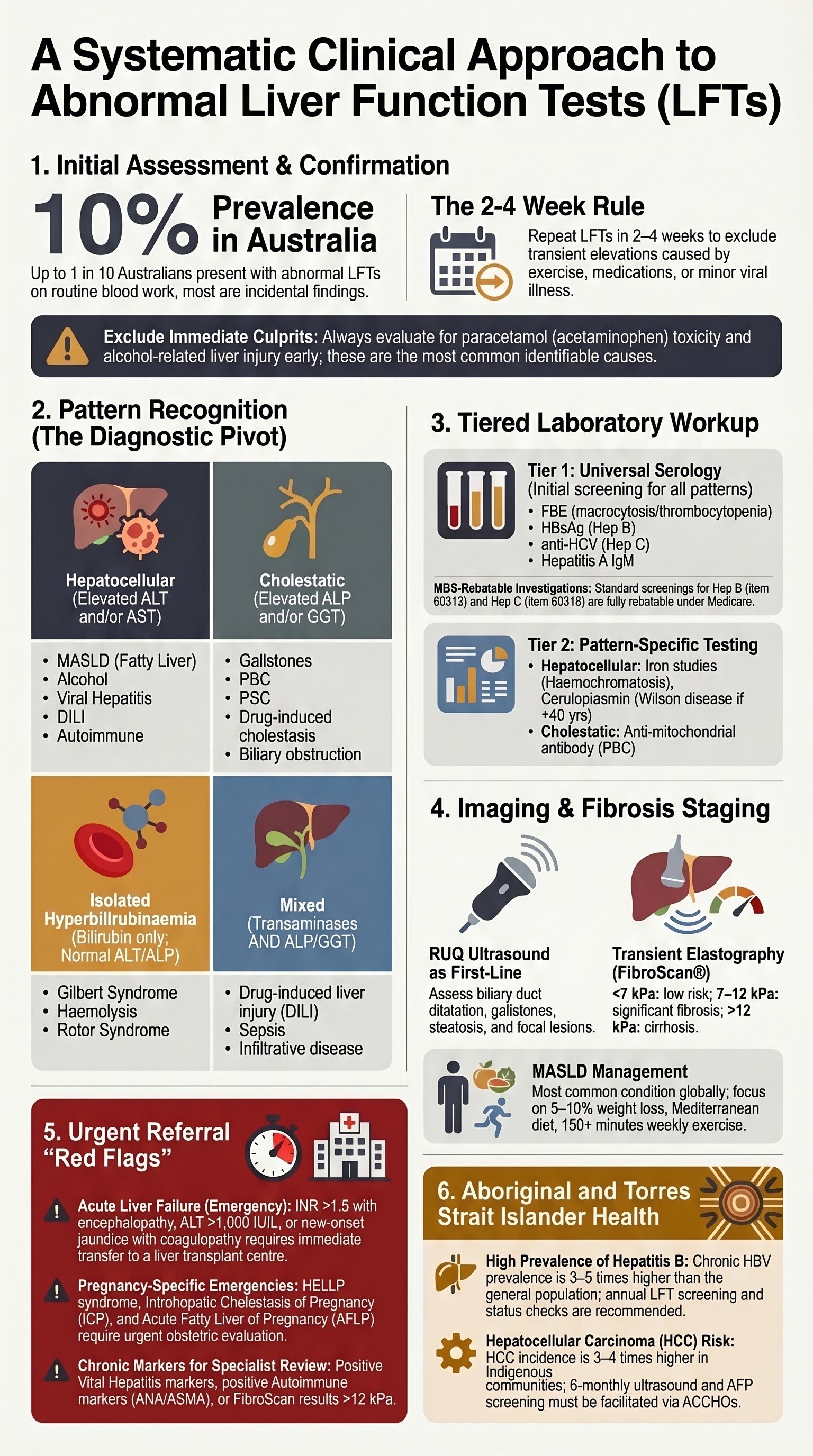
When to Refer
Timely referral to gastroenterology or hepatology is critical when the clinical picture suggests significant or progressive liver disease. The following referral indications should be applied in Australian primary care settings.
Urgent Referral (Within 1–2 Weeks)
- Suspected acute liver failure: INR >1.5 with encephalopathy, or ALT >1,000 IU/L with coagulopathy
- Severe acute hepatitis (ALT >10× ULN) of any cause
- New-onset jaundice with coagulopathy in the absence of pre-existing liver disease
- Suspected paracetamol overdose — initiate N-acetylcysteine (NAC) immediately; do not wait for referral
- Pregnancy with suspected HELLP syndrome or acute fatty liver of pregnancy
Semi-Urgent Referral (Within 4–6 Weeks)
- Positive hepatitis B (HBsAg positive) — for assessment of viral load and consideration of antiviral therapy (entecavir or tenofovir disoproxil, PBS Authority Required)
- Positive hepatitis C (HCV RNA positive) — for direct-acting antiviral (DAA) therapy initiation (PBS-listed; GPs with s100 prescriber status can initiate)
- Positive autoimmune markers (ANA/ASMA or AMA) with elevated LFTs
- Suspected Wilson disease or haemochromatosis on screening tests
- FibroScan LSM >12 kPa suggesting cirrhosis
Routine Referral (Within 2–3 Months)
- Persistently elevated LFTs >6 months despite addressing modifiable risk factors (weight loss, alcohol cessation, medication review)
- Unexplained hepatocellular pattern with normal Tier 1 and Tier 2 investigations
- Unexplained cholestatic pattern with normal imaging
- Progressive elevation of LFTs over serial measurements (rising trend)
- Suspected advanced fibrosis or cirrhosis on imaging or clinical grounds (palpable liver edge, spider naevi, palmar erythema, gynaecomastia)
- Focal liver lesion requiring characterisation beyond ultrasound capability
Referral Pathways in Australia
Public hospital hepatology/gastroenterology outpatient clinics accept referrals via standard referral letters (minimum requirement under the Health Insurance Act). Many states now use centralised referral triaging (e.g., NSW eReferral, Victorian TAS referral system). Private gastroenterology referrals can be made directly; typical out-of-pocket costs should be discussed with patients.
For patients in rural and remote areas, the following telehealth and outreach options are available:
- Hepatology telehealth consultations via state-based liver services (e.g., WA Country Health Service hepatology outreach)
- Royal Flying Doctor Service specialist outreach clinics
- GP/specialist shared-care models for hepatitis B and C management
- Liver nurse specialists available in most tertiary centres for coordination of care
Monitoring Prior to Referral
While awaiting specialist review, continue monitoring LFTs in primary care:
- Mildly deranged (1–2× ULN): Repeat every 3 months
- Moderately deranged (2–5× ULN): Repeat every 4–6 weeks
- Severely deranged (>5× ULN): Repeat weekly to fortnightly; consider earlier referral
Management of Common Causes Identified During Workup
While definitive management of identified liver conditions often requires specialist input, initiation of first-line therapy may occur in primary care for several conditions.
MASLD / Metabolic Dysfunction-Associated Steatotic Liver Disease
Management is centred on addressing metabolic risk factors. No PBS-listed pharmacotherapy currently exists specifically for MASLD/MASH, though resmetirom (Rezdiffra®) has received FDA approval in the United States and may become available in Australia.
- Weight loss target: 5–10% of body weight (improves steatosis at 5%; fibrosis improvement at ≥10%)
- Structured exercise: ≥150 minutes/week of moderate-intensity activity
- Dietary modification: Mediterranean diet pattern; reduce ultra-processed foods, added sugars (especially fructose), and saturated fats
- Manage associated conditions: optimise glycaemic control (GLP-1 receptor agonists such as semaglutide [Ozempic®/Wegovy®] show hepatic benefits), treat dyslipidaemia (statins are safe in liver disease — do not withhold), address hypertension
- Avoid alcohol completely or limit to ≤10 standard drinks/week (NHMRC 2020 guidelines)
- Vitamin E 800 IU/day (off-label) may be considered in non-diabetic patients with biopsy-proven NASH — discuss with hepatologist
Alcohol-Related Liver Disease
Hepatitis B — Initial Management in Primary Care
All HBsAg-positive patients require specialist hepatology assessment for staging and treatment eligibility. However, primary care plays a vital role in:
- Confirming the diagnosis and ordering initial viral serology (HBV DNA, HBeAg, anti-HBe, anti-HBc IgM)
- Screening for hepatocellular carcinoma: 6-monthly AFP and liver ultrasound (MBS-rebatable for chronic HBV)
- Vaccinating household contacts and sexual partners (HBV vaccine — funded under NIP for at-risk groups)
- Counselling regarding alcohol avoidance, medication safety (avoid hepatotoxic agents), and safe sex practices
Hepatitis C — DAA Therapy
Direct-acting antiviral (DAA) therapy for hepatitis C is PBS-listed and can be initiated by GPs with s100 prescriber endorsement, as well as by gastroenterologists, hepatologists, and infectious disease physicians. Treatment is curative in >95% of cases.
Special Populations
Pregnancy
Paediatrics
Elderly (>65 Years)
Renal Impairment
Hepatic Impairment
Immunocompromised
Aboriginal and Torres Strait Islander Australians experience significantly higher rates of liver disease, liver-related hospitalisation, and liver-related mortality compared with non-Indigenous Australians. Culturally safe, trauma-informed care and awareness of the unique epidemiological and social determinants are essential.
📚 References
- 1. European Association for the Study of the Liver (EASL). EASL Clinical Practice Guidelines on drug-induced liver injury. J Hepatol. 2019;70(6):1222–1261.
- 2. Royal Australian College of General Practitioners (RACGP). Guidelines for Preventive Activities in General Practice (Red Book). 9th ed. Melbourne: RACGP; 2018.
- 3. Australian Institute of Health and Welfare (AIHW). Liver and Biliary Cancer in Australia. Cat. no. CAN 133. Canberra: AIHW; 2023.
- 4. National Health and Medical Research Council (NHMRC). Australian Guidelines to Reduce Health Risks from Drinking Alcohol. Canberra: NHMRC; 2020.
- 5. Chalasani N, Younossi Z, Lavine JE, et al. The diagnosis and management of nonalcoholic fatty liver disease: Practice guidance from the American Association for the Study of Liver Diseases. Hepatology. 2018;67(1):328–357.
- 6. Hepatitis B Virus Living Guidelines. Australian National HBV Testing and Clinical Advisory Group. ASHM (Australasian Society for HIV, Viral Hepatitis and Sexual Health Medicine). Sydney; 2023. Available at: www.ashm.org.au
- 7. World Health Organization (WHO). Guidelines for the Prevention, Care and Treatment of Persons with Chronic Hepatitis B Infection. Geneva: WHO; 2015 (updated 2024).
- 8. Australian Government Department of Health and Aged Care. National Hepatitis C Strategy 2023–2025. Canberra: Commonwealth of Australia; 2023.
- 9. Gastroenterological Society of Australia (GESA). Liver Guidelines: Investigation of Abnormal Liver Function Tests. Melbourne: GESA; 2020 (updated 2023).
- 10. EASL-ALEH Clinical Practice Guidelines on non-invasive tests for evaluation of liver disease severity and prognosis. J Hepatol. 2015;63(1):237–264.
- 11. Aboriginal and Torres Strait Islander Hepatitis B Mapping Project. Kirby Institute, UNSW Sydney, in partnership with ASHM. Sydney; 2022.
- 12. Drugs and Therapeutics Advisory Committee (DTAC). Northern Territory Medicines List: Hepatitis B and C Treatment Guidelines for Remote Practice. Darwin: NT Department of Health; 2023.
- 13. Wong VW, Adams LA, de Ledinghen V, Wong GL, Sookoian S. Non-invasive biomarkers in NAFLD and NASH — current progress and future promise. Nat Rev Gastroenterol Hepatol. 2018;15(8):461–478.
- 14. Torres DM, Harrison SA. Diagnosis and therapy of nonalcoholic steatohepatitis. Gastroenterology. 2008;134(6):1682–1698.
- 15. National Aboriginal Community Controlled Health Organisation (NACCHO). Position Statement: Aboriginal and Torres Strait Islander Health Checks and Chronic Disease Management. Canberra: NACCHO; 2023.