Home Gastrointestinal Pancreatitis – Primary Care Interface

Pancreatitis – Primary Care Interface

🎧 Pancreatitis – Primary Care Interface — deep-dive podcast

📋 Key Information Summary

📋
  • Acute pancreatitis presents with severe epigastric pain radiating to the back, often with nausea and vomiting; serum lipase ≥3× the upper limit of normal is the diagnostic threshold.
  • The two leading aetiologies in Australia are gallstones (~40%) and alcohol (~30%); medications and hypertriglyceridaemia account for a further 10–15%.
  • Most suspected acute pancreatitis requires emergency department assessment — primary care role is early recognition, initial analgesia, nil by mouth, and prompt referral.
  • Aggressive IV fluid resuscitation (lactated Ringer's solution, 5–10 mL/kg/hr initial bolus then target-guided) within the first 24 hours reduces organ failure and mortality.
  • The Revised Atlanta Classification stratifies severity into mild (no organ failure, no local complications), moderately severe (transient organ failure <48 h), and severe (persistent organ failure ≥48 h).
  • BISAP score (BUN >8.2 mmol/L, impaired mental status, SIRS, age >60, pleural effusion) ≥3 predicts high mortality and guides escalation of care.
  • Cholangitis or persistent biliary obstruction requires urgent ERCP within 24–72 hours; magnetic resonance cholangiopancreatography (MRCP) or endoscopic ultrasound can confirm choledocholithiasis.
  • Primary care follow-up must address the underlying cause: alcohol cessation counselling, triglyceride management (target <1.7 mmol/L with fibrates), and elective cholecystectomy for biliary pancreatitis.
  • Post-discharge monitoring includes repeat lipase/amylase if symptomatic, HbA1c/fasting glucose for new-onset diabetes, and faecal elastase for exocrine insufficiency after recurrent episodes.
  • Drug-induced pancreatitis requires immediate cessation of the offending agent; high-risk drugs include azathioprine, valproate, GLP-1 receptor agonists, and thiazide diuretics.
  • Aboriginal and Torres Strait Islander Australians have significantly higher rates of gallstone disease, alcohol-related pancreatitis, and hypertriglyceridaemia, compounded by rural and remote access barriers.
  • Patients with recurrent acute pancreatitis (≥2 episodes) should be referred to a gastroenterologist for consideration of genetic testing (PRSS1, SPINK1, CFTR), autoimmune workup, and CT/MRI pancreas protocol imaging.
🎬 Pancreatitis – Primary Care Interface — clinical explainer

Introduction & Australian Epidemiology

Acute pancreatitis is an inflammatory condition of the pancreas that accounts for a significant and increasing burden on Australian emergency departments and hospitals. It ranges from a self-limiting disease (approximately 80% of cases) to a life-threatening condition with multi-organ failure and mortality rates of 20–30% in severe cases. The primary care clinician plays a critical role in early recognition, initial management, timely referral, and long-term follow-up to prevent recurrence.

In Australia, acute pancreatitis results in approximately 15,000–20,000 hospital admissions per year, with an age-standardised incidence of 30–40 per 100,000 population. Incidence has been rising over the past two decades, driven by increasing gallstone disease (linked to obesity), alcohol consumption patterns, and metabolic syndrome-related hypertriglyceridaemia. The disease carries an overall mortality of 2–5%, but this rises sharply in the elderly and those with systemic inflammatory response syndrome (SIRS) or organ failure.

Gallstone pancreatitis accounts for approximately 40% of cases nationally and is more prevalent in women over 40 years. Alcoholic pancreatitis, representing 30–35% of cases, is more common in men aged 30–50 and is disproportionately over-represented in Aboriginal and Torres Strait Islander communities. Idiopathic pancreatitis, diagnosed when no clear cause is identified after initial workup, accounts for 15–25% of presentations; a proportion of these have a genetic or anatomical basis (e.g., pancreas divisum, SPINK1/PRSS1 mutations) identifiable with specialist evaluation.

⚠️
Rising incidence in Australia: AIHW data show hospitalisations for acute pancreatitis have increased by approximately 30% over the past decade, with the highest rates in the Northern Territory and Queensland, reflecting both alcohol burden and gallstone prevalence in Indigenous populations.
Aetiology Proportion (%) Key Associations
Gallstones 40% Female sex, obesity, age >40, Aboriginal and Torres Strait Islander populations
Alcohol 30–35% Chronic heavy use (>50 g/day), binge drinking, male predominance
Idiopathic 15–25% Requires further workup; consider genetic, anatomical, microlithiasis
Hypertriglyceridaemia 5–7% Triglycerides >11.3 mmol/L; diabetes, metabolic syndrome, familial
Drug-induced 2–5% Azathioprine, valproate, GLP-1 RAs, thiazides, mesalazine
Other 3–5% Post-ERCP, trauma, autoimmune (IgG4), hereditary, malignancy

Recognition & Risk Factors

Clinical Presentation

Acute pancreatitis classically presents with the acute onset of severe, constant epigastric pain that radiates to the back (present in 50–60% of cases). The pain is typically worse after eating and may be partially relieved by sitting forward. Associated symptoms include nausea, vomiting (often persistent and not relieving pain), anorexia, and abdominal distension.

On examination, patients may be tachycardic (reflecting SIRS and hypovolaemia), febrile (low-grade; high fever suggests infected necrosis or cholangitis), and have marked tenderness in the epigastrium with or without guarding. Cullen's sign (periumbilical ecchymosis) and Grey Turner's sign (flank ecchymosis) are rare but indicate severe haemorrhagic pancreatitis.

🚨
Red flags requiring immediate hospital referral: Haemodynamic instability (SBP <90 mmHg, HR >120 bpm), signs of peritonitis, fever >38.5°C with rigors (consider cholangitis or infected necrosis), altered mental status, respiratory distress, or inability to tolerate oral intake with persistent vomiting.

Diagnostic Criteria

The diagnosis of acute pancreatitis requires two of the following three criteria (Revised Atlanta Classification 2012):

  1. Characteristic abdominal pain (acute onset, severe, epigastric, often radiating to the back)
  2. Serum lipase (or amylase) ≥3 times the upper limit of normal
  3. Characteristic findings on cross-sectional imaging (CT, MRI, or ultrasound)
ℹ️
Lipase vs Amylase: Serum lipase is the preferred biomarker. It is more sensitive (85–100%) and specific (85–99%) than amylase, remains elevated for 8–14 days (vs 3–5 days for amylase), and is not confounded by salivary gland pathology. Amylase may be normal in alcohol-related pancreatitis and in patients presenting late. Lipase >3× ULN in the context of characteristic pain is sufficient for diagnosis without imaging.

Risk Factors for Acute Pancreatitis

Mechanical
Gallstones & Structural
Choledocholithiasis, microlithiasis, pancreas divisum, sphincter of Oddi dysfunction, pancreatic duct obstruction by tumour or stricture.
Most common cause in Australia; cholecystectomy definitive treatment
Metabolic
Alcohol & Hypertriglyceridaemia
Chronic heavy alcohol use (>50 g/day), binge drinking, fasting triglycerides >11.3 mmol/L, uncontrolled diabetes mellitus, metabolic syndrome, pregnancy-related hypertriglyceridaemia.
Second most common cause; risk increases with cumulative exposure
Iatrogenic / Other
Drug-Induced & Post-ERCP
Azathioprine/6-mercaptopurine, valproate, GLP-1 receptor agonists (exenatide, liraglutide, semaglutide — though risk is very low), thiazides, furosemide, tetracyclines, mesalazine, didanosine, simvastatin (rare).
Post-ERCP pancreatitis in 3–10% of procedures; risk stratification with indomethacin prophylaxis

Medications Implicated in Drug-Induced Pancreatitis

Drug Class Examples Evidence Strength Typical Latency
Immunosuppressants Azathioprine, 6-mercaptopurine Definite (Class Ia) Days to months
Antiepileptics Valproate Definite (Class Ia) Weeks to months
GLP-1 Receptor Agonists Exenatide, liraglutide, semaglutide, dulaglutide Probable (Class II) Weeks to years
Diuretics Thiazides, furosemide Probable (Class II) Weeks to months
5-ASA compounds Mesalazine, sulfasalazine Probable (Class II) Days to months
NRTIs Didanosine, stavudine Definite (Class Ia) Weeks to years
Statins Simvastatin, atorvastatin Possible (Class III) Months

Pathophysiology

Acute pancreatitis is initiated by premature intracellular activation of trypsinogen to trypsin within pancreatic acinar cells. This triggers a cascade of digestive enzyme activation (elastase, phospholipase A2, kallikrein) leading to autodigestion of the pancreas and peripancreatic tissues. The resultant injury activates nuclear factor-κB (NF-κB) signalling, amplifying local and systemic inflammatory responses.

In gallstone pancreatitis, obstruction of the common bile duct or pancreatic duct by a migrating stone causes ductal hypertension and enzyme reflux. In alcohol-related disease, ethanol metabolites (fatty acid ethyl esters) are directly toxic to acinar cells, promote intracellular calcium overload, and impair autophagy. In hypertriglyceridaemia, free fatty acids released by pancreatic lipase from excess triglycerides cause direct lipotoxic injury to acinar cells and vascular endothelium.

Two-Phase Model of Severe Pancreatitis

1
Early Phase (Days 1–7)
Systemic inflammatory response syndrome (SIRS) driven by release of cytokines (IL-1, IL-6, TNF-α, IL-8). This phase determines whether organ failure (pulmonary, renal, cardiovascular) develops. Organ failure that resolves within 48 hours is classified as "transient"; persistence beyond 48 hours defines "persistent" organ failure and severe disease.
2
Late Phase (Days 7+)
Driven by local complications — pancreatic necrosis, walled-off necrosis (WON), pseudocyst formation, and secondary infection. Infected necrosis (present in 30–40% of necrotising pancreatitis) is the leading cause of late mortality. This phase may require percutaneous or endoscopic drainage, and step-up approaches (catheter drainage → minimally invasive necrosectomy → open surgery).

The Revised Atlanta Classification (2012) classifies local complications based on time course and content: acute peripancreatic fluid collection (APFC, <4 weeks, homogeneous), pseudocyst (>4 weeks, encapsulated, no solid debris), acute necrotic collection (ANC, <4 weeks, heterogeneous), and walled-off necrosis (>4 weeks, encapsulated, heterogeneous).

Investigations

Initial Laboratory Assessment

Investigations should be performed in the emergency department or hospital setting. The primary care clinician should request these if the patient presents to general practice acutely, alongside arranging transfer.

Essential Serum lipase Preferred diagnostic test; ≥3× ULN is diagnostic. MBS Item 66680. Remains elevated 8–14 days. More sensitive and specific than amylase. Available in all Australian laboratories.
Essential Serum amylase Supportive but less reliable; may be normal in alcohol-related pancreatitis and late presentations. MBS Item 66675. Confounded by salivary gland disease and macroamylasaemia.
Essential Full blood count (FBC) Haemoconcentration (Hct >44%) predicts severe disease. Leucocytosis >12×10⁹/L is a SIRS criterion. MBS Item 66512.
Essential Urea, electrolytes, creatinine (UEC) Elevated BUN (>8.2 mmol/L) is a BISAP criterion. Rising creatinine indicates renal failure. Guide fluid resuscitation targets. MBS Item 66515.
Essential Liver function tests (LFTs) ALT >150 U/L has 95% positive predictive value for gallstone aetiology. Elevated ALP/GGT suggests biliary obstruction. Bilirubin >40 µmol/L suggests persistent common bile duct stone. MBS Item 66553.
Essential Serum calcium (corrected) Hypocalcaemia (corrected Ca <2.0 mmol/L) is a Ranson's criterion at 48 hours and indicates severe disease. Hypercalcaemia suggests hyperparathyroidism as a cause. MBS Item 66556.
Essential Fasting lipid profile Triglycerides >11.3 mmol/L as a cause. Note: lipaemic sample may interfere with amylase measurement; lipase remains reliable. MBS Item 66575.
Essential Blood glucose / HbA1c Hyperglycaemia (>10 mmol/L) is a Ranson's and BISAP criterion. New-onset diabetes occurs in 15–25% after acute pancreatitis. MBS Item 66554.
Essential CRP CRP >150 mg/L at 48 hours predicts severe pancreatitis and necrosis. Serial CRP useful for monitoring. MBS Item 66542.
Available Arterial blood gas (ABG) Hypoxaemia (PaO₂ <60 mmHg) indicates ARDS; metabolic acidosis (pH <7.35, lactate >2 mmol/L) indicates organ failure and tissue hypoperfusion. MBS Item 11704.

Imaging

Essential Transabdominal ultrasound First-line imaging for all patients. Identifies gallstones (sensitivity 95% for gallbladder stones, 50–70% for CBD stones), dilated CBD (>6 mm), and peripancreatic fluid. Should be performed within 24 hours of admission. MBS Item 55304. Operator-dependent for CBD assessment.
Available Contrast-enhanced CT (CECT) abdomen Not required for diagnosis. Best performed at 72–96 hours if clinical deterioration, suspected necrosis, or diagnostic uncertainty. Assesses extent of necrosis (CT severity index). MBS Item 55066. Avoid early CT (<72 h) as necrosis may not be evident.
Referral MRCP (Magnetic Resonance Cholangiopancreatography) Non-invasive assessment of bile duct stones when ultrasound is inconclusive and clinical suspicion of choledocholithiasis persists. Sensitivity 85–95% for CBD stones. No radiation. MBS Item 63524. Available at major metropolitan and regional centres.
Specialist Endoscopic Ultrasound (EUS) Highest sensitivity (95–100%) for small CBD stones and microlithiasis. Also used for fine-needle aspiration of pancreatic lesions. Requires specialist endoscopist. MBS Item 30624. Available at tertiary centres.
Specialist ERCP (Endoscopic Retrograde Cholangiopancreatography) Therapeutic intervention for persistent biliary obstruction, cholangitis, or confirmed choledocholithiasis. Not for diagnostic use alone. MBS Item 30624. Risk of post-ERCP pancreatitis (3–10%); rectal indomethacin 100 mg prophylaxis recommended for high-risk procedures.

Risk Stratification & Severity Scoring

Risk stratification should be performed at presentation and at 48 hours. The Revised Atlanta Classification is the standard framework, and the BISAP score is a practical bedside tool for primary care and emergency settings.

Revised Atlanta Classification — Severity

Mild
~80% of Cases
No organ failure. No local or systemic complications. Resolves within 5–7 days. Mortality <1%. Early oral feeding usually possible.
Setting: General ward or short-stay unit
Moderately Severe
~15% of Cases
Transient organ failure (resolving within 48 hours) OR local complications (peripancreatic fluid collections, pseudocyst, necrosis) without persistent organ failure. Mortality 1–5%.
Setting: HDU / monitored bed; gastroenterology consultation
Severe
~5% of Cases
Persistent organ failure (>48 hours) — single or multi-organ. Respiratory failure (PaO₂/FiO₂ ≤300), renal failure (creatinine ≥170 µmol/L), cardiovascular failure (SBP <90 mmHg, fluid unresponsive). Mortality 20–36%.
Setting: ICU; interventional radiology/endoscopy on standby

BISAP Score (Bedside Index for Severity in Acute Pancreatitis)

Score calculated within first 24 hours. Score ≥3 indicates high risk of mortality (5–20%).

Parameter Criterion Points
BUN >8.2 mmol/L (25 mg/dL) 1
Impaired mental status GCS <15 or disorientation 1
SIRS ≥2 of: T <36 or >38°C, HR >90, RR >20, WCC <4 or >12×10⁹/L, PaCO₂ <32 mmHg 1
Age >60 years 1
Pleural effusion Present on imaging or CXR 1
⚠️
Primary care utility: The BISAP score can be calculated from initial ED bloods and bedside assessment. A score of 0–1 predicts very low mortality (<1%); score of 2 carries 2% mortality; score ≥3 carries 5–20% mortality and warrants ICU-level monitoring. Persistent SIRS at 48 hours is the strongest predictor of organ failure progression.

Ranson's Criteria (Revised)

At Admission At 48 Hours
Age >55 years HCT fall >10%
WCC >16×10⁹/L BUN rise >1.8 mmol/L
Glucose >10 mmol/L Ca²⁺ <2.0 mmol/L
LDH >350 IU/L PaO₂ <60 mmHg
AST >250 IU/L Base deficit >4 mEq/L
Fluid sequestration >6 L

Gallstone pancreatitis (non-operated): ≥3 criteria = severe. Alcoholic pancreatitis: ≥6 criteria = severe. Mortality rises significantly with increasing scores.
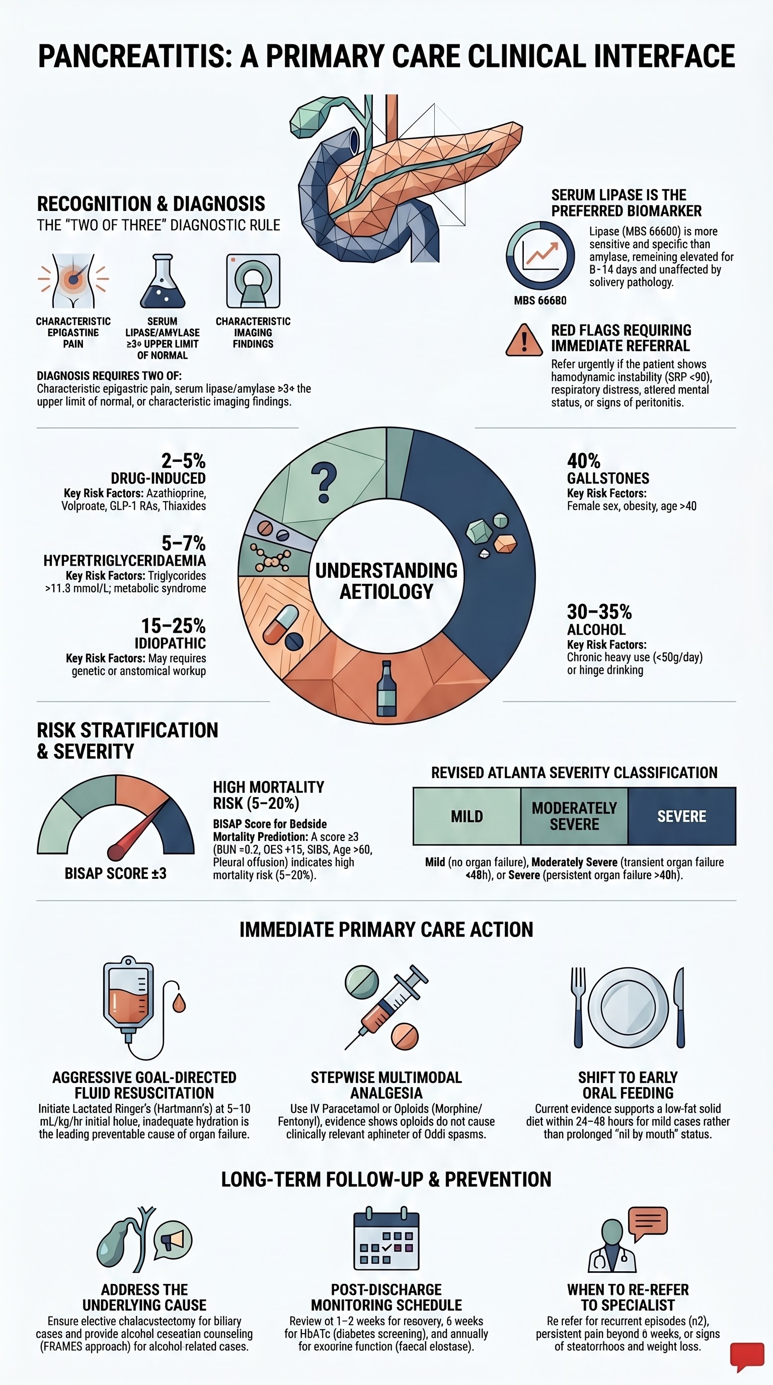
🖼️ Pancreatitis – Primary Care Interface — visual summary
Pancreatitis – Primary Care Interface visual summary infographic

Immediate Management

Most patients with suspected acute pancreatitis require emergency department assessment and hospital admission. The primary care clinician's role is early recognition, initial resuscitation measures, and rapid referral. The following management should be initiated in the ED and continued on the ward.

🚨
Primary care action: If you suspect acute pancreatitis in your rooms, do NOT delay transfer for imaging or bloods beyond basic tests. Ensure the patient is nil by mouth, provide IV access if possible, administer parenteral analgesia (paracetamol IV or opioid if available), and arrange immediate transfer to the nearest ED. Document the time of symptom onset, pain character, and last oral intake.

1. Fluid Resuscitation

Aggressive goal-directed fluid therapy is the single most important intervention in the first 24–48 hours. Inadequate fluid resuscitation is the most common preventable cause of organ failure.

💧
Lactated Ringer's Solution (Hartmann's)
Hartmann's® · Balanced crystalloid · First-line fluid
Adult dose Initial bolus 20 mL/kg (or 1–1.5 L) over 1–2 hours, then 3 mL/kg/hr (approximately 200–250 mL/hr) guided by clinical targets
Paediatric dose 10–20 mL/kg bolus over 1 hour, then 1.5× maintenance rate
Route IV
Duration First 24–48 hours; taper when oral intake tolerated
Renal adjustment Reduce rate in renal impairment; monitor potassium (LR contains 4 mmol/L K⁺)
PBS status ✔ PBS General Benefit
💧
0.9% Sodium Chloride (Normal Saline)
NaCl 0.9% · Isotonic crystalloid · Alternative
Adult dose Same rate as lactated Ringer's; acceptable alternative if LR unavailable
Caution High chloride load may cause hyperchloraemic metabolic acidosis; prefer LR when available. Evidence suggests LR associated with reduced SIRS at 24 hours vs NS.
PBS status ✔ PBS General Benefit

Fluid resuscitation targets (goal-directed): Target BUN <7.1 mmol/L, decreasing haematocrit, urine output >0.5 mL/kg/hr, improving heart rate, and normalising lactate. Avoid aggressive fluid overload in elderly patients and those with cardiac or renal comorbidities — reassess volume status every 4–6 hours using clinical, biochemical, and point-of-care ultrasound parameters.

2. Analgesia

Pain management is a priority. Pancreatitis pain can be severe and undertreated. A stepwise multimodal approach is recommended. There is no evidence that opioids cause sphincter of Oddi spasm clinically relevant to pancreatitis outcomes — pain relief should not be withheld due to this theoretical concern.

💊
Paracetamol
Panadol® · Analgesic · First-line
Adult dose 1 g PO/IV QID (max 4 g/day; 2 g/day if hepatic impairment or weight <50 kg)
Paediatric dose 15 mg/kg QID (max 60 mg/kg/day)
Route IV preferred initially (nil by mouth); PO when tolerated
Renal adjustment No specific adjustment; use with caution if eGFR <30
PBS status ✔ PBS General Benefit
💊
Morphine
Ordine® · Opioid analgesic · Second-line / severe pain
Adult dose 2.5–5 mg IV/SC Q4H PRN; titrate to effect. Oral: 5–10 mg Q4H when tolerated. PCA (patient-controlled analgesia) for severe pain.
Paediatric dose 0.1–0.2 mg/kg IV Q4H PRN
Renal adjustment Reduce dose by 50% if eGFR 10–50; avoid active metabolites (M6G accumulation) — consider fentanyl in severe renal impairment
PBS status ⚠ PBS Authority Required
💊
Fentanyl
Sublimaze® · Opioid analgesic · For renal impairment
Adult dose 25–50 mcg IV Q1–2H PRN; or 25–75 mcg/hr transdermal patch (for chronic pain after recovery)
Renal adjustment Preferred opioid in renal failure (no active metabolites)
PBS status ⚠ PBS Authority Required
💊
Ketorolac
Toradol® · NSAID · Adjunct for opioid-sparing
Adult dose 10–30 mg IV stat, then 10–15 mg IV Q6H (max 5 days, max 90 mg/day)
Caution Avoid if eGFR <30, active GI bleeding, or coagulopathy. Short courses only. NSAIDs may have a role in reducing pancreatic inflammation.
PBS status ✔ PBS General Benefit

3. Antiemetic Therapy

💊
Ondansetron
Zofran® · 5-HT₃ antagonist · First-line antiemetic
Adult dose 4–8 mg IV/PO Q8H PRN
Paediatric dose 0.1–0.15 mg/kg IV/PO Q8H (max 4 mg/dose)
Renal adjustment No adjustment required
PBS status ✔ PBS General Benefit

4. Nil by Mouth & Nutritional Support

Traditional management mandated nil by mouth until pain resolved and enzymes normalised. Current evidence supports early oral feeding (within 24–48 hours) in mild pancreatitis with a low-fat solid diet — there is no benefit of prolonged fasting or clear-fluid diet stepping. Nasogastric feeding should be initiated if the patient cannot eat by 48–72 hours. Enteral nutrition (via nasojejunal tube if NG feeding not tolerated) is preferred over parenteral nutrition, which increases infectious complications.

Key practice change: Early oral feeding (low-fat solid diet) within 24 hours in mild pancreatitis is safe, reduces hospital stay, and does not increase complications or readmissions (PANTHER trial, Dutch Pancreatitis Study Group). Parenteral nutrition should be avoided unless enteral route is not feasible.

5. Gallstone Pancreatitis — Urgent ERCP

Urgent ERCP (within 24–72 hours) is indicated for patients with concurrent cholangitis (fever, jaundice, right upper quadrant pain — Charcot's triad) or persistent biliary obstruction (bilirubin >40 µmol/L, dilated CBD on imaging). ERCP is not indicated for uncomplicated gallstone pancreatitis without cholangitis or persistent obstruction — early cholecystectomy during the same admission is the standard of care.

6. Prophylactic Antibiotics

Prophylactic antibiotics are not recommended for sterile necrotising pancreatitis (multiple RCTs, including the landmark Dutch and German trials, show no mortality benefit). Antibiotics are indicated for confirmed or strongly suspected infected necrosis (gas in necrosis on CT, positive FNA culture, persistent sepsis ≥7–10 days after onset) and for concurrent infections (cholangitis, pneumonia, UTI, line sepsis).

⚠️
When infected necrosis is suspected: Start empiric carbapenem (meropenem 1 g IV TDS) which achieves adequate pancreatic tissue penetration. Add antifungal cover (fluconazole 400 mg IV daily) if fungal infection suspected. Adjust based on CT-guided FNA culture results. PBS: Meropenem — Authority Required.

7. Specific Interventions by Aetiology (Acute Phase)

Aetiology Acute Intervention Timing
Gallstone with cholangitis Urgent ERCP + sphincterotomy ± stone extraction Within 24 hours
Gallstone without cholangitis Same-admission cholecystectomy (laparoscopic) Before discharge (once pain resolved, enzymes trending down)
Hypertriglyceridaemia (>11.3 mmol/L) Insulin infusion 0.1–0.3 U/kg/hr ± heparin 500 U bolus; plasmapheresis if TG >22.6 mmol/L or refractory Immediate
Alcohol-related Supportive care; alcohol withdrawal protocol if at risk; thiamine 100–300 mg IV/PO daily Immediate; ongoing
Drug-induced Immediate cessation of suspected agent; document and counsel As soon as identified
Post-ERCP Aggressive IV fluids, standard analgesia; usually self-limiting Immediate
💊
Thiamine
Vitamine B1® · Vitamin · For alcohol-related pancreatitis
Adult dose 100 mg IV daily for 3–5 days, then 100 mg PO daily ongoing
Indication All patients with alcohol-related pancreatitis; give BEFORE glucose to prevent Wernicke's encephalopathy
PBS status ✔ PBS General Benefit

Primary Care Follow-Up

After discharge from hospital, the patient's ongoing care should be managed in close coordination with their general practitioner. The primary care clinician is central to addressing the underlying cause, preventing recurrence, monitoring for complications, and managing long-term sequelae such as pancreatic exocrine insufficiency and new-onset diabetes.

First Visit After Discharge (Within 1–2 Weeks)

1
Review Discharge Summary
Confirm aetiology, severity, complications, imaging results, operative notes (if cholecystectomy performed), and follow-up plan. Note any pending investigations (genetic testing, autoimmune markers).
2
Assess Clinical Recovery
Resolution of pain, tolerance of oral diet, weight recovery, bowel habit. Check vital signs, abdominal examination. Order repeat bloods if symptoms persistent (lipase, LFTs, glucose, calcium).
3
Address the Underlying Cause
This is the most critical long-term intervention — see specific aetiology management below.
4
Screen for Complications
New-onset diabetes (HbA1c, fasting glucose), exocrine insufficiency (faecal elastase if steatorrhoea or weight loss), pseudocyst symptoms (persistent pain, early satiety, gastric outlet obstruction).

Addressing Underlying Causes — Long-Term Management

Alcohol Cessation

For patients with alcohol-related pancreatitis, complete abstinence is the single most effective intervention to prevent recurrence. Even moderate alcohol resumption (1–2 standard drinks/day) significantly increases recurrence risk. The GP should:

  • Provide brief intervention and motivational interviewing (using the FRAMES approach or 5As framework)
  • Refer to community alcohol and drug services, counselling, or rehabilitation as appropriate
  • Consider pharmacotherapy for alcohol dependence: naltrexone (Revia®) 50 mg PO daily or acamprosate (Campral®) 666 mg PO TDS
  • Screen for alcohol withdrawal risk at discharge; ensure community-based withdrawal management if not completed in hospital
  • Monitor LFTs (GGT trend), CDT (carbohydrate-deficient transferrin) as objective markers of abstinence
💊
Naltrexone
Revia® · Opioid antagonist · Alcohol relapse prevention
Adult dose 50 mg PO daily; start after 7–10 days of abstinence (avoid precipitating withdrawal)
Contraindications Active opioid use, acute hepatitis, liver failure (AST/ALT >3× ULN)
PBS status ⚠ PBS Authority Required
💊
Acamprosate
Campral® · Glutamate modulator · Alcohol relapse prevention
Adult dose 666 mg PO TDS with meals
Renal adjustment Contraindicated if eGFR <30 mL/min; reduce to 333 mg TDS if eGFR 30–50
PBS status ⚠ PBS Authority Required

Gallstone Management

  • Same-admission cholecystectomy: Current evidence (PONCHO trial) supports laparoscopic cholecystectomy during the index admission for mild gallstone pancreatitis. This reduces readmission for recurrent biliary events from 17% (delayed cholecystectomy at 2–4 weeks) to 5%.
  • If cholecystectomy was not performed: Ensure the patient is on the surgical waiting list. Urgent referral if there is recurrent biliary colic or pancreatitis. A low-fat diet should be advised in the interim.
  • In patients unfit for surgery: Endoscopic sphincterotomy reduces recurrence risk but is less effective than cholecystectomy. Ursodeoxycholic acid (UDCA) may reduce gallstone recurrence.
💊
Ursodeoxycholic acid
Ursofalk® · Bile acid · Gallstone dissolution / recurrence prevention
Adult dose 8–10 mg/kg/day PO in 2–3 divided doses
Indication Patients unfit for cholecystectomy with radiolucent gallstones; post-ERCP stone clearance
Duration 6–12 months; ultrasound at 6 months to assess response
PBS status ⚠ PBS Authority Required

Hypertriglyceridaemia Management

Patients with pancreatitis secondary to hypertriglyceridaemia require aggressive lipid management to prevent recurrence. Target triglyceride level: <1.7 mmol/L (ideally), <5.6 mmol/L (absolute minimum to prevent recurrence).

  • Dietary modification: Very low-fat diet (<20% of calories from fat), eliminate simple sugars and alcohol, Mediterranean-style diet. Dietitian referral essential.
  • Weight loss: If overweight/obese, even 5–10% weight reduction significantly lowers triglycerides.
  • Diabetes optimisation: Tight glycaemic control (HbA1c target <53 mmol/mol / <7%).
  • Fibrate therapy: First-line pharmacotherapy. Fenofibrate reduces triglycerides by 30–50%.
  • Icosapent ethyl (Vascepa®): EPA-based omega-3 fatty acid; reduces triglycerides by 20–30%. PBS listed for severe hypertriglyceridaemia.
  • Combination therapy: Fibrates + omega-3s ± niacin for refractory cases. Avoid statin + fibrate combinations (increased myopathy risk) — if needed, fenofibrate is the preferred fibrate with statins.
💊
Fenofibrate
Lipidil® · Fibrate · First-line triglyceride reduction
Adult dose 145 mg PO daily (Lipidil EZ) or 160 mg PO daily (Lipidil Supra); take with food
Expected effect Reduces triglycerides 30–50%; raises HDL-C 5–15%
Renal adjustment Contraindicated if eGFR <15; dose reduction if eGFR 15–30
Key interactions Warfarin (increased INR — monitor); statins (myopathy risk — use fenofibrate, not gemfibrozil)
PBS status ✔ PBS General Benefit
💊
Icosapent ethyl
Vascepa® · Purified EPA omega-3 · Adjunct triglyceride reduction
Adult dose 2 g PO BD with food (4 g/day total)
Expected effect Reduces triglycerides 20–30%; major cardiovascular event reduction (REDUCE-IT trial)
Renal adjustment Use with caution in severe renal impairment
PBS status ⚠ PBS Authority Required — for severe hypertriglyceridaemia (TG ≥5.6 mmol/L) not controlled on fibrates alone

Medication Review

If drug-induced pancreatitis is suspected, permanently discontinue the offending agent and document the reaction. If the medication is essential (e.g., azathioprine for IBD or transplant), specialist discussion regarding rechallenge or alternative is required. Document pancreatitis as an adverse drug reaction in the patient's record and the Australian Adverse Drug Reaction Reporting System (ADRAC/TGA) if appropriate.

Monitoring Schedule in Primary Care

Timepoint Assessment Investigations
1–2 weeks post-discharge Clinical review, pain, diet tolerance, wound check (if surgical), medication reconciliation Repeat lipase if symptomatic, LFTs, glucose, renal function
6 weeks Weight, alcohol status, lipid profile review, diabetes screening, specialist follow-up compliance HbA1c, fasting lipids, faecal elastase if symptoms of steatorrhoea
3 months Ongoing cause-specific management (alcohol abstinence, triglycerides), psychological wellbeing Lipids (if hypertriglyceridaemia), CDT (if alcohol-related), HbA1c
6 months Long-term disease burden assessment, ongoing specialist liaison, recurrence risk review Full metabolic panel, lipids, HbA1c, faecal elastase if indicated
Annually (ongoing) Preventive care, recurrence vigilance, diabetes screening, exocrine function, cholecystectomy follow-up HbA1c, lipids, faecal elastase, nutritional markers (albumin, B12, fat-soluble vitamins)

When to Re-Refer

  • Recurrent acute pancreatitis (≥2 episodes) — for specialist evaluation including genetic testing (PRSS1, SPINK1, CFTR, CTRC), autoimmune serology (IgG4), and cross-sectional imaging
  • Persistent pain beyond 4–6 weeks — consider chronic pancreatitis, pseudocyst, or ductal complications
  • Steatorrhoea, weight loss, or nutritional deficiency — consider exocrine pancreatic insufficiency (faecal elastase <200 µg/g); initiate pancreatic enzyme replacement therapy (PERT)
  • New-onset diabetes mellitus — endocrinology referral if insulin-dependent or difficult to control
  • Pancreatic mass or cystic lesion discovered on imaging — urgent gastroenterology/oncology referral
💊
Pancreatin (Pancreatic Enzyme Replacement Therapy)
Creon® · Enzyme replacement · For exocrine insufficiency
Adult dose 25,000–50,000 units of lipase with each meal; titrate to symptoms (reduce steatorrhoea, maintain weight)
Paediatric dose Age-dependent: infants 2,000–4,000 units lipase per feed; children >4 years same as adult dosing
Route Oral; take at start of meal (open capsule onto acidic food if unable to swallow)
Renal adjustment None required
PBS status ⚠ PBS Authority Required

Recurrence Prevention — Patient Counselling Points

Lifestyle Modification
  • Complete alcohol abstinence (if alcohol-related aetiology) or moderation (<2 standard drinks/day, <10/week)
  • Low-fat diet with gradual liberalisation; avoid high-fat meals and fried foods
  • Smoking cessation — smoking is an independent risk factor for recurrent and chronic pancreatitis
  • Regular exercise and weight management (BMI target 18.5–25)
  • Maintain hydration, especially during illness or fasting
When to Seek Emergency Care
  • Return of severe epigastric pain radiating to the back
  • Persistent vomiting preventing oral intake for >12 hours
  • Fever (>38.5°C) with abdominal pain
  • Jaundice (yellow skin/eyes, dark urine, pale stools)
  • Signs of dehydration (dizziness, reduced urine output, confusion)

Special Populations

🤰

Pregnancy

Incidence
Approximately 1 in 1,000–3,000 pregnancies. Most common in the third trimester. Gallstones account for ~70% of pregnancy-related pancreatitis.
Diagnosis
Lipase remains the preferred biomarker in pregnancy — amylase is physiologically elevated (from placenta and amniotic fluid) and less reliable. Ultrasound is first-line imaging (no radiation). MRI without gadolinium is safe if further imaging needed.
Management
Supportive care as per standard protocols. ERCP is safe in pregnancy with appropriate shielding and fetal monitoring. Cholecystectomy can be performed in the second trimester if indicated (laparoscopic approach preferred). Avoid statins and fibrates in pregnancy.
Analgesia
Paracetamol first-line. Opioids (morphine, fentanyl) can be used short-term. Avoid NSAIDs, especially in the third trimester (risk of premature closure of ductus arteriosus). Panadol Osteo® is not recommended in pregnancy.
Hypertriglyceridaemia
Pregnancy can precipitate severe hypertriglyceridaemia in women with underlying familial disorders. Insulin infusion and plasmapheresis are the mainstays of acute treatment. Omega-3 fatty acids may be continued. Lipid-lowering drugs should be stopped — resume post-partum.
👶

Paediatrics

Epidemiology
Less common than in adults but increasing incidence. Common causes include blunt abdominal trauma, systemic illness, medications (L-asparaginase, valproate), and hereditary conditions (PRSS1, SPINK1, CFTR mutations). Gallstones are less common but increasing with childhood obesity.
Diagnosis
Same criteria as adults (2 of 3). Paediatric-specific lipase reference ranges apply. Imaging with ultrasound first-line; MRI for diagnostic uncertainty.
Fluid resuscitation
10–20 mL/kg bolus of lactated Ringer's, then 1.5× maintenance rate. Use weight-based charts. Monitor strict fluid balance and urine output (>1 mL/kg/hr).
Analgesia
Paracetamol 15 mg/kg QID first-line. Morphine 0.1–0.2 mg/kg IV Q4H for severe pain. Avoid codeine in children <12 years (TGA warning). PCA for adolescents.
Nutrition
Early enteral feeding (within 24–48 hours) is safe in paediatric pancreatitis. Nasogastric route preferred if oral intake not tolerated. Monitor growth parameters if recurrent episodes.
Special considerations
Recurrent episodes warrant genetic testing and specialist gastroenterology referral. Hereditary pancreatitis (PRSS1 mutations) carries a lifetime pancreatic cancer risk of 40–50% and requires surveillance from age 40 (or 10 years after diagnosis).
👴

Elderly (≥65 Years)

Higher mortality
Mortality 10–15% in patients >65 years (vs 1–3% in younger adults), driven by comorbidities, reduced physiological reserve, and delayed presentation. Age >60 is a BISAP criterion.
Aetiology
Gallstones are the dominant cause (~60%). Drug-induced pancreatitis is more common due to polypharmacy. Malignancy (pancreatic or ampullary) must be considered, especially with recurrent episodes.
Fluid resuscitation caution
Aggressive fluid resuscitation may cause pulmonary oedema or heart failure in elderly patients with cardiac comorbidities. Use clinical targets (urine output, lactate) and frequent reassessment. Point-of-care ultrasound for IVC collapsibility and lung B-lines may guide therapy.
Analgesia
Start with paracetamol; opioids at lower doses with careful titration (reduced renal clearance). Avoid morphine in renal impairment — fentanyl preferred. Avoid NSAIDs (GI bleeding, renal impairment, cardiovascular risk).
Cholecystectomy decision
Operative risk must be weighed against recurrence risk. FIT elderly patients should still have cholecystectomy. For frail patients, endoscopic sphincterotomy + UDCA may be more appropriate. MDT discussion recommended.
🫘

Renal Impairment

Diagnosis
Serum lipase is reliable in renal impairment (amylase is elevated in renal failure due to reduced clearance, making it less specific). Interpret BUN cautiously for BISAP scoring.
Fluid management
Goal-directed but cautious; monitor for fluid overload. Lactated Ringer's is preferred (lower potassium content than normal saline). May need renal replacement therapy if acute kidney injury develops.
Analgesia adjustments
Paracetamol: no significant dose adjustment. Morphine: reduce dose 50%, avoid in severe renal failure (active metabolite accumulation). Fentanyl: preferred opioid in renal failure (no active metabolites). Ketorolac: contraindicated if eGFR <30.
Medication review
Review all renally cleared medications. Fenofibrate contraindicated if eGFR <15. Acamprosate contraindicated if eGFR <30. Adjust allopurinol dose if gout present (associated with pancreatitis risk).
🫁

Hepatic Impairment

Overlapping pathology
Alcoholic liver disease frequently co-exists with alcoholic pancreatitis. Elevated LFTs may reflect underlying liver disease rather than biliary obstruction. Differentiate using imaging (ultrasound, MRCP).
Analgesia
Paracetamol: max 2 g/day (not 4 g) if significant hepatic impairment (Child-Pugh B or C). Avoid NSAIDs (coagulopathy, GI bleeding, hepatorenal risk). Opioids at reduced doses with careful monitoring (impaired metabolism).
Drug metabolism
Naltrexone is hepatotoxic — contraindicated if AST/ALT >3× ULN. If alcohol cessation pharmacotherapy needed, use acamprosate (not hepatically metabolised). Statins should be reviewed — generally safe if ALT <3× ULN.
🛡️

Immunocompromised

Drug-induced risk
Azathioprine and 6-mercaptopurine are definite causes of drug-induced pancreatitis (5–10% of patients). Occurs at any point during therapy. If pancreatitis develops, the drug must be permanently discontinued — rechallenge carries a 60–100% recurrence risk. Switch to alternative immunosuppressant (e.g., methotrexate, mycophenolate).
Infection risk
Immunocompromised patients with pancreatic necrosis have a higher risk of fungal infection (Candida species). Consider empiric antifungal therapy if necrotising pancreatitis. CMV and opportunistic infections may cause pancreatitis in transplant recipients.
HIV
Didanosine and stavudine are established causes of pancreatitis in HIV patients. Other antiretrovirals (atazanavir, ritonavir) are less commonly implicated. Hypertriglyceridaemia from protease inhibitors increases risk.

Aboriginal and Torres Strait Islander Health Considerations

Aboriginal and Torres Strait Islander Health

Aboriginal and Torres Strait Islander Australians experience a disproportionately high burden of acute pancreatitis, with hospitalisation rates approximately 2–3 times those of the non-Indigenous population, and significantly higher mortality. This disparity is driven by a convergence of higher rates of gallstone disease, harmful alcohol use, metabolic syndrome, hypertriglyceridaemia, and barriers to timely healthcare access in rural and remote communities.

Higher gallstone prevalence
Gallstone disease is significantly more prevalent in Aboriginal and Torres Strait Islander populations, particularly in remote communities. Obesity, metabolic syndrome, and dietary factors contribute. Gallstone pancreatitis is the leading cause of acute pancreatitis in this population.
Alcohol-related disease burden
While harmful alcohol use affects many Australians, Aboriginal and Torres Strait Islander populations bear a higher burden of alcohol-related pancreatitis and chronic pancreatitis. Culturally appropriate alcohol rehabilitation programmes, including residential programmes, are essential. Dry community policies in some remote areas have shown benefit but require community-led implementation.
Hypertriglyceridaemia
Type 2 diabetes and metabolic syndrome are 3–4 times more prevalent in Aboriginal and Torres Strait Islander populations (AIHW data). This drives higher rates of severe hypertriglyceridaemia-related pancreatitis. Triglyceride levels should be checked in all Aboriginal and Torres Strait Islander patients presenting with pancreatitis.
Remote access barriers
Patients in remote communities may present late due to distance from emergency services, limited transport (Royal Flying Doctor Service delays), and initial assessment by Aboriginal health workers or remote area nurses. Telehealth support from ED/critical care specialists is available through RFDS and NT/QLD Telehealth services.
Delayed cholecystectomy
Patients in remote areas often face prolonged waiting times for cholecystectomy. Same-admission cholecystectomy may not be possible if transfer is required to a surgical centre. Ensure these patients are placed on priority surgical lists and have ongoing biliary symptom monitoring via local health service. Consider endoscopic sphincterotomy as a temporising measure.
Cultural safety in care delivery
Use Aboriginal health workers and liaison officers as cultural brokers. Address shame and stigma associated with alcohol-related disease non-judgementally. Acknowledge kinship obligations and community decision-making. Provide health education in plain language and local languages where possible. Follow the principles of the National Agreement on Closing the Gap.
Follow-up and chronic disease management
Aboriginal Community Controlled Health Organisations (ACCHOs) are best placed to provide long-term follow-up for pancreatitis patients. Link pancreatitis management with existing chronic disease programmes (diabetes, renal, cardiovascular). Ensure follow-up appointments are facilitated with transport assistance where needed.
Preventive health and screening
Primary prevention through the MBS Item 715 health assessment (Aboriginal and Torres Strait Islander health check) should include alcohol screening (AUDIT-C), lipid profile, HbA1c, and weight monitoring. Early intervention for metabolic risk factors and harmful alcohol use can prevent pancreatitis episodes.
📊 Pancreatitis – Primary Care Interface — slide deck

Open slides PDF in new tab

📚 References

  1. 1. Banks PA, Bollen TL, Dervenis C, et al. Classification of acute pancreatitis — 2012: revision of the Atlanta classification and definitions by international consensus. Gut. 2013;62(1):102-111.
  2. 2. Tenner S, Baillie J, DeWitt J, Vege SS. American College of Gastroenerology guideline: management of acute pancreatitis. Am J Gastroenterol. 2013;108(9):1400-1415.
  3. 3. Working Group IAP/APA Acute Pancreatitis Guidelines. IAP/APA evidence-based guidelines for the management of acute pancreatitis. Pancreatology. 2013;13(4 Suppl 2):e1-e15.
  4. 4. Crockett SD, Wani S, Gardner TB, Falck-Ytter Y, Barkun AN; American Gastroenterological Association Institute Clinical Guidelines Committee. American Gastroenterological Association Institute guideline on initial management of acute pancreatitis. Gastroenterology. 2018;154(4):1096-1101.
  5. 5. Bakker OJ, van Brunschot S, van Santvoort HC, et al. Early versus on-demand nasenteric feeding in acute pancreatitis. N Engl J Med. 2014;371(21):1983-1993. (PANTHER trial)
  6. 6. van Santvoort HC, Besselink MG, Bakker OJ, et al. A step-up approach or open necrosectomy for necrotizing pancreatitis. N Engl J Med. 2010;362(16):1491-1502. (PANTER trial)
  7. 7. da Costa DW, Bouwense SA, Schepers NJ, et al. Same-admission versus interval cholecystectomy for mild gallstone pancreatitis (PONCHO): a multicentre randomised controlled trial. Lancet. 2015;386(10000):1261-1268.
  8. 8. Yadav D, Lowenfels AB. The epidemiology of pancreatitis and pancreatic cancer. Gastroenterology. 2013;144(6):1252-1261.
  9. 9. Australian Institute of Health and Welfare (AIHW). Aboriginal and Torres Strait Islander Health Performance Framework: Summary report 2023. Canberra: AIHW; 2023.
  10. 10. Navarro S, Cubiella J, Feu F, et al. Hypertriglyceridemic acute pancreatitis. Diagnosis and treatment. Pancreatology. 2020;20(4):571-577.
  11. 11. Working Group of the International Association of Pancreatology (IAP). IAP guidelines for the surgical management of acute pancreatitis. Pancreatology. 2013;13(4 Suppl 2):e16-e22.
  12. 12. Garg PK, Singh VP. Organ failure due to systemic injury in acute pancreatitis. Gastroenterology. 2019;156(7):2008-2023.
  13. 13. Talukdar R, Vege SS. Recent developments in acute pancreatitis. Clin Gastroenterol Hepatol. 2017;15(6):799-806.
  14. 14. Buxbaum JL, Quezada M, Chong B, et al. Early aggressive hydration hastens clinical improvement in mild acute pancreatitis. Am J Gastroenterol. 2017;112(5):797-803.
  15. 15. Australian Commission on Safety and Quality in Health Care (ACSQHC). National Safety and Quality Health Service Standards. 2nd ed. Sydney: ACSQHC; 2021.