Home Haematology Disseminated Intravascular Coagulation (DIC)

Disseminated Intravascular Coagulation (DIC)

📋 Key Information Summary

📋
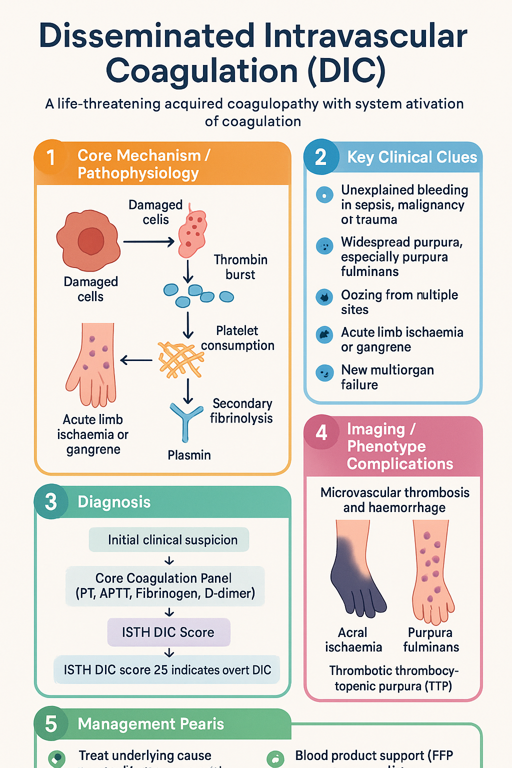
  • Disseminated intravascular coagulation (DIC) is a life-threatening acquired coagulopathy characterised by systemic activation of coagulation leading to simultaneous microvascular thrombosis and consumptive haemorrhage.
  • The most common trigger in Australia is severe sepsis (bacterial, viral, or fungal), followed by major trauma, obstetric emergencies, and advanced malignancy (especially acute promyelocytic leukaemia).
  • The ISTH DIC score (platelets, fibrinogen, D-dimer, PT prolongation) is the recommended bedside scoring system — a score ≥5 is compatible with overt DIC and carries high mortality.
  • Laboratory hallmarks: prolonged PT/APTT, falling fibrinogen, elevated D-dimer, schistocytes on blood film, and progressive thrombocytopaenia.
  • Treat the underlying cause aggressively — source control in sepsis, delivery in placental abruption, chemotherapy in APL — as no blood product alone will resolve DIC.
  • Transfuse FFP (15 mL/kg) for active bleeding with prolonged PT/APTT; cryoprecipitate (1 unit per 5–10 kg) to maintain fibrinogen ≥1.0 g/L (≥1.5 g/L if bleeding or pre-procedure).
  • Transfuse platelets when count <50 × 10⁹/L with active bleeding, or <10 × 10⁹/L prophylactically; aim <100 × 10⁹/L before invasive procedures.
  • Anticoagulation (heparin) may be considered in thrombotic-predominant DIC (e.g., purpura fulminans, APL) but is contraindicated in active haemorrhage.
  • In septic DIC, early broad-spectrum antibiotics within 1 hour, haemodynamic resuscitation, and ICU admission are essential components of care.
  • Obstetric DIC (abruption, amniotic fluid embolism, HELLP) requires coordinated obstetric–haematology–ICU management and early consideration of massive transfusion protocol activation.
  • Aboriginal and Torres Strait Islander peoples have higher rates of sepsis-related DIC; ensure culturally safe care and timely transfer from remote settings.
  • Serial laboratory monitoring (every 4–6 hours in acute DIC) guides blood product therapy; avoid prophylactic transfusion in non-bleeding patients with compensated DIC.

Introduction & Australian Epidemiology

Disseminated intravascular coagulation (DIC) is a serious acquired coagulopathy characterised by the systemic activation of coagulation pathways within the microvasculature. This leads to the paradoxical combination of widespread fibrin deposition (thrombosis) and consumption of clotting factors and platelets (haemorrhage). DIC is never a primary diagnosis — it is always secondary to an underlying condition that triggers the coagulation cascade.

In Australia, DIC is most frequently encountered in the context of severe sepsis and septic shock, accounting for approximately 30–50% of all DIC presentations to tertiary intensive care units. Other significant triggers include major polytrauma (particularly relevant given Australia's high burden of road traffic and agricultural injuries), obstetric emergencies, and haematological malignancies — notably acute promyelocytic leukaemia (APL, FAB M3).

The incidence of DIC in Australian ICUs is estimated at 8–12 per 1,000 ICU admissions, with in-hospital mortality ranging from 40–80% depending on the underlying aetiology and severity of organ dysfunction. Sepsis-related DIC carries a mortality of approximately 45–65%, while DIC associated with solid tumours or trauma has somewhat lower but still significant mortality (25–50%). The Australian Institute of Health and Welfare (AIHW) reports that sepsis-related hospitalisations have been rising at approximately 3–5% per year, which has implications for the frequency of DIC in clinical practice.

Early recognition, aggressive treatment of the triggering condition, and judicious blood product support remain the cornerstones of DIC management in Australian hospitals. This guideline provides a framework for the identification and management of DIC in primary haematology, emergency medicine, and intensive care settings across metropolitan, regional, and remote Australian healthcare facilities.

Disseminated Intravascular Coagulation (DIC) clinical infographic — pathophysiology, clinical clues, diagnosis, imaging, and management
Tap or click image to enlarge — Disseminated Intravascular Coagulation (DIC): pathophysiology, clinical clues, diagnosis, imaging, and management.
Disseminated Intravascular Coagulation (DIC) infographic, full size

Pathogenesis & Triggers

DIC results from the unregulated systemic activation of the coagulation cascade, overwhelming the natural anticoagulant systems (antithrombin, protein C, tissue factor pathway inhibitor) and fibrinolytic capacity. The pathophysiology can be conceptualised in four overlapping phases.

Pathophysiological Mechanism

  • Tissue factor exposure: Damaged or activated cells (monocytes, endothelium, tumour cells) express tissue factor (TF), which activates the extrinsic coagulation pathway via factor VII.
  • Thrombin burst: Massive thrombin generation leads to widespread fibrin formation in the microvasculature, consuming fibrinogen, factor V, factor VIII, and other procoagulant factors.
  • Platelet consumption: Platelets are activated and consumed in microthrombi, leading to progressive thrombocytopaenia.
  • Secondary fibrinolysis: Plasminogen activation generates plasmin, cleaving fibrin and producing fibrin degradation products (FDPs) and D-dimer. In advanced DIC, excess plasmin can paradoxically worsen haemorrhage.

Major Triggers in Australian Practice

Trigger Category Common Causes Mechanism Approximate Frequency
Sepsis / Infection Gram-negative bacteraemia, meningococcaemia, Staphylococcus aureus, Clostridium perfringens, malaria, COVID-19 Endotoxin/cytokine-mediated TF expression on monocytes and endothelium 30–50%
Malignancy Acute promyelocytic leukaemia (APL), adenocarcinoma (pancreas, lung, prostate), mucin-secreting tumours Cancer cell TF expression, chemotherapy-induced tumour lysis 15–25%
Trauma Major polytrauma, crush injuries, burns, traumatic brain injury, snake envenomation Tissue damage, fat embolism, endothelial injury, shock-induced hypoperfusion 10–20%
Obstetric Placental abruption, amniotic fluid embolism, HELLP syndrome, retained dead fetus, eclampsia Amniotic fluid/thromboplastin entry into maternal circulation 5–10%
Vascular Giant haemangiomas (Kasabach–Merritt), aortic aneurysm, large vessel aneurysms Stasis, turbulent flow, endothelial activation 2–5%
Other Heat stroke, severe transfusion reaction, pancreatitis, liver failure, envenomation (brown snake, taipan) Variable — toxin-mediated, immune-mediated 5–10%
⚠️
Australian envenomation note: Brown snake (Pseudonaja spp.) and taipan (Oxyuranus spp.) envenomation can cause rapid-onset consumptive coagulopathy mimicking DIC. Australian snake-specific antivenom and serial coagulation monitoring are essential. Contact the National Poisons Information Centre (13 11 26) for expert advice.

Clinical Features — Bleeding & Thrombosis

DIC presents a clinical paradox: patients may bleed, thrombose, or — most commonly — exhibit features of both simultaneously. The dominant phenotype depends on the trigger and the balance between procoagulant activation and fibrinolysis.

Predominantly Haemorrhagic
Bleeding Phenotype
  • Oozing from venepuncture sites, cannulae, wounds
  • Petechiae and purpura
  • Gingival bleeding, epistaxis
  • GI haemorrhage, haematuria
  • Menorrhagia (in premenopausal women)
Common in: sepsis, APL, obstetric DIC
Predominantly Thrombotic
Thrombosis Phenotype
  • Acral (digital) ischaemia and gangrene
  • Purpura fulminans (skin necrosis)
  • Deep vein thrombosis, pulmonary embolism
  • Renal cortical necrosis (oliguria/anuria)
  • Hepatic infarction, adrenal haemorrhagic necrosis
Common in: meningococcaemia, purpura fulminans, malignancy
Mixed / Organ Dysfunction
Multiorgan Failure
  • Acute kidney injury (microvascular thrombosis)
  • ARDS / respiratory failure
  • Altered consciousness, seizures
  • Shock liver (hepatic dysfunction)
  • Adrenal haemorrhagic infarction (Waterhouse–Friderichsen syndrome)
Common in: severe sepsis, massive trauma, amniotic fluid embolism

Clinical Clues to DIC

  • Unexplained bleeding in a patient with known sepsis, malignancy, or major trauma.
  • Widespread purpura, especially purpura fulminans with skin necrosis.
  • Oozing from multiple sites simultaneously (cannulae, wounds, mucosal surfaces).
  • Acute limb ischaemia or gangrene in a patient with systemic illness.
  • New multiorgan failure unresponsive to standard resuscitation.
  • Schistocytes on peripheral blood film (microangiopathic haemolytic anaemia).
🚨
Purpura fulminans is a medical emergency presenting with rapidly progressive haemorrhagic skin necrosis, typically in the setting of sepsis (especially meningococcal) or inherited protein C/S deficiency. Requires immediate ICU admission, anticoagulation consideration, and replacement of protein C if available.

Investigations

Laboratory investigation is the cornerstone of DIC diagnosis. Serial testing is essential, as DIC is a dynamic process and a single set of results may not capture the full picture.

Core Coagulation Panel

Essential
Prothrombin Time (PT) / INR
Prolonged in DIC due to consumption of factors II, V, VII, X. INR >1.5 is significant. Available in all Australian pathology laboratories. MBS Item 65070.
Essential
Activated Partial Thromboplastin Time (APTT)
Prolonged due to consumption of intrinsic pathway factors. May be paradoxically shortened early in DIC due to excess thrombin. MBS Item 65070.
Essential
Fibrinogen (Clauss method)
Low fibrinogen (<1.5 g/L) is a hallmark of DIC. It is an acute-phase reactant — a falling trend is more informative than an absolute value. Fibrinogen <1.0 g/L indicates severe consumption. MBS Item 65090.
Essential
D-dimer
Markedly elevated (>4× upper limit of normal) due to fibrinolysis of cross-linked fibrin. Very sensitive but not specific. Useful for trend monitoring. MBS Item 65104.
Essential
Full Blood Count (FBC) with Film
Thrombocytopaenia (progressive fall); schistocytes on blood film indicate microangiopathic haemolytic anaemia (MAHA). MBS Item 65060.

Additional Investigations

Available
Thrombin Time
Prolonged if fibrinogen is low or FDPs are elevated. Available in major laboratories.
Available
Fibrin Degradation Products (FDPs)
Elevated in DIC. Used in some scoring systems alongside D-dimer.
Available
Antithrombin Level
Consumed in DIC; low levels (<70%) correlate with worse prognosis. Available in tertiary centres.
Specialist
Soluble Fibrin Monomers / Thrombin-Antithrombin Complex
Sensitive markers of thrombin generation. Available in specialist coagulation laboratories; not routinely required.
Specialist
Thromboelastography (TEG/ROTEM)
Point-of-care viscoelastic testing. Increasingly used in Australian trauma and cardiac surgery ICUs for real-time coagulation assessment. Not yet standard in DIC.

ISTH DIC Scoring System

The International Society on Thrombosis and Haemostasis (ISTH) scoring system is the most widely validated tool for diagnosing overt DIC. It is simple, reproducible, and recommended by the Australian and New Zealand Society of Blood Transfusion (ANZSBT).

Parameter 0 Points 1 Point 2 Points 3 Points
Platelet count (× 10⁹/L) ≥100 <100 <50
D-dimer (fibrin-related marker) No increase Moderate increase Strong increase
Prolonged PT (seconds) <3 s 3–6 s >6 s
Fibrinogen (g/L) ≥1.0 <1.0
Interpretation: Score <5 = not consistent with overt DIC (repeat in 6–8 hours if clinical suspicion remains). Score ≥5 = compatible with overt DIC — initiate management and monitor serially.

Supportive Tests

  • Blood cultures (at least 2 sets, including peripheral and any central lines) — essential in sepsis-triggered DIC.
  • Lactate, blood gas, renal function, liver function — for organ dysfunction assessment.
  • Peripheral blood film — look for schistocytes, which confirm microangiopathic process.
  • Urinalysis — haematuria and proteinuria may indicate renal microvascular thrombosis.
  • Imaging (CT, ultrasound) — as indicated for source control (abscess, effusion, thrombosis).

Risk Stratification & Severity Scoring

Beyond the ISTH score, several clinical and laboratory factors help predict outcome and guide the intensity of monitoring and intervention.

Risk Factor Poor Prognostic Indicator Implication
ISTH DIC score ≥7 (severe DIC) Mortality >60%; requires ICU admission
SOFA score ≥10 Severe multiorgan dysfunction; consider goals-of-care discussion
Platelet trend Falling >50% in 24 h or <20 × 10⁹/L Active consumption; high bleeding risk
Fibrinogen <0.5 g/L Severe consumption; cryoprecipitate urgently required
Lactate >4 mmol/L Severe tissue hypoperfusion; aggressive resuscitation needed
Underlying cause APL, septic shock, amniotic fluid embolism Highest-mortality triggers; early specialist consultation essential

When to Escalate

  • ICU referral: All patients with overt DIC (ISTH ≥5) and organ dysfunction; any patient with DIC and haemodynamic instability.
  • Haematology referral: APL or other haematological malignancy; unexplained DIC; DIC unresponsive to initial management; need for anticoagulation in DIC.
  • Massive transfusion protocol (MTP): Activate when anticipated need exceeds 5 units of packed red blood cells within 4 hours, or any patient with DIC and life-threatening haemorrhage.

Management

DIC management has two pillars: (1) treat the underlying cause, and (2) supportive blood product replacement guided by laboratory results and clinical bleeding. No single therapy resolves DIC without addressing the trigger.

Step 1 — Treat the Underlying Cause

1
Sepsis
Empirical IV antibiotics within 1 hour. Source control (drain abscess, remove infected device). Haemodynamic resuscitation with IV fluids and vasopressors. Refer to sepsis guideline.
2
Trauma
Damage control surgery. Massive transfusion protocol (1:1:1 PRBC:FFP:platelets). Tranexamic acid 1 g IV over 10 min within 3 hours of injury (CRASH-2 protocol). Avoid hypothermia and acidosis ("lethal triad").
3
Malignancy
APL: All-trans retinoic acid (ATRA) + arsenic trioxide is standard induction. Initiate ATRA at first suspicion (do not wait for molecular confirmation). Manage differentiation syndrome with dexamethasone.
4
Obstetric
Delivery of fetus and placenta is definitive treatment. Coordinate with obstetric team. Activate MTP early in abruption or amniotic fluid embolism.

Step 2 — Blood Product Support

Transfusion in DIC should be goal-directed, guided by laboratory parameters and clinical bleeding. Prophylactic transfusion in non-bleeding patients does not improve outcomes and may worsen thrombosis.

🩸
Fresh Frozen Plasma (FFP)
Human plasma derivative · Procoagulant factors
Indication Active bleeding with prolonged PT/APTT (>1.5× control); pre-procedure; massive transfusion
Dose 12–15 mL/kg (typically 4 units for 70 kg adult). Repeat based on coagulation results.
Paediatric dose 10–15 mL/kg IV
Caution Volume overload risk — consider 4-factor prothrombin complex concentrate (PCC) if contraindicated
PBS status ✔ PBS General Benefit
🩸
Cryoprecipitate
Human plasma derivative · Fibrinogen, factor VIII, vWF
Indication Fibrinogen <1.0 g/L (or <1.5 g/L if actively bleeding or pre-procedure)
Dose 1 pooled unit (from 5 donors, ~300 mg fibrinogen) per 5–10 kg body weight. Typically 5–10 pooled units for an adult.
Paediatric dose 1 unit per 5–10 kg, or 10–20 mL/kg
Target Fibrinogen ≥1.0 g/L (≥1.5 g/L if bleeding)
PBS status ✔ PBS General Benefit
🩸
Platelet Concentrate
Apheresis or pooled · Haemostatic
Indication Count <50 × 10⁹/L with active bleeding; <10 × 10⁹/L prophylactically; <100 × 10⁹/L pre-procedure
Dose 1 adult therapeutic dose (1 apheresis unit or 5–6 pooled buffy coat units). Expect rise of ~30 × 10⁹/L.
Paediatric dose 10–15 mL/kg (1 unit per 10 kg)
Caution In DIC, platelet survival is shortened; repeated transfusions may be required. Avoid in APL until ATRA initiated (risk of thrombosis).
PBS status ✔ PBS General Benefit
💊
Tranexamic Acid
Cyklokapron® · Generic · Antifibrinolytic
Adult dose 1 g IV over 10 minutes, then 1 g over 8 hours (trauma: within 3 hours of injury). In DIC, only use if evidence of excessive fibrinolysis.
Paediatric dose 15 mg/kg IV (max 1 g) bolus, then 2 mg/kg/h infusion
Caution Do not use empirically in DIC without evidence of hyperfibrinolysis (may worsen thrombosis). Avoid in renal cortical necrosis, DIC with thrombosis, or APL.
Renal adjustment eGFR <30: reduce dose or avoid. Dose not well established; use with caution.
PBS status ✔ PBS General Benefit
💊
Unfractionated Heparin (UFH)
Heparin sodium · Anticoagulant
Adult dose 50–100 units/kg IV bolus, then 10–20 units/kg/h infusion. Titrate to APTT 1.5–2.5× baseline.
Indication Thrombotic-predominant DIC (purpura fulminans, venous thrombosis); APL induction; chronic compensated DIC
Contraindications Active haemorrhage; platelets <50 × 10⁹/L (unless thrombotic indication is compelling); recent CNS surgery
Caution HIT risk — monitor platelets. APTT unreliable in DIC; may require anti-Xa monitoring.
PBS status ✔ PBS General Benefit

Step 3 — Adjunctive & Emerging Therapies

ℹ️
Recombinant Thrombomodulin (ART-123): Currently under investigation (SCARLET-3 trial). Not yet approved or available in Australia. Antithrombin concentrate: may be considered when levels <60% in septic DIC, but evidence of clinical benefit is inconsistent. Available in some Australian centres via BloodSTAR. Protein C concentrate: used in purpura fulminans with severe protein C deficiency (congenital or acquired). Limited availability — contact haematology.
⚠️
Massive Transfusion Protocol (MTP): In patients with DIC and life-threatening haemorrhage, activate the hospital MTP immediately. Standard MTP ratios (1:1:1 PRBC:FFP:platelets) may not correct DIC-specific deficits — ensure cryoprecipitate is included early to correct hypofibrinogenaemia. Most Australian tertiary centres have a defined MTP pathway — consult local protocols.

Transfusion Triggers — Quick Reference

Component
Trigger
Target
Notes
FFP
PT/APTT >1.5× control + bleeding
PT/APTT <1.5× control
15 mL/kg; caution in TACO risk
Cryoprecipitate
Fibrinogen <1.0 g/L
≥1.0 g/L (≥1.5 if bleeding)
1 unit per 5–10 kg
Platelets
<50 + bleeding; <10 prophylactic
≥50 (≥100 pre-procedure)
Avoid in APL before ATRA
PRBCs
Hb <70 g/L (or <80 if ACS)
Hb ≥70 g/L
Restrictive strategy preferred

Monitoring

DIC is a dynamic process. Serial laboratory and clinical monitoring is essential to guide therapy and detect deterioration early.

Recommended Monitoring Schedule

Baseline
PT/INR, APTT, fibrinogen, D-dimer, FBC, blood film, blood cultures, lactate, biochemistry. ISTH DIC score.
Every 4–6 hours
Repeat coagulation panel (PT, APTT, fibrinogen, FBC) in active DIC. Recalculate ISTH score.
Every 8–12 hours
D-dimer, blood film review (schistocyte count), renal and liver function, lactate.
Daily
Comprehensive reassessment. Document bleeding assessment, transfusion volumes, fluid balance, organ function (SOFA score). Re-evaluate goals of care.
Resolution
DIC resolved when ISTH score <5 on two consecutive occasions, platelets stable or rising, fibrinogen normalised, D-dimer trending down. Continue monitoring for 48 h after resolution.
ℹ️
APTT monitoring with heparin: In DIC, the APTT is unreliable for heparin monitoring due to concurrent factor consumption. Anti-Xa activity assay is the preferred method when available. If anti-Xa is not accessible, use clinical assessment (bleeding/thrombosis) alongside APTT trends.

Special Populations

🤰 Pregnancy
Triggers: Placental abruption, HELLP, amniotic fluid embolism, eclampsia, septic abortion, retained dead fetus.
Key principle: Delivery of fetus and placenta is the definitive treatment for obstetric DIC. Coordinate with obstetric team early.
Blood products: All standard components are safe. Maintain fibrinogen ≥1.5 g/L (pregnancy has higher baseline). Activate MTP early.
Cryoprecipitate: Preferred over fibrinogen concentrate in pregnancy (Australian Blood Service availability).
Heparin: Unfractionated heparin is safe in pregnancy (does not cross placenta). Consider in thrombotic DIC. LMWH may also be used.
Tranexamic acid: Use with caution. Emerging evidence supports use postpartum haemorrhage (WOMAN trial), but data in DIC-specific obstetric settings limited. Avoid antepartum without haematology guidance.
👶 Paediatrics
Triggers: Meningococcaemia (most common in children), severe viral infections (influenza, dengue), haemolytic uraemic syndrome (HUS — distinct from DIC), trauma.
Scoring: ISTH DIC score can be applied in children but has lower sensitivity in neonates. Use clinical judgement.
FFP: 10–15 mL/kg IV.
Cryoprecipitate: 1 unit per 5–10 kg, or 10–20 mL/kg.
Platelets: 10–15 mL/kg (1 unit per 10 kg).
Neonates: DIC in neonates is challenging (physiological coagulation differences). Involve neonatal haematology early. Avoid heparin unless clearly thrombotic DIC. Consider congenital thrombophilias if DIC unresponsive.
👴 Elderly
Higher risk: Sepsis-related DIC more common; worse outcomes due to reduced physiological reserve and comorbidities.
Volume overload: FFP and cryoprecipitate volumes may cause transfusion-associated circulatory overload (TACO). Consider diuretics prophylactically.
Goals of care: Early discussion with patient/family regarding escalation limits, especially with high-mortality triggers.
🫘 Renal Impairment
AKI is common in DIC: Microvascular thrombosis in renal cortex (renal cortical necrosis) is a severe complication — persistent anuria with normal renal ultrasound may be the clue.
Tranexamic acid: Reduce dose or avoid if eGFR <30 mL/min (accumulation risk).
CRRT: Continuous renal replacement therapy is preferred over intermittent haemodialysis in haemodynamically unstable patients. Citrate anticoagulation may be used but requires careful monitoring in DIC.
🫁 Hepatic Impairment
Diagnostic difficulty: Chronic liver disease causes baseline coagulopathy (low fibrinogen, prolonged PT) which can mimic DIC. Serial trends (falling platelets, rising D-dimer) are key.
Heparin: Reduced hepatic clearance — monitor more frequently. Use anti-Xa if available.
🛡️ Immunocompromised
Higher risk of fungal/viral DIC: Consider invasive fungal sepsis, CMV, or EBV in immunocompromised patients with DIC.
APL: Patients on chemotherapy with DIC should be managed in conjunction with haematology. ATRA must be started immediately if APL is suspected.
Bone marrow transplant: DIC may complicate veno-occlusive disease (VOD/SOS) and graft-versus-host disease (GVHD). Specialised haematology management is essential.

Aboriginal and Torres Strait Islander Health Considerations

Aboriginal and Torres Strait Islander Health
Higher sepsis burden
Aboriginal and Torres Strait Islander peoples experience sepsis at 2–3 times the rate of non-Indigenous Australians (AIHW, 2023). Sepsis is the most common cause of DIC, making this a significant health disparity.
Remote access
Many Indigenous Australians live in remote communities where rapid access to ICU, haematology, and blood product services is limited. Aeromedical retrieval (RFDS, CareFlight) is critical — early activation of retrieval services improves outcomes.
Blood product availability
Fresh frozen plasma and cryoprecipitate may not be available at remote health centres. The Australian Red Cross Lifeblood service maintains regional blood banks, but transport delays can be significant. Remote clinicians should discuss with haematology early and consider empirical antibiotic and fluid resuscitation while arranging transfer.
CA-MRSA & tropical infections
Community-associated methicillin-resistant Staphylococcus aureus (CA-MRSA) and tropical infections (melioidosis in northern Australia, dengue, Ross River virus) are more prevalent in Indigenous communities. Empirical antibiotics should cover local resistance patterns (e.g., IV vancomycin or flucloxacillin as per local antibiograms).
Cultural safety
Provide culturally safe care: use Aboriginal Health Workers and interpreters, involve family in decision-making, and respect sorry business and cultural protocols. The National Safety and Quality Health Service (NSQHS) Standards require culturally appropriate care for Aboriginal and Torres Strait Islander peoples.
Discharge planning
Post-DIC survivors may require ongoing monitoring, follow-up coagulation testing, and rehabilitation. Ensure clear communication with remote health services and arrange follow-up through Aboriginal Community Controlled Health Organisations (ACCHOs) where possible.

📚 References

  1. 1. Levi M, Toh CH, Thachil J, Watson HG. Guidelines for the diagnosis and management of disseminated intravascular coagulation. British Journal of Haematology. 2009;145(1):24–33.
  2. 2. Taylor FB Jr, Toh CH, Hoots WK, Wada H, Levi M. Towards definition, clinical and laboratory criteria, and a scoring system for disseminated intravascular coagulation. Thrombosis and Haemostasis. 2001;86(5):1327–1330.
  3. 3. Wada H, Thachil J, Di Nisio M, et al. Guidance for diagnosis and treatment of DIC from harmonization of the recommendations from three guidelines. Journal of Thrombosis and Haemostasis. 2013;11(4):761–767.
  4. 4. Australian Institute of Health and Welfare (AIHW). Sepsis in Australia. Cat. no. PHE 312. Canberra: AIHW; 2023.
  5. 5. National Blood Authority (NBA). Patient Blood Management Guidelines: Module 1 — Critical Bleeding / Massive Transfusion. Canberra: NBA; 2011.
  6. 6. National Blood Authority (NBA). Patient Blood Management Guidelines: Module 2 — Perioperative. Canberra: NBA; 2012.
  7. 7. CRASH-2 Collaborators. Effects of tranexamic acid on death, vascular occlusive events, and blood transfusion in trauma patients with significant haemorrhage (CRASH-2): a randomised, placebo-controlled trial. The Lancet. 2010;376(9734):23–32.
  8. 8. WOMAN Trial Collaborators. Effect of early tranexamic acid administration on mortality, hysterectomy, and other morbidities in women with post-partum haemorrhage (WOMAN): an international, randomised, double-blind, placebo-controlled trial. The Lancet. 2017;389(10084):2105–2116.
  9. 9. Squizzato A, Hunt BJ, Kinasewitz GT, et al. Supportive management strategies for disseminated intravascular coagulation. Thrombosis and Haemostasis. 2016;115(5):896–904.
  10. 10. Australian Commission on Safety and Quality in Health Care (ACSQHC). National Safety and Quality Health Service Standards. 2nd ed. Sydney: ACSQHC; 2021.
  11. 11. Thachil J, Wada H, Gando S. DIC in the new millennium. Current Opinion in Hematology. 2022;29(5):230–237.
  12. 12. ISBT (International Society of Blood Transfusion) / ANZSBT. Guidelines for the administration of blood products. 3rd ed. 2022.
  13. 13. Brown AF, Lane DA. Anticoagulation in disseminated intravascular coagulation: current evidence and future directions. Internal Medicine Journal. 2020;50(4):401–408.
  14. 14. RHDAustralia (Australian Government Department of Health). Recommendations for managing acute rheumatic fever and rheumatic heart disease in Australia. 3rd ed. Darwin: Menzies School of Health Research; 2020.