📋 Key Information Summary
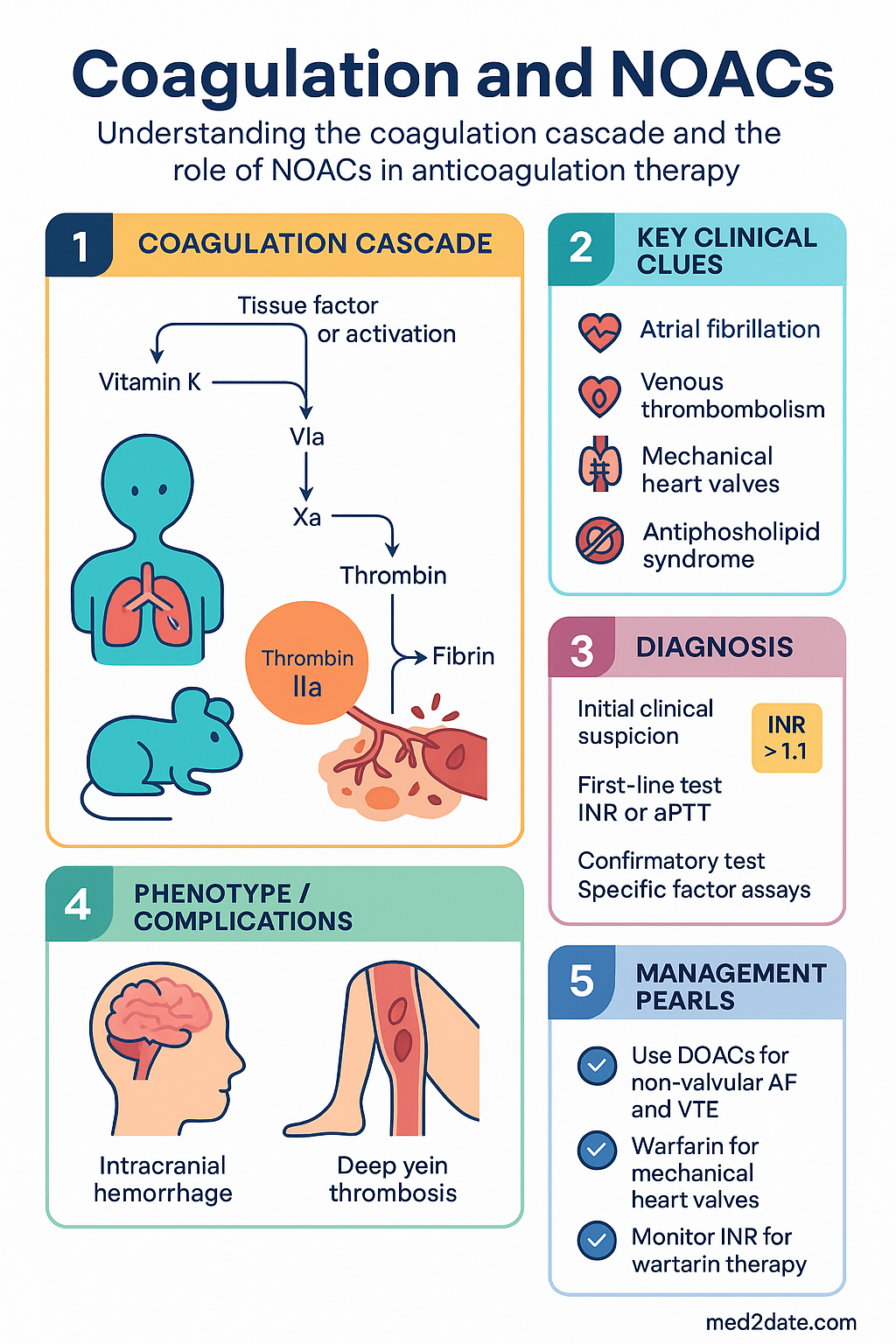
- The coagulation cascade comprises intrinsic (contact-activated), extrinsic (tissue factor–triggered), and common pathways; modern anticoagulants target specific factors (IIa, Xa) rather than the entire cascade.
- Warfarin inhibits vitamin K–dependent factors (II, VII, IX, X) and requires INR monitoring with a target range of 2.0–3.0 for most indications and 2.5–3.5 for mechanical heart valves.
- Warfarin has a narrow therapeutic index, extensive drug–food interactions, and a slow onset/offset; it remains the preferred agent for mechanical heart valves and antiphospholipid syndrome in Australia.
- Warfarin over-anticoagulation is managed with vitamin K₁ (phytomenadione) 1–2.5 mg PO for INR 4.5–8.0 without bleeding; IV prothrombin complex concentrate (PCC) for life-threatening haemorrhage.
- UFH is the anticoagulant of choice for cardiopulmonary bypass, ECMO, and haemodialysis; it requires aPTT or anti-Xa monitoring and is immediately reversible with protamine sulfate.
- LMWH (enoxaparin, dalteparin) provides predictable pharmacokinetics, is weight-based dosed, and requires no routine monitoring in most patients — anti-Xa levels guide dosing in obesity, renal impairment, and pregnancy.
- DOACs (apixaban, rivaroxaban, edoxaban, dabigatran) are first-line for non-valvular atrial fibrillation and VTE treatment/prophylaxis in Australia, supported by PBS listing and Phase III trial data.
- Dabigatran (direct thrombin inhibitor) is the only DOAC with a specific reversal agent available in Australia — idarucizumab (Praxbind®), PBS Authority Required.
- Andexanet alfa (Ondexxya®) is PBS-listed for apixaban and rivaroxaban reversal in life-threatening haemorrhage; PCC 4-factor (Octaplex®) is an alternative non-specific reversal strategy.
- DOACs are contraindicated in mechanical heart valves and antiphospholipid syndrome; dose reduction criteria vary by agent and must be applied per TGA-approved indications to avoid under-dosing.
- Renal function (eGFR/creatinine clearance) is critical for DOAC dosing: dabigatran is contraindicated if CrCl <30 mL/min; dose adjustments apply to all DOACs with declining renal function.
- Aboriginal and Torres Strait Islander peoples experience higher rates of AF and VTE with lower anticoagulant utilisation; culturally safe prescribing, medication access, and monitoring support are essential.
Introduction & Australian Epidemiology
Anticoagulant therapy is fundamental to the management of venous thromboembolism (VTE), atrial fibrillation (AF), mechanical heart valves, and various hypercoagulable states. The main classes — vitamin K antagonists (warfarin), heparins (UFH and LMWH), and direct oral anticoagulants (DOACs) — differ markedly in their mechanisms of action, monitoring requirements, drug interactions, and reversal strategies. Understanding these differences is essential for safe prescribing in Australian clinical practice.
In Australia, VTE affects approximately 14,000–20,000 people annually, with an age-adjusted incidence of 83 per 100,000 population. Atrial fibrillation prevalence increases with age, affecting 4–5% of Australians over 60 years and approximately 10% of those aged ≥80 years. The 2023 AIHW National Hospital Morbidity Database reports over 60,000 hospitalisations with a primary or secondary diagnosis of AF and more than 30,000 with VTE per year.
The shift from warfarin to DOACs has been transformative. Australian PBS dispensing data (2023) demonstrate that apixaban and rivaroxaban now account for approximately 75% of anticoagulant prescriptions for non-valvular AF. However, warfarin retains important niches — mechanical prosthetic heart valves, antiphospholipid syndrome, and severe renal impairment — and UFH/LMWH remain critical for acute inpatient management, bridging therapy, and pregnancy.
Coagulation Cascade Overview
The coagulation cascade is a series of enzymatic reactions involving serine proteases (clotting factors) that ultimately convert fibrinogen to a stable fibrin clot. The cascade is traditionally divided into three pathways, though in vivo haemostasis is best understood through the cell-based model of coagulation.
Classical Three-Pathway Model
| Pathway | Trigger | Key Factors | Laboratory Test |
|---|---|---|---|
| Intrinsic (contact) | Contact with negatively charged surface (collagen, glass in vitro) | XII → XI → IX → VIII | aPTT (activated partial thromboplastin time) |
| Extrinsic (tissue factor) | Tissue factor (TF) exposure from damaged vessel wall | TF + VII | PT / INR |
| Common | Convergence of intrinsic and extrinsic pathways | X → V → II (prothrombin) → I (fibrinogen) → XIII | Both PT and aPTT |
Cell-Based Model (Current Understanding)
The cell-based model describes three overlapping phases occurring on cell surfaces:
- Initiation: Tissue factor exposed on subendothelial cells binds factor VIIa, generating small amounts of thrombin (IIa) and activating factors V, VIII, IX, and X.
- Amplification: Trace thrombin activates platelets and cofactors (V, VIII, XI) on the platelet surface, setting the stage for large-scale thrombin generation.
- Propagation: The tenase complex (IXa/VIIIa) and prothrombinase complex (Xa/Va) on activated platelet surfaces generate a "thrombin burst," cleaving fibrinogen to fibrin and activating factor XIII for clot stabilisation.
Natural Anticoagulant Mechanisms
| Mechanism | Target | Clinical Relevance |
|---|---|---|
| Antithrombin (AT) | Inhibits thrombin (IIa) and factor Xa | Heparin cofactor; AT deficiency → thrombophilia |
| Protein C / Protein S | Inactivate factors Va and VIIIa | Protein C/S deficiency → warfarin-induced skin necrosis risk |
| Tissue factor pathway inhibitor (TFPI) | Inhibits TF–VIIa–Xa complex | Limiting initiation phase |
Fibrinolysis
The fibrinolytic system dissolves clots through plasmin, generated from plasminogen by tissue plasminogen activator (tPA). Alpha-2-antiplasmin and plasminogen activator inhibitor-1 (PAI-1) regulate fibrinolysis. Pharmacological fibrinolysis (e.g., alteplase) is used in acute ST-elevation myocardial infarction, massive pulmonary embolism, and acute ischaemic stroke.
Warfarin: Mechanism, Monitoring & Reversal
Mechanism of Action
Warfarin inhibits vitamin K epoxide reductase (VKORC1), preventing recycling of vitamin K to its reduced (hydroquinone) form. This blocks the gamma-carboxylation of vitamin K–dependent clotting factors (II, VII, IX, X) and anticoagulant proteins (C and S), resulting in non-functional, partially carboxylated factors.
Warfarin has near-complete oral bioavailability, is highly protein-bound (>95% to albumin), and is metabolised primarily by CYP2C9 (S-warfarin, the more potent enantiomer) and CYP3A4 (R-warfarin). The CYP2C9*2 and *3 polymorphisms reduce metabolism and increase bleeding risk. The VKORC1 -1639G→A polymorphism increases warfarin sensitivity. Pharmacogenomic dosing algorithms (e.g., IWPC algorithm) exist but are not yet standard of care in Australia.
Pharmacokinetics
- Onset: Anticoagulant effect begins in 24–72 hours (dependent on factor VII t½ ~6 hours); full effect at 5–7 days (factor II t½ ~60 hours).
- Duration: Effect lasts 3–5 days after cessation.
- Absorption: Complete; peak plasma at 1–4 hours.
- Protein binding: >95% albumin.
- Metabolism: Hepatic CYP2C9 (major), CYP3A4, CYP1A2.
- Half-life: 20–60 hours (mean ~40 hours).
Australian Indications
| Indication | Target INR | Duration |
|---|---|---|
| Atrial fibrillation (valvular or mechanical valve) | 2.0–3.0 (non-valvular AF: DOACs preferred) | Long-term |
| Mechanical prosthetic heart valve | 2.5–3.5 (aortic: 2.0–3.0 if bileaflet + no risk factors) | Lifelong |
| Antiphospholipid syndrome (APS) | 2.0–3.0 (triple-positive: 3.0–4.0) | Lifelong |
| VTE treatment & secondary prophylaxis | 2.0–3.0 | 3–12 months or indefinite |
| Bioprosthetic valve (first 3 months post-op) | 2.0–3.0 | 3 months |
Dosing & Initiation
Standard initiation in Australia: 5 mg daily for most adults. Lower starting doses (2–3 mg) recommended for elderly patients (>70 years), hepatic impairment, malnutrition, heart failure, concomitant interacting medications, or CYP2C9 variant carriers.
Monitoring
- INR target: 2.0–3.0 for most indications (see table above).
- Initiation: Check INR at day 3, day 5, then daily until stable.
- Stable: INR every 4 weeks (may extend to 12 weeks in highly stable patients per RACGP guidance).
- Dose changes: Adjust by 5–20%; recheck INR in 1–2 weeks.
- Point-of-care (POC) INR: CoaguChek® devices validated for community use; ensure quality assurance programme compliance.
Drug Interactions (Key Examples)
| Interaction Type | Drugs | Mechanism |
|---|---|---|
| Potentiate (↑ INR) | Amiodarone, fluconazole, metronidazole, erythromycin, ciprofloxacin, NSAIDs, SSRIs | CYP2C9 inhibition, protein displacement, antiplatelet effect |
| Reduce effect (↓ INR) | Rifampicin, carbamazepine, phenytoin, St John's wort | CYP enzyme induction |
| Vitamin K–rich foods | Green leafy vegetables, broccoli, Brussels sprouts | Antagonise warfarin; maintain consistent intake |
Reversal Strategies
Heparin (UFH & LMWH): Indications & Reversal
Mechanism of Action
Heparin is a glycosaminoglycan that potentiates antithrombin (AT) by approximately 1,000-fold. The heparin–AT complex inactivates thrombin (factor IIa) and factor Xa. UFH requires a minimum pentasaccharide chain of 18 saccharides to catalyse both anti-IIa and anti-Xa activity, while LMWH fragments (mean MW 4,500 Da) primarily catalyse anti-Xa activity with minimal anti-IIa effect.
UFH vs LMWH — Comparative Features
| Feature | Unfractionated Heparin (UFH) | Low Molecular Weight Heparin (LMWH) |
|---|---|---|
| Molecular weight | 3,000–30,000 Da (mean ~15,000) | 2,000–9,000 Da (mean ~4,500) |
| Anti-Xa:Anti-IIa ratio | 1:1 | 2:1 to 4:1 |
| Bioavailability | 30% SC (unpredictable) | 90% SC (predictable) |
| Half-life | 1–2 hours (IV) | 3–7 hours (SC) |
| Monitoring | Required — aPTT (or anti-Xa) | Not routine — anti-Xa if indicated (obesity, renal, pregnancy) |
| Reversal agent | Protamine sulfate (full neutralisation) | Protamine sulfate (~60% neutralisation of anti-Xa) |
| HIT risk | Higher (~1–5%) | Lower (~0.1–1%) |
| Renal clearance | Reticuloendothelial | Renal — dose adjust if CrCl <30 mL/min |
| Route | IV infusion or SC | SC injection |
Indications
| UFH Preferred | LMWH Preferred |
|---|---|
| Cardiopulmonary bypass | VTE prophylaxis (medical/surgical) |
| ECMO | VTE treatment (outpatient eligible) |
| Haemodialysis | Cancer-associated thrombosis |
| Acute massive PE with haemodynamic instability | Pregnancy (enoxaparin preferred) |
| Severe renal impairment (CrCl <15 mL/min) | Bridging therapy for warfarin |
| When rapid reversal anticipated | ACS (STEMI/NSTEMI — enoxaparin) |
LMWH Agents Available in Australia
UFH Dosing & Monitoring
UFH for treatment of VTE: initial bolus 80 IU/kg IV (max 5,000 IU), followed by infusion 18 IU/kg/hour (max 1,560 IU/hour). Adjust per institution nomogram targeting aPTT 1.5–2.5× control (typically 60–85 seconds, assay-dependent). Anti-Xa levels (target 0.3–0.7 IU/mL) are preferred in pregnancy and lupus anticoagulant–positive patients where aPTT is unreliable.
Reversal — Protamine Sulfate
Heparin-Induced Thrombocytopenia (HIT)
| 4T Score Criterion | 0 Points | 1 Point | 2 Points |
|---|---|---|---|
| Thrombocytopenia | <30% fall or <10 × 10⁹/L | 30–50% fall or 10–19 × 10⁹/L | >50% fall and ≥20 × 10⁹/L |
| Timing | <4 days without recent exposure | Day 5–10 or ≤1 day if exposure 30–100 days ago | Day 5–10 (clear) or ≤1 day (recent exposure) |
| Thrombosis | None | Progressive/recurrent thrombosis; skin necrosis | Confirmed thrombosis or necrosis |
| Other causes | Definite alternative | Possible alternative | None apparent |
Interpretation: Score 0–3: low probability (HIT unlikely); 4–5: intermediate (test for HIT antibodies — PF4/heparin ELISA and serotonin release assay); 6–8: high probability (cease heparin, start alternative anticoagulant immediately pending results).
Direct Oral Anticoagulants (DOACs) & Reversal Agents
Overview & Advantages
DOACs represent a paradigm shift in anticoagulation, offering fixed dosing, predictable pharmacokinetics, fewer drug interactions, and no routine laboratory monitoring. Four DOACs are PBS-listed in Australia: apixaban, rivaroxaban, edoxaban (factor Xa inhibitors), and dabigatran (direct thrombin inhibitor).
DOAC Pharmacology Comparison
| Feature | Apixaban | Rivaroxaban | Edoxaban | Dabigatran |
|---|---|---|---|---|
| Target | Factor Xa | Factor Xa | Factor Xa | Factor IIa (thrombin) |
| Prodrug | No | No | No | Yes (dabigatran etexilate) |
| Bioavailability | ~50% | ~80% (with food) | ~62% | ~6.5% |
| t½ (normal renal) | ~12 hours | ~5–9 hours (young); 11–13 hrs (elderly) | ~10–14 hours | ~12–17 hours |
| Renal clearance | ~27% | ~33% (active drug); 66% total | ~50% | ~80% |
| Food effect | Minimal | Take with food (↑ absorption 39%) | Minimal | Minimal |
| CYP metabolism | CYP3A4 (minor) | CYP3A4/2J2 (major) | Minimal CYP | None (ester hydrolysis) |
| P-gp substrate | Yes | Yes | Yes | Yes (major) |
| Dialysable | No | No | No | Yes (~60% in 2–3 hrs) |
Australian Indications (PBS-listed)
| Indication | Apixaban | Rivaroxaban | Edoxaban | Dabigatran |
|---|---|---|---|---|
| Non-valvular AF | ✔ | ✔ | ✔ | ✔ |
| VTE treatment | ✔ | ✔ | ✔ | ✔ |
| VTE prophylaxis (hip/knee) | ✔ (hip only) | ✔ (hip & knee) | ✘ | ✔ (hip & knee) |
| ACS (with antiplatelet) | ✘ | ✔ (2.5 mg BD + DAPT) | ✘ | ✘ |
| CAD/PAD | ✘ | ✔ (2.5 mg BD + ASA) | ✘ | ✘ |
Dosing — Atrial Fibrillation
Key Drug Interactions
| Interaction | Affected DOACs | Management |
|---|---|---|
| Strong CYP3A4 + P-gp inhibitors (ketoconazole, itraconazole, ritonavir, clarithromycin) | Apixaban, rivaroxaban | Avoid combination or reduce dose |
| Strong CYP3A4 + P-gp inducers (rifampicin, carbamazepine, phenytoin, St John's wort) | All DOACs | Avoid — significantly reduces DOAC levels |
| P-gp inhibitors (amiodarone, verapamil, dronedarone, ciclosporin, ticagrelor) | Dabigatran (major), edoxaban | Dose reduction (dabigatran 110 mg BD with verapamil); caution with others |
| Dual antiplatelet therapy | All | Increased bleeding risk; minimise triple therapy duration (see AF + ACS guidance) |
| NSAIDs | All | Avoid if possible; increased GI bleeding risk |
Laboratory Assessment of DOAC Effect
DOACs do not require routine monitoring. However, laboratory assessment may be necessary in specific clinical scenarios:
| DOAC | Preferred Assay | Routine Coagulation | Clinical Scenarios for Testing |
|---|---|---|---|
| Apixaban, Rivaroxaban, Edoxaban | Calibrated anti-Xa assay | PT: mildly prolonged (rivaroxaban most). aPTT: insensitive. | Acute bleeding, emergency surgery, extremes of weight, renal impairment, suspected non-adherence, overdose |
| Dabigatran | dTT (dilute thrombin time) or ECT | aPTT: prolonged (non-linear). TT: very prolonged/saturating (qualitative, not quantitative). | As above; also to confirm absence of drug prior to surgery |
DOAC Reversal Strategies
Specific Reversal Agents — Detail
Monitoring
Monitoring by Anticoagulant Class
| Agent | Routine Monitoring | Laboratory Test | Special Monitoring |
|---|---|---|---|
| Warfarin | Required — INR every 4 weeks | PT/INR (target 2.0–3.0) | LFTs at baseline; FBC for occult bleeding |
| UFH | Required — aPTT or anti-Xa | aPTT 1.5–2.5× control (or anti-Xa 0.3–0.7 IU/mL) | FBC (platelets) every 2–3 days for HIT surveillance |
| LMWH | Not routine | Anti-Xa (peak 4 hrs post-dose; target 0.5–1.0 IU/mL BD, 1.0–2.0 IU/mL OD) | Renal function, anti-Xa in obesity, pregnancy, CrCl <30 |
| DOACs | Not required | Calibrated anti-Xa (Xa inhibitors) or dTT/ECT (dabigatran) | Renal function every 6–12 months (more often if CrCl <60). FBC annually. LFTs at baseline. |
Renal Function Monitoring for DOACs
- CrCl ≥60 mL/min: Monitor annually.
- CrCl 30–59 mL/min: Monitor every 6 months.
- CrCl 15–29 mL/min: Monitor every 3 months (dabigatran contraindicated; Xa inhibitors — limited data, specialist oversight recommended).
- Use Cockcroft–Gault equation (not CKD-EPI) for DOAC dosing decisions, as trial data were based on Cockcroft–Gault–derived CrCl.
MBS Items Relevant to Anticoagulant Monitoring
Special Populations
Aboriginal and Torres Strait Islander Health Considerations
Aboriginal and Torres Strait Islander Australians experience a higher burden of conditions requiring anticoagulation, including atrial fibrillation, venous thromboembolism, rheumatic heart disease, and cardiovascular disease, often at younger ages and with greater comorbidity than non-Indigenous Australians. Despite this, anticoagulant utilisation rates remain lower, and outcomes are poorer.
📚 References
- 1. National Blood Authority Australia. Australian Patient Blood Management Guidelines — Module 2: Perioperative. Canberra: NBA; 2012 (updated 2022).
- 2. Lip GYH, Banerjee A, Boriani G, et al. Antithrombotic therapy for atrial fibrillation: CHEST guideline and expert panel report. Chest. 2018;154(5):1121–1201.
- 3. Granger CB, Alexander JH, McMurray JJV, et al. Apixaban versus warfarin in patients with atrial fibrillation (ARISTOTLE). N Engl J Med. 2011;365(11):981–992.
- 4. Patel MR, Mahaffey KW, Garg J, et al. Rivaroxaban versus warfarin in nonvalvular atrial fibrillation (ROCKET AF). N Engl J Med. 2011;365(10):883–891.
- 5. Connolly SJ, Ezekowitz MD, Yusuf S, et al. Dabigatran versus warfarin in patients with atrial fibrillation (RE-LY). N Engl J Med. 2009;361(12):1139–1151.
- 6. Pollack CV Jr, Reilly PA, van Ryn J, et al. Idarucizumab for dabigatran reversal — full cohort analysis (RE-VERSE AD). N Engl J Med. 2017;377(5):431–441.
- 7. Connolly SJ, Crowther M, Eikelboom JW, et al. Full study report of andexanet alfa for bleeding associated with factor Xa inhibitors (ANNEXA-4). N Engl J Med. 2019;380(14):1326–1335.
- 8. RACGP. Guidelines for preventive activities in general practice (Red Book), 10th edn. Melbourne: RACGP; 2024.
- 9. Australian Commission on Safety and Quality in Health Care (ACSQHC). National Safety and Quality Health Service Standards, 2nd edn. Sydney: ACSQHC; 2021.
- 10. Australian Institute of Health and Welfare (AIHW). Atrial fibrillation in Australia. Cat. no. CVD 90. Canberra: AIHW; 2024.
- 11. RHDAustralia. The Australian guideline for prevention, diagnosis and management of acute rheumatic fever and rheumatic heart disease, 3rd edn. Darwin: Menzies School of Health Research; 2020.
- 12. Hindricks G, Potpara T, Dagres N, et al. 2020 ESC guidelines for the diagnosis and management of atrial fibrillation. Eur Heart J. 2021;42(5):373–498.
- 13. Konstantinides SV, Meyer G, Becattini C, et al. 2019 ESC guidelines for the diagnosis and management of acute pulmonary embolism. Eur Heart J. 2020;41(4):543–603.
- 14. Warkentin TE, Greinacher A. Heparin-induced thrombocytopenia: recognition, treatment, and prevention. Chest. 2004;126(3 Suppl):311S–337S.
- 15. Man-Son-Hing M, Nichol G, Lau A, Laupacis A. Choosing antithrombotic therapy for elderly patients with atrial fibrillation who are at risk for falls. Arch Intern Med. 1999;159(7):677–685.