Home Haematology Thrombophilia

Thrombophilia

๐Ÿ“‹ Key Information Summary

๐Ÿ“‹
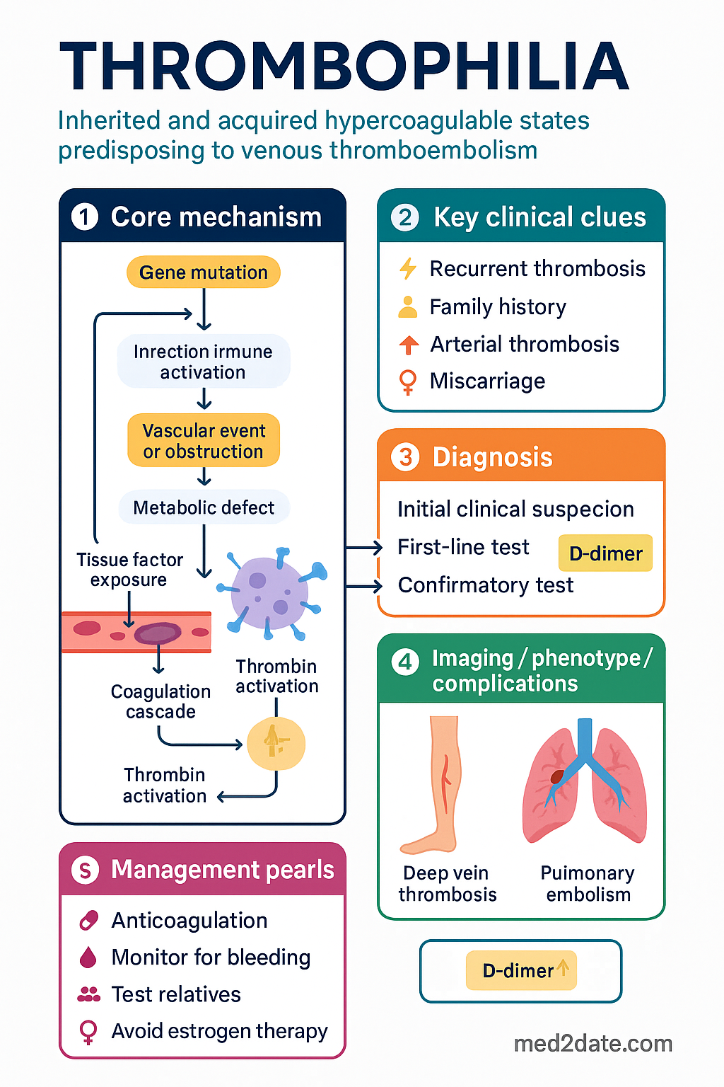
  • Thrombophilia encompasses inherited and acquired hypercoagulable states predisposing to venous thromboembolism (VTE), with the majority presenting as deep vein thrombosis (DVT) or pulmonary embolism (PE).
  • Factor V Leiden (FVL) is the most common inherited thrombophilia in Australian populations, with a heterozygous prevalence of approximately 5โ€“8% in people of European descent.
  • Prothrombin G20210A mutation is the second most common inherited thrombophilia, with a heterozygous prevalence of approximately 2โ€“3% in Europeans.
  • Protein C, Protein S, and antithrombin (AT) deficiency are rarer but confer significantly higher thrombotic risk (especially homozygous compound states).
  • Routine thrombophilia screening after a first unprovoked VTE is generally not recommended โ€” test only when results will change management (e.g., anticoagulation duration, family screening in first-degree relatives).
  • Testing should be deferred for at least 2โ€“4 weeks after completing anticoagulation to avoid false-negative results from consumption or warfarin interference.
  • Anticoagulation duration is determined by the balance of provoked vs unprovoked VTE, recurrence risk (VTE-DASH or Vienna Prediction Model score), and bleeding risk โ€” not by thrombophilia genotype alone.
  • Heterozygous FVL or PT G20210A alone is not an indication for extended anticoagulation after provoked VTE.
  • Antithrombin deficiency, combined thrombophilias, or homozygous/mixed states generally warrant extended or indefinite anticoagulation after VTE.
  • Direct oral anticoagulants (DOACs) are first-line for most patients; warfarin remains preferred for antithrombin deficiency and antiphospholipid syndrome (APS).
  • Pre-procedural and peri-partum bridging with low-molecular-weight heparin (LMWH) is essential in high-risk thrombophilias (AT deficiency, Protein C/S deficiency, combined defects).
  • Aboriginal and Torres Strait Islander peoples may have under-diagnosed thrombophilia; access to specialist haematology and thrombophilia clinics is limited in remote Australia, increasing the need for GP-initiated testing frameworks.

Introduction & Australian Epidemiology

Thrombophilia refers to an inherited or acquired predisposition to venous thromboembolism (VTE), encompassing deep vein thrombosis (DVT) and pulmonary embolism (PE). The concept encompasses genetic mutations affecting coagulation factor function, natural anticoagulant deficiency, and acquired hypercoagulable states such as antiphospholipid syndrome (APS) and malignancy.

VTE affects approximately 1โ€“2 per 1,000 Australian adults annually, with incidence rising sharply with age (โ‰ฅ6 per 1,000 in those >70 years). Thrombophilias modify recurrence risk rather than being the primary driver of first VTE events, which remain most commonly provoked by surgery, immobilisation, oestrogen-containing medications, pregnancy, and malignancy.

The overall prevalence of inherited thrombophilias in the Australian population is as follows:

Thrombophilia Heterozygous Prevalence Homozygous Prevalence Relative Risk (first VTE)
Factor V Leiden 5โ€“8% (European) ~1 in 5,000 ร—3โ€“8 (het) / ร—50โ€“80 (hom)
Prothrombin G20210A 2โ€“3% (European) ~1 in 10,000 ร—2โ€“5 (het) / ร—20+ (hom)
Antithrombin deficiency 0.02โ€“0.2% Rare (often lethal) ร—5โ€“50
Protein C deficiency 0.2โ€“0.5% ~1 in 500,000โ€“1,000,000 ร—7โ€“10 (het)
Protein S deficiency 0.1โ€“1% Very rare ร—5โ€“10 (het)
โš ๏ธ
Key principle: A positive thrombophilia result does not automatically change management. Clinicians must evaluate the clinical context โ€” provoked vs unprovoked VTE, recurrence risk, bleeding risk โ€” before altering anticoagulation duration.

In Australia, inherited thrombophilias are predominantly identified in people of European descent. Factor V Leiden and prothrombin G20210A are exceedingly rare in East Asian, African, and First Nations populations, while Protein C/S and antithrombin deficiency occur across all ancestries. Thrombophilia testing patterns vary widely across Australian states and territories, with significant practice variation between metropolitan and rural/remote settings.

Thrombophilia clinical infographic โ€” pathophysiology, clinical clues, diagnosis, imaging, and management
Tap or click image to enlarge โ€” Thrombophilia: pathophysiology, clinical clues, diagnosis, imaging, and management.
Thrombophilia infographic, full size

Inherited Thrombophilias โ€” Factor V Leiden & Prothrombin G20210A

Factor V Leiden (FVL)

Factor V Leiden results from a point mutation (G1691A) in the F5 gene causing an arginine-to-glutamine substitution at position 506 (R506Q). This renders activated Factor V resistant to cleavage by activated Protein C (APC resistance), thereby sustaining the prothrombotic state.

๐Ÿงช
Factor V Leiden โ€” Clinical Features
F5 gene ยท R506Q ยท APC resistance
Inheritance Autosomal dominant
VTE risk (het) ร—3โ€“8 baseline; ร—30+ with OCP
VTE risk (hom) ร—50โ€“80 baseline
Recurrence (5-yr, unprovoked) ~15โ€“20% (het); ~40%+ (hom)
Australian prevalence 5โ€“8% European descent; <1% ATSI/Asian

Prothrombin Gene Mutation (PT G20210A)

The prothrombin G20210A mutation is a gain-of-function variant in the 3โ€ฒ untranslated region of the F2 gene, leading to increased prothrombin (Factor II) plasma levels (approximately 30% elevation). Elevated prothrombin enhances thrombin generation and promotes a prothrombotic phenotype.

๐Ÿงช
Prothrombin G20210A โ€” Clinical Features
F2 gene ยท Gain-of-function ยท โ†‘ prothrombin
Inheritance Autosomal dominant
VTE risk (het) ร—2โ€“5 baseline
VTE risk (hom) ร—20+ baseline
Recurrence (5-yr, unprovoked) ~10โ€“15% (het)
Synergy Combined FVL + PT G20210A โ†’ ร—20+ risk
โ„น๏ธ
Compound heterozygosity: Patients who carry both FVL and PT G20210A (compound heterozygotes) have a substantially elevated VTE risk (~ร—20) and should be managed similarly to homozygous states.

Protein C, Protein S & Antithrombin Deficiency

These natural anticoagulant deficiencies are less common than FVL or PT G20210A but confer significantly higher thrombotic risk and are more likely to influence anticoagulation decisions. All three follow autosomal dominant inheritance with variable penetrance.

Antithrombin (AT) Deficiency

Antithrombin is the primary physiological inhibitor of thrombin (Factor IIa) and Factor Xa. Deficiency may be quantitative (Type I โ€” reduced synthesis) or qualitative (Type II โ€” functional defect in the reactive site, heparin-binding site, or pleiotropic). AT deficiency confers the highest thrombotic risk of all inherited thrombophilias.

Type II โ€” Heparin Binding Site
Moderate Risk
AT antigen preserved but heparin affinity reduced. Lower thrombotic risk than other Type II variants. May be missed on heparin cofactor assays.
Consider extended anticoagulation after unprovoked VTE
Type I (Quantitative)
High Risk
~50% of normal AT activity and antigen. Lifetime VTE risk of 50โ€“80%. Often presents in young adulthood. Heparin resistance may occur.
Extended/indefinite anticoagulation after VTE; LMWH bridging peri-procedurally
Homozygous / Compound
Very High Risk
Usually lethal in utero (Type I homozygous). Type II homozygotes (pleiotropic) may survive with neonatal purpura fulminans. Requires lifelong AT concentrate and anticoagulation.
Tertiary centre management; AT concentrate (Thrombate IIIยฎ); lifelong warfarin
๐Ÿšจ
Heparin resistance: Patients with antithrombin deficiency may exhibit heparin resistance (failure to achieve therapeutic aPTT despite escalating heparin doses) because heparin requires AT as a cofactor. Management: IV AT concentrate (if available) or switch to direct thrombin inhibitors in refractory cases.

Protein C Deficiency

Protein C is a vitamin K-dependent zymogen that, when activated by the thrombin-thrombomodulin complex, proteolytically inactivates Factors Va and VIIIa. Heterozygous deficiency confers a ร—7โ€“10 increased VTE risk. Critically, warfarin initiation can precipitate warfarin-induced skin necrosis (WISN) due to the more rapid fall in Protein C relative to procoagulant factors.

๐Ÿšจ
Warfarin-induced skin necrosis: In Protein C (or S) deficiency, never start warfarin without adequate heparin/LMWH bridging for โ‰ฅ5 days AND until INR โ‰ฅ2.0 for โ‰ฅ24 hours. Consider low-dose initiation (2โ€“3 mg) for the first 3โ€“5 days. If skin necrosis develops, withhold warfarin, administer vitamin K, and provide fresh frozen plasma or Protein C concentrate.

Protein S Deficiency

Protein S is a cofactor for activated Protein C and also has independent anticoagulant activity via direct inhibition of prothrombinase. Three types are recognised:

Type Free PS Total PS PS Activity
Type I โ†“ โ†“ โ†“
Type II Normal Normal โ†“
Type III โ†“ Normal โ†“
โš ๏ธ
Measurement pitfalls: Protein S is an acute-phase reactant and levels fall during acute thrombosis, pregnancy, and oestrogen use โ€” leading to false-positive diagnoses. Always repeat testing when the patient is well, off anticoagulation, and not pregnant. Free PS (antigen) is the most reliable assay.

Testing Indications & Interpretation

When to Test

โš ๏ธ
Do not test routinely: Thrombophilia testing after a first provoked VTE, or when results will not change management, is not recommended by the RACGP, BSH, or ISTH. Testing is most useful when results will influence anticoagulation duration, family counselling, or pre-procedural planning.

Appropriate indications for thrombophilia testing in Australia:

  • Unprovoked VTE in a patient <50 years, especially if considering stopping anticoagulation
  • Recurrent VTE (โ‰ฅ2 episodes)
  • VTE in unusual sites (cerebral venous sinus, hepatic, mesenteric, portal veins)
  • First-degree relative with high-risk thrombophilia (AT deficiency, homozygous FVL, combined defects)
  • Neonatal purpura fulminans or warfarin-induced skin necrosis (test for Protein C/S deficiency)
  • Recurrent pregnancy loss (โ‰ฅ3 first-trimester or โ‰ฅ1 second-trimester losses) โ€” after exclusion of APS
  • Heparin resistance (suggestive of AT deficiency)

When NOT to Test

  • First provoked VTE with a strong transient risk factor (surgery, plaster cast, >3 days immobility)
  • During acute VTE (consumption lowers Protein C/S/AT levels)
  • While on anticoagulation (warfarin lowers Protein C/S; DOACs interfere with clot-based assays)
  • During pregnancy or oestrogen use (falsely low Protein S)
  • Routine screening in the general population or pre-employment

Interpretation Framework

Test Method Timing Requirements Interference
Factor V Leiden DNA-based (PCR); APTT-based APC-R screening Any time (genetic test โ€” constant) APC-R assay unreliable if on warfarin or lupus anticoagulant present
Prothrombin G20210A DNA-based (PCR); prothrombin level screening Any time (genetic test โ€” constant) Prothrombin levels unreliable on warfarin
Antithrombin Chromogenic activity assay ยฑ antigen โ‰ฅ2 weeks off anticoagulation; not during acute thrombosis Heparin (in vitro); acute thrombosis (consumption); DIC
Protein C Chromogenic or clot-based activity ยฑ antigen โ‰ฅ2โ€“4 weeks off warfarin (Vit K-dependent); can test on heparin Warfarin (falsely โ†“); acute thrombosis; liver disease; DIC
Protein S Free PS antigen (immunological) + activity โ‰ฅ2โ€“4 weeks off warfarin; not during pregnancy or OCP Warfarin, pregnancy, OCP, acute thrombosis, liver disease โ€” all falsely โ†“

Australian Laboratory Considerations

MBS Available
Factor V Leiden (DNA)
MBS Item 73302. Available at major pathology laboratories (Sullivan Nicolaides, Douglass Hanly Moir, Melbourne Pathology). Turnaround: 5โ€“10 business days. Bulk-billed in most settings.
MBS Available
Prothrombin G20210A (DNA)
MBS Item 73302 (often co-tested with FVL). Same turnaround and availability as FVL.
MBS Available
Protein C activity, Protein S (free Ag), Antithrombin activity
MBS Items 71108, 71110, 71106. Available at major metropolitan and regional labs. Not all remote/regional centres offer these; refer blood to tertiary lab if local turnaround is prolonged.
Referral Recommended
Specialist Haematology / Thrombophilia Clinic
Complex cases (AT deficiency, combined defects, recurrent VTE despite anticoagulation) should be referred to a hospital-based thrombophilia or haemostasis clinic. Available at most tertiary centres (Royal Melbourne, Westmead, Royal Adelaide, Princess Alexandra, Royal Perth).

Management โ€” Anticoagulation Duration

Anticoagulation decisions in thrombophilia should be based on the clinical context of the VTE (provoked vs unprovoked), validated recurrence risk scores, bleeding risk, and patient preference โ€” not the thrombophilia genotype alone.

Anticoagulation Duration Decision Framework

1
Classify the VTE
Provoked (major transient risk: surgery, trauma, โ‰ฅ3 days immobility) vs unprovoked vs weakly provoked (OCP, long-haul travel, minor illness).
2
Assess Recurrence Risk
Use VTE-DASH score, Vienna Prediction Model, or HERDOO2. Unprovoked VTE: 5-year recurrence ~30% without anticoagulation.
3
Evaluate Thrombophilia
High-risk (AT def, combined, homozygous) โ†’ strongly favour extended anticoagulation. Low-risk (het FVL/PT) โ†’ manage by clinical context.
4
Assess Bleeding Risk
HAS-BLED score. Age โ‰ฅ65, renal impairment, prior bleeding, antiplatelet therapy increase risk.
5
Shared Decision-Making
Discuss recurrence vs bleeding risk. Annual VTE recurrence on treatment ~2% vs off treatment ~5โ€“10% (unprovoked). Patient preference is crucial.

Recommended Duration by Thrombophilia Type

Thrombophilia Provoked VTE Unprovoked VTE
No thrombophilia 3 months โ‰ฅ3 months; reassess for extended
Heterozygous FVL or PT G20210A 3 months โ‰ฅ3 months; reassess โ€” genotype alone does not mandate extended
Homozygous FVL or PT G20210A 3โ€“6 months; reassess Extended/indefinite
Compound heterozygous (FVL + PT) 3โ€“6 months; reassess Extended/indefinite
Protein C deficiency (het) 3โ€“6 months Extended/indefinite
Protein S deficiency (het) 3โ€“6 months Extended/indefinite
Antithrombin deficiency (het) Extended/indefinite Extended/indefinite
Combined thrombophilias Extended/indefinite Extended/indefinite

Pharmacological Agents โ€” Australian PBS

๐Ÿ’Š
Apixaban
Eliquisยฎ ยท Factor Xa inhibitor ยท DOAC
Adult dose โ€” acute 10 mg PO BD ร— 7 days, then 5 mg PO BD ร— 6 months
Extended prophylaxis 2.5 mg PO BD (PBS-listed for extended VTE prevention)
Paediatric dose Weight-based (โ‰ฅ12 months); see product PI
Renal adjustment CrCl <25 mL/min: avoid. No dose change CrCl 25โ€“50
Hepatic adjustment Avoid in severe hepatic impairment (Child-Pugh C)
PBS status โœ” PBS General Benefit
๐Ÿ’Š
Rivaroxaban
Xareltoยฎ ยท Factor Xa inhibitor ยท DOAC
Adult dose โ€” acute 15 mg PO BD ร— 21 days, then 20 mg PO OD with food
Extended prophylaxis 10 mg PO OD (PBS-listed for extended VTE prevention)
Renal adjustment CrCl <15 mL/min: avoid. CrCl 15โ€“49: use 15 mg for treatment (not 10 mg prophylaxis)
PBS status โœ” PBS General Benefit
๐Ÿ’Š
Warfarin
Marevanยฎ ยท Coumarin ยท Vitamin K antagonist
Adult dose Individualised; target INR 2.0โ€“3.0. Start 2โ€“5 mg PO daily
Preferred when Antithrombin deficiency, APS, mechanical heart valve
Renal adjustment No specific dose change; monitor INR closely
Key interaction Numerous drug-food interactions; INR monitoring essential
PBS status โœ” PBS General Benefit
๐Ÿ’Š
Enoxaparin
Clexaneยฎ ยท LMWH ยท Antithrombin-dependent
Adult dose โ€” treatment 1 mg/kg SC BD (or 1.5 mg/kg SC OD)
Adult dose โ€” prophylaxis 40 mg SC OD (or 20 mg SC OD if CrCl <30)
Paediatric dose 0.75 mg/kg SC BD (<2 months); 0.5 mg/kg SC BD (โ‰ฅ2 months)
Key role Bridging anticoagulation; pregnancy; peri-procedural in high-risk thrombophilias
PBS status โœ” PBS General Benefit
โš ๏ธ
DOACs vs Warfarin โ€” choice in thrombophilia: DOACs (apixaban, rivaroxaban) are first-line for most thrombophilia patients with VTE. However, warfarin is preferred in: (1) antithrombin deficiency (DOACs are AT-independent and may be less effective), (2) antiphospholipid syndrome (warfarin superior to rivaroxaban per TRAPS trial), and (3) mechanical heart valves.

Monitoring

Week 1โ€“2
Initial VTE treatment phase. Ensure therapeutic anticoagulation achieved. If on warfarin: commence bridging with LMWH, monitor INR daily until stable.
Month 3
First reassessment point. For provoked VTE with low-risk thrombophilia: consider stopping anticoagulation. For unprovoked VTE or high-risk thrombophilia: reassess for extended therapy.
Month 6
Second reassessment if on extended anticoagulation. Check renal function (affects DOAC dosing). Review bleeding risk (HAS-BLED). Reconfirm patient preference.
Annually
Annual review of extended anticoagulation. FBC, renal function, LFTs. Reassess bleeding risk. Discuss de-escalation if recurrence risk has declined. Document shared decision.

Warfarin Monitoring

  • Target INR: 2.0โ€“3.0 for most VTE. Target INR 2.5โ€“3.5 for APS with recurrent thrombosis.
  • INR monitoring every 1โ€“2 weeks initially, then every 4โ€“6 weeks when stable.
  • Time in therapeutic range (TTR) should be >65% โ€” if lower, consider DOAC switch if appropriate.
  • CYP2C9 and VKORC1 genotyping (MBS Item 73302) may guide initial dosing in complex cases.

DOAC Monitoring

  • DOACs do not require routine coagulation monitoring.
  • Check renal function (eGFR) every 6โ€“12 months โ€” essential for dose adjustment.
  • Anti-Xa levels (calibrated for specific DOAC) can be measured in clinical emergencies (major bleeding, urgent surgery, suspected treatment failure) โ€” available at tertiary centres.

Special Populations

๐Ÿคฐ Pregnancy
DOACs (apixaban, rivaroxaban, edoxaban, dabigatran)
Contraindicated in pregnancy (teratogenic risk, especially first trimester). Discontinue and switch to LMWH before conception or immediately upon pregnancy diagnosis.
Enoxaparin (Clexaneยฎ)
First-line anticoagulation in pregnancy. Dose: treatment 1 mg/kg SC BD; prophylactic 40 mg SC OD. Adjust for weight. Safe in all trimesters. Does not cross placenta.
Warfarin
Teratogenic (warfarin embryopathy) weeks 6โ€“12. Avoid in first trimester. May use weeks 13โ€“36 in exceptional cases (e.g., mechanical valve). Switch to LMWH before delivery.
AT concentrate (Thrombate IIIยฎ)
For AT-deficient pregnant women: AT replacement peri-partum to maintain activity >80%. Coordinate with obstetric haematology. Not PBS-listed โ€” hospital supply.
๐Ÿ‘ถ Paediatrics
VTE in children
Childhood VTE is rare (~0.07โ€“0.14 per 10,000 children/year). Most are provoked (central venous catheters, malignancy). Inherited thrombophilia testing should be guided by paediatric haematology.
Neonatal purpura fulminans
Homozygous Protein C or S deficiency. Emergent: fresh frozen plasma 10โ€“20 mL/kg, Protein C concentrate (Ceprotinยฎ), transition to warfarin with careful LMWH bridging.
๐Ÿ‘ด Elderly (โ‰ฅ65 years)
Anticoagulation considerations
Higher bleeding risk (HAS-BLED โ‰ฅ3). Renal decline affects DOAC dosing. Annual risk-benefit reassessment is essential. DOACs preferred over warfarin (fewer monitoring visits, lower ICH risk).
๐Ÿฉบ Renal Impairment
CrCl 30โ€“50 mL/min
Rivaroxaban: use 15 mg for treatment. Apixaban: no dose change. Monitor renal function every 3โ€“6 months.
CrCl 15โ€“29 mL/min
Apixaban may be used with caution (limited data). Rivaroxaban: 15 mg treatment dose. Avoid dabigatran. Consider warfarin as alternative.
CrCl <15 mL/min / Dialysis
Warfarin is preferred. DOACs not recommended. LMWH: dose-reduce and monitor anti-Xa levels.
๐Ÿซ Hepatic Impairment
Child-Pugh Aโ€“B
DOACs may be used with caution in mild-moderate hepatic impairment. Monitor LFTs. Synthetic liver function affects Protein C/S levels โ€” interpret with caution.
Child-Pugh C
Avoid DOACs. Warfarin with careful INR monitoring. Coagulopathy may mimic or mask thrombophilia.
๐Ÿฆ  Immunocompromised
Cancer-associated VTE
LMWH (enoxaparin 1 mg/kg SC BD) was traditional standard; DOACs (rivaroxaban, apixaban) now PBS-listed alternatives in cancer. GI/GU malignancies: higher bleeding risk with DOACs โ€” assess carefully.

Aboriginal and Torres Strait Islander Health Considerations

Aboriginal and Torres Strait Islander Health
Epidemiological context
VTE incidence in Aboriginal and Torres Strait Islander peoples is not well characterised in the literature, but cardiovascular and metabolic risk factors are more prevalent. Factor V Leiden and PT G20210A are rare (<1%) in First Nations populations; Protein C, S, and AT deficiency occur at rates comparable to non-Indigenous Australians. Thrombophilia may be under-diagnosed due to limited testing in remote settings.
Remote & rural access
Specialist haematology and thrombophilia clinics are concentrated in metropolitan tertiary centres. Aboriginal Community Controlled Health Services (ACCHS) may lack access to specialised coagulation testing (Protein C/S, AT). Telehealth consultations with haematologists can facilitate testing interpretation and management planning.
Pathology access
DNA-based tests (FVL, PT G20210A) are stable for transport and can be performed on dried blood spots or posted samples. Protein C, S, and AT require venepuncture with rapid transport to reference laboratory โ€” challenging in remote communities. Pre-analytical sample handling is critical (avoid haemolysis, timely centrifugation).
Anticoagulation adherence
DOACs (no INR monitoring) may improve adherence compared to warfarin in settings where regular pathology access is difficult. Ensure PBS co-payment affordability. Address health literacy around anticoagulation risks and adherence with culturally appropriate education resources.
Pregnancy care
Pregnant Aboriginal and Torres Strait Islander women with known or suspected thrombophilia should be co-managed with obstetric medicine and haematology. LMWH (enoxaparin) self-injection training should be provided with culturally safe support. Telehealth follow-up can supplement antenatal visits in remote areas.
Cultural safety
Engage Aboriginal Health Workers and Liaison Officers in thrombophilia education and anticoagulation counselling. Respect kinship obligations when discussing family screening. Utilise AIHW Closing the Gap frameworks and RHDAustralia resources for venous thromboembolism awareness in First Nations communities.

๐Ÿ“š References

  1. 1. Connors JM. Thrombophilia testing and venous thrombosis. N Engl J Med. 2017;377(12):1177โ€“1187.
  2. 2. Middeldorp S, Nieuwlaat R, Baumann Kreuziger L, et al. American Society of Hematology 2023 guidelines for management of venous thromboembolism: thrombophilia testing. Blood Adv. 2023;7(22):7101โ€“7138.
  3. 3. Baglin T, Gray E, Greaves M, et al. Clinical guidelines for testing for heritable thrombophilia. Br J Haematol. 2010;149(2):209โ€“220.
  4. 4. Kearon C, Akl EA, Ornelas J, et al. Antithrombotic therapy for VTE disease: CHEST guideline and expert panel report. Chest. 2016;149(2):315โ€“352.
  5. 5. Tran HA, Gibbs H, Merriman E, et al. New guidelines from the Thrombosis and Haemostasis Society of Australia and New Zealand for the diagnosis and management of venous thromboembolism. Med J Aust. 2019;210(5):227โ€“235.
  6. 6. Australian Institute of Health and Welfare (AIHW). Cardiovascular disease in Aboriginal and Torres Strait Islander people. AIHW; 2023. Available from: https://www.aihw.gov.au
  7. 7. Bates SM, Greer IA, Middeldorp S, et al. VTE, thrombophilia, antithrombotic therapy, and pregnancy: ACCP evidence-based clinical practice guidelines (9th ed). Chest. 2012;141(2 Suppl):e691Sโ€“e736S.
  8. 8. Pengo V, Lensing AWA, Prins MH, et al. Incidence of chronic thromboembolic pulmonary hypertension after pulmonary embolism (for the Thromboembolic Hypertension Study Group). N Engl J Med. 2004;350(22):2257โ€“2264.
  9. 9. Dentali F, Ageno W, Bozzato S, et al. Role of Factor V Leiden and prothrombin G20210A in patients with pulmonary embolism: systematic review and meta-analysis. Thromb Haemost. 2012;107(2):251โ€“258.
  10. 10. Crowther MA, Kelton JG. Congenital thrombophilic states associated with venous thrombosis: a qualitative overview and proposed classification system. Ann Intern Med. 2003;138(2):128โ€“134.
  11. 11. Monagle P, Cuello CA, Augustine C, et al. American Society of Hematology 2018 guidelines for management of venous thromboembolism: treatment of pediatric venous thromboembolism. Blood Adv. 2018;2(22):3292โ€“3316.
  12. 12. RHDAustralia. Australian guideline for prevention, diagnosis and management of acute rheumatic fever and rheumatic heart disease (3rd ed). Menzies School of Health Research; 2020. [Referenced for ATSI health frameworks].