📋 Key Information Summary
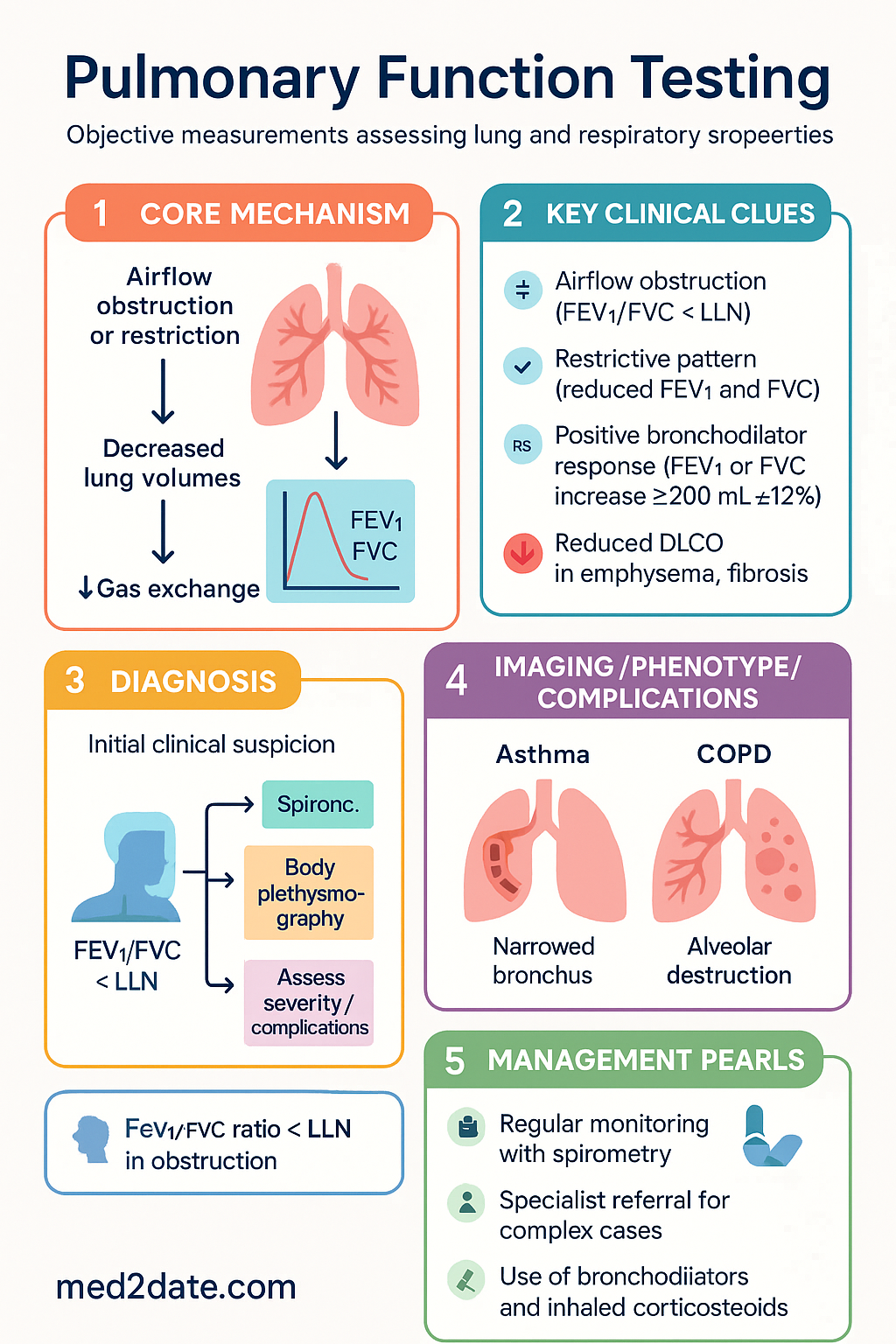
- Spirometry is the cornerstone pulmonary function test (PFT); an FEV1/FVC ratio below the lower limit of normal (LLN) defines airflow obstruction; fixed ratio <0.70 alone may overdiagnose in older adults and underdiagnose in younger adults.
- Quality criteria per ATS/ERS 2019: ≥3 acceptable manoeuvres, reproducibility of FEV1 and FVC within 150 mL (≥200 mL in adults), back-extrapolated volume <5% of FVC or <100 mL.
- A positive bronchodilator response is an increase in FEV1 or FVC ≥200 mL AND ≥12% from baseline after 400 µg salbutamol MDI via spacer.
- Lung volumes by body plethysmography are the gold standard; gas dilution (helium dilution or nitrogen washout) underestimates trapped gas. TLC <LLN confirms restriction; elevated RV/TLC ratio suggests air trapping.
- DLCO measures gas transfer across the alveolar-capillary membrane; correct for haemoglobin (anaemia lowers DLCO), carboxyhaemoglobin (smoking), and altitude. Reduced DLCO is seen in emphysema, pulmonary fibrosis, pulmonary vascular disease, and anaemia.
- An isolated reduction in DLCO with normal spirometry and lung volumes raises suspicion for pulmonary vascular disease (e.g., pulmonary hypertension, pulmonary embolism).
- Methacholine challenge (bronchoprovocation) is indicated when spirometry is normal but asthma is clinically suspected; a fall in FEV1 ≥20% at ≤8 mg/mL (PC20) supports airway hyperresponsiveness.
- Cardiopulmonary exercise testing (CPET) differentiates cardiac, pulmonary, deconditioning, and musculoskeletal causes of exertional dyspnoea using VO2max, anaerobic threshold, and ventilatory equivalents.
- Respiratory muscle strength is assessed by maximal inspiratory pressure (MIP/SNIP) and maximal expiratory pressure (MEP); reduced values suggest neuromuscular causes of respiratory failure.
- Spirometry is available in most Australian general practices and respiratory clinics; lung volumes, DLCO, bronchoprovocation, and CPET require specialised respiratory laboratory referral (MBS items available for spirometry and DLCO).
- In Aboriginal and Torres Strait Islander populations, spirometry access is limited in remote communities; interpreter use, culturally safe technique coaching, and use of appropriate reference equations are essential.
- Always interpret PFTs in clinical context — pattern recognition (obstructive, restrictive, mixed, isolated gas transfer defect) guides differential diagnosis and further investigation.
Introduction & Australian Epidemiology
Pulmonary function testing (PFT) encompasses a range of objective, non-invasive measurements that assess the mechanical and physiological properties of the lungs and respiratory system. PFTs are essential for diagnosing respiratory disease, quantifying severity, monitoring disease progression, evaluating treatment response, assessing pre-operative risk, and determining disability.
In Australia, chronic respiratory diseases affect approximately 7 million people and are the third leading cause of disease burden. Chronic obstructive pulmonary disease (COPD) affects 1 in 13 Australians aged ≥40 years, while asthma affects approximately 11% of the population. Despite this burden, spirometry is underutilised in Australian primary care — studies suggest fewer than 30% of patients with newly diagnosed COPD undergo confirmatory spirometry.
The Australian Commission on Safety and Quality in Health Care (ACSQHC) and the Thoracic Society of Australia and New Zealand (TSANZ) recommend spirometry as the standard for diagnosing and monitoring obstructive airway diseases. Medicare Benefits Schedule (MBS) item numbers support spirometry (MBS 11503, 11505, 11506) and diffusing capacity testing (MBS 11506) when performed in accredited facilities.
This guideline covers the four principal domains of pulmonary function testing: spirometry, lung volume measurement, diffusing capacity, and specialised testing including bronchoprovocation, exercise testing, and respiratory muscle strength assessment.
Spirometry
Technique
Spirometry measures the volume of air an individual can inhale and exhale as a function of time. The forced vital capacity (FVC) manoeuvre is the fundamental measurement, yielding the key parameters: FEV1 (forced expiratory volume in 1 second), FVC, and the FEV1/FVC ratio.
Quality Criteria (ATS/ERS 2019 Standards)
| Criterion | Requirement |
|---|---|
| Good start | Back-extrapolated volume (BEV) <5% of FVC or <100 mL (whichever is greater); time to peak expiratory flow <120 ms (ideally <75 ms in adults) |
| Free from artefact | No cough (in first 1 second), glottis closure, early termination, leak, or obstructed mouthpiece |
| Satisfactory end-of-test | Plateau in volume-time curve: volume change <25 mL in final 1 second (≥1 second exhalation); exhalation time ≥6 seconds (adults), ≥3 seconds (children ≥10 years) |
| Reproducibility | Largest two FEV1 values within 150 mL; largest two FVC values within 150 mL |
| Minimum acceptable curves | ≥3 acceptable curves; report best FEV1 and best FVC (from any curve) |
Key Parameters
| Parameter | Definition | Clinical Significance |
|---|---|---|
| FEV1 | Volume exhaled in the first second of a forced manoeuvre | Marker of airflow limitation severity; used for COPD staging (GOLD), asthma monitoring, pre-operative assessment |
| FVC | Total volume exhaled during forced manoeuvre | Reduced in restriction, air trapping, and submaximal effort |
| FEV1/FVC ratio | Proportion of FVC exhaled in 1 second | Ratio <LLN (or <0.70 for COPD diagnosis per GOLD) indicates obstruction; ratio ≥LLN with low TLC confirms restriction |
| FEF25–75% | Forced expiratory flow during middle 50% of FVC | Sensitive to small airway disease; highly variable; low specificity in isolation |
| PEF | Peak expiratory flow rate | Useful for asthma self-monitoring; effort-dependent; poor for diagnosis |
Interpretation Framework
Use the lower limit of normal (LLN) (5th percentile of predicted) rather than fixed ratios for interpretation. The GLI-2012 (Global Lung Function Initiative) reference equations are recommended for Australian populations, incorporating age, sex, height, and ethnicity.
| Pattern | FEV1/FVC | FEV1 | FVC | Common Causes |
|---|---|---|---|---|
| Normal | ≥LLN | ≥LLN | ≥LLN | No significant ventilatory defect |
| Obstructive | <LLN | Reduced | Normal or reduced | Asthma, COPD, bronchiectasis, cystic fibrosis |
| Restrictive | ≥LLN (or elevated) | Reduced | Reduced proportionally | IPF, ILD, chest wall disease, obesity, neuromuscular |
| Mixed | <LLN | Reduced | Reduced | Combined COPD + fibrosis, advanced disease |
| Non-specific | ≥LLN | Reduced | ≥LLN | Suboptimal effort, early disease; requires lung volumes for confirmation |
Severity Classification (FEV1 % Predicted)
Severity grading uses FEV1 as a percentage of predicted (GLI-2012 equations). This applies to both obstructive and restrictive patterns once the pattern is confirmed.
Bronchodilator Response
Bronchodilator reversibility testing assesses the degree of acute improvement in airflow following administration of a short-acting bronchodilator. It is performed after baseline spirometry.
Lung Volumes
Measurement Techniques
Lung volume measurement provides essential information beyond spirometry. While spirometry measures dynamic volumes (FEV1, FVC), it cannot measure total lung capacity (TLC), residual volume (RV), or functional residual capacity (FRC) — these require body plethysmography or gas dilution techniques.
| Technique | Principle | Measures | Advantages | Limitations |
|---|---|---|---|---|
| Body plethysmography | Boyle's law (P1V1=P2V2) applied during panting manoeuvres in a sealed box | FRC (thoracic gas volume), then TLC and RV calculated | Gold standard; measures all lung volumes including trapped gas; fast (~3 min) | Expensive equipment; claustrophobic patients; artefact from glottic closure or shallow panting |
| Helium dilution | Equilibration of known helium concentration in a closed circuit | FRC (gas-communicating volume); TLC and RV derived | Widely available; less expensive than plethysmography | Underestimates FRC when significant air trapping (non-communicating gas); equilibration time 3–7 min |
| Nitrogen washout | Washout of endogenous nitrogen using 100% O2 | FRC; also provides distribution of ventilation (closing volume) | Additional ventilation distribution information | Underestimates FRC with air trapping; requires leak-free system; O2 washout contraindicated in some patients |
Lung Volume Compartments
| Volume | Definition | Measurement |
|---|---|---|
| TLC (Total Lung Capacity) | Volume of air in lungs at maximal inspiration | FRC + IC (inspiratory capacity) or RV + VC |
| RV (Residual Volume) | Volume remaining after maximal expiration | TLC − VC or FRC − ERV |
| FRC (Functional Residual Capacity) | Volume at end of normal tidal expiration (relaxation equilibrium) | Directly measured by plethysmography or gas dilution |
| RV/TLC ratio | Proportion of TLC that is residual volume | Calculated; elevated in air trapping and hyperinflation |
| IC (Inspiratory Capacity) | Volume from end-tidal expiration to maximal inspiration | TLC − FRC |
Restrictive Pattern Confirmation
Common causes of confirmed restriction (TLC <LLN):
- Pulmonary: Idiopathic pulmonary fibrosis (IPF), hypersensitivity pneumonitis, sarcoidosis, pneumoconiosis, radiation pneumonitis
- Extrapulmonary — chest wall: Severe kyphoscoliosis, ankylosing spondylitis (advanced), flail chest, pectus excavatum (severe)
- Extrapulmonary — pleural: Pleural effusion, pleural thickening, mesothelioma
- Extrapulmonary — neuromuscular: Motor neurone disease, muscular dystrophy, myasthenia gravis, diaphragmatic paralysis
- Extrapulmonary — abdominal: Obesity (BMI >30), massive ascites, pregnancy (late)
Air Trapping and Hyperinflation
Elevated RV and RV/TLC ratio indicate air trapping, a hallmark of obstructive lung disease. In COPD:
- RV >120% predicted and/or RV/TLC >40% suggests significant air trapping
- Static hyperinflation (elevated FRC and IC reduction) correlates with dyspnoea severity and exercise limitation
- Dynamic hyperinflation during exercise is an important therapeutic target (e.g., with LAMA/LABA therapy, lung volume reduction surgery)
- A markedly elevated TLC (>120% predicted) with elevated RV suggests emphysema (loss of elastic recoil leading to overdistension)
Diffusing Capacity (DLCO)
Technique — Single-Breath Method
The diffusing capacity for carbon monoxide (DLCO, also known as transfer factor TLCO) is measured using the single-breath technique, which is the standard method in Australian respiratory laboratories.
Quality Requirements
- Inspired volume ≥85% of largest FVC (to ensure adequate alveolar sampling)
- Breath-hold time 8–12 seconds
- Inspiratory time <2.5 seconds (rapid inhalation to TLC)
- No evidence of Valsalva or Müller manoeuvre during breath hold
- Alveolar sample collected within 3 seconds of exhalation onset
- Two measurements within 10% of each other or 3 mL/min/mmHg
Interpretation
DLCO is expressed as a percentage of predicted (using GLI-2017 reference equations). Interpretation requires correction for confounding variables:
| DLCO Level | Interpretation |
|---|---|
| ≥LLN (usually ≥80% predicted) | Normal gas transfer |
| 60–79% predicted | Mildly reduced |
| 40–59% predicted | Moderately reduced |
| <40% predicted | Severely reduced |
| >140% predicted | Elevated (consider obesity correction, polycythaemia, left-to-right shunt, early pulmonary haemorrhage) |
Causes of Altered DLCO
- Emphysema — loss of alveolar-capillary surface area (hallmark of COPD with emphysema phenotype)
- Interstitial lung disease — IPF, NSIP, hypersensitivity pneumonitis, sarcoidosis, drug-induced ILD
- Pulmonary vascular disease — pulmonary hypertension, chronic pulmonary embolism
- Pneumonectomy/lobectomy — reduced lung volume
- Anaemia — reduced haemoglobin for CO binding (must correct)
- Restrictive chest wall/pleural disease — severe obesity, kyphoscoliosis, pleural effusion
- Early interstitial disease — DLCO may fall before spirometry or volumes change
- Polycythaemia — increased haemoglobin mass
- Left-to-right intracardiac shunt — increased pulmonary blood flow
- Pulmonary haemorrhage — CO uptake by intra-alveolar blood (Goodpasture, vasculitis)
- Obesity — increased pulmonary blood volume (corrected by indexing to alveolar volume: DLCO/VA or KCO)
- Asthma — mildly elevated DLCO may occur during acute exacerbation (increased pulmonary blood volume)
- Exercise — increased cardiac output and pulmonary blood flow
Corrections
- Haemoglobin correction (mandatory): DLCOcorrected = DLCOmeasured × factor. For males: factor = 10.22/(Hb + 1.72); for females: factor = 9.38/(Hb + 1.72). Using standard ATS/ERS equations. Significant in anaemia (Hb <100 g/L) where uncorrected DLCO may falsely suggest parenchymal disease.
- Carboxyhaemoglobin (COHb) correction: Smokers may have COHb 5–15%, which reduces the CO driving force and lowers measured DLCO. Correct using: DLCOcorrected = DLCOmeasured × (1.0 + 0.0136 × COHb%).
- Altitude correction: Not typically required for Australian sites (most facilities at sea level or moderate altitude). At altitude, inspired PO2 is lower; DLCO may be elevated due to compensatory mechanisms.
- KCO (DLCO/VA): The transfer coefficient (KCO) is DLCO indexed to alveolar volume (VA). A normal or elevated KCO with reduced DLCO and reduced VA suggests restriction (extrapulmonary). A low KCO with low DLCO suggests intrinsic alveolar-capillary disease (emphysema, fibrosis).
Specialized Testing
Bronchoprovocation Testing (Methacholine Challenge)
Bronchoprovocation testing assesses airway hyperresponsiveness (AHR) and is indicated when clinical suspicion for asthma is high but spirometry is normal (FEV1 ≥70% predicted) and bronchodilator reversibility is negative.
| Protocol | Details |
|---|---|
| Agent | Methacholine (Provocholine®) — cholinergic agonist causing bronchoconstriction; or mannitol (Aridol®) — osmotic stimulus (TGA-approved in Australia) |
| Method (5-breath dosimeter) | Doubling concentrations from 0.03 mg/mL to 16 mg/mL (or 32 mg/mL); 5 breaths per concentration via dosimeter; spirometry 60–90 seconds after each dose |
| Endpoint | 20% fall in FEV1 from post-diluent baseline (PC20) or cumulative dose (PD20) |
| Positive result | PC20 ≤16 mg/mL (or PD20 ≤200 µg cumulative dose) |
| Interpretation | PC20 <1 mg/mL = highly suggestive of active asthma; 1–4 mg/mL = moderate AHR; 4–16 mg/mL = mild AHR (may be seen in allergic rhinitis, post-viral, COPD); >16 mg/mL = negative |
| Mannitol (Aridol®) | Capsule-based dry powder inhalation; dose escalation from 0 mg to 160 mg; PD15 ≤635 mg = positive; available on PBS (authority required) in some settings; simpler to administer than methacholine in non-specialist labs |
Exercise Testing
Exercise testing evaluates the physiological response to exertion and is indicated for unexplained dyspnoea, exercise-induced bronchoconstriction (EIB), pre-operative assessment, and disability evaluation.
Exercise-Induced Bronchoconstriction (EIB) Testing
- Protocol: 6–8 minutes of high-intensity exercise (≥85% predicted maximum heart rate) on a treadmill or cycle ergometer in dry air (relative humidity <50%)
- Spirometry performed at baseline, then 5, 10, 15, 20, and 30 minutes post-exercise
- Positive: ≥10% fall in FEV1 from baseline (some guidelines use ≥15%)
- Eucapnic voluntary hyperpnoea (EVH) is an alternative provocation — breathing dry air containing 5% CO2 at 85% maximal voluntary ventilation for 6 minutes; sensitive for elite athlete assessment
- EIB is a major concern in Australian elite athletes; sports-specific screening using EVH is recommended by the Australian Institute of Sport (AIS) and World Anti-Doping Agency (WADA)
Cardiopulmonary Exercise Testing (CPET)
CPET is the gold standard for evaluating exertional dyspnoea and exercise capacity. It integrates cardiovascular, pulmonary, and musculoskeletal responses during incremental exercise.
| Parameter | Definition | Clinical Interpretation |
|---|---|---|
| VO2max (peak VO2) | Maximum oxygen consumption at peak exercise | Overall exercise capacity marker; <80% predicted = reduced; used for transplant listing, disability assessment |
| Anaerobic threshold (AT) | O2 consumption at which anaerobic metabolism supplements aerobic (V-slope method) | <40% predicted = severe deconditioning or cardiac limitation; critical for pre-operative risk assessment (pneumonectomy candidacy: AT >15 mL/kg/min) |
| VE/VCO2 slope | Ventilatory equivalent for CO2 (efficiency of ventilation) | Elevated (>34) suggests pulmonary vascular disease, heart failure, or respiratory muscle weakness |
| O2 pulse | VO2 ÷ heart rate (stroke volume × arteriovenous O2 difference) | Low or flat O2 pulse = cardiac limitation (reduced stroke volume) |
| Respiratory exchange ratio (RER) | VCO2 ÷ VO2 at peak exercise | RER ≥1.10 confirms maximal effort; <1.0 may indicate submaximal test |
| SpO2 at peak exercise | Oxygen saturation during maximal exertion | Desaturation >4% from baseline or SpO2 <88% = exercise-induced hypoxaemia |
| Breathing reserve | 1 − (peak VE / MVV); MVV = FEV1 × 35–40 | <20% suggests ventilatory limitation (pulmonary cause of dyspnoea) |
CPET Differential Patterns
| Pattern | VO2max | AT | Breathing Reserve | Heart Rate Reserve | Cause |
|---|---|---|---|---|---|
| Normal | Normal | Normal | Normal | Normal | Deconditioning or non-cardiopulmonary cause of dyspnoea |
| Cardiac | ↓ | ↓ | Preserved | Exhausted early | Heart failure, valvular disease, CAD, pulmonary hypertension |
| Pulmonary (ventilatory) | ↓ | ↓ or normal | Exhausted | Preserved | COPD, ILD, bronchiectasis |
| Pulmonary vascular | ↓ | ↓ | Preserved or mild↓ | Exhausted early | Pulmonary hypertension; ↑ VE/VCO2, flat O2 pulse, exercise desaturation |
| Deconditioning | ↓ | ↓ | Preserved | Preserved | Physical inactivity; responds to training |
Respiratory Muscle Strength
Respiratory muscle testing evaluates the strength of inspiratory and expiratory muscles and is indicated when neuromuscular disease, unexplained dyspnoea, or hypercapnic respiratory failure is suspected.
| Test | Method | Normal Values | Abnormal Suggests |
|---|---|---|---|
| MIP (PImax) | Maximal inspiratory pressure from RV against occluded airway; sustained for ≥1 second | Males: −120 to −80 cmH2O; Females: −90 to −60 cmH2O (age-dependent) | MIP <−80 cmH2O (males) or <−60 cmH2O (females) = inspiratory muscle weakness; MIP <−30 cmH2O = severe weakness, may predict weaning failure |
| SNIP | Sniff nasal inspiratory pressure from FRC through occluded nostril; more reproducible and comfortable than MIP | Males: ≥80 cmH2O; Females: ≥70 cmH2O | Preferred over MIP in many centres; SNIP <70 cmH2O (males) or <60 cmH2O (females) = inspiratory weakness; particularly useful in neuromuscular disease monitoring |
| MEP (PEmax) | Maximal expiratory pressure from TLC against occluded airway | Males: 200–150 cmH2O; Females: 150–100 cmH2O | MEP <60 cmH2O = expiratory muscle weakness (impaired cough); critical for secretion management risk |
Clinical applications:
- Diagnosis and monitoring of neuromuscular diseases (MND/ALS, myasthenia gravis, muscular dystrophy, post-polio syndrome)
- Predicting weaning failure in mechanically ventilated patients (MIP more negative than −20 to −30 cmH2O suggests ability to wean)
- Assessing eligibility for non-invasive ventilation (NIV) in neuromuscular disease (typically when FVC <50% or MIP <−60 cmH2O)
- Pre-operative assessment for major thoracic or upper abdominal surgery
- Monitoring disease progression in MND (serial SNIP and FVC measurements every 2–3 months)
Monitoring & Follow-Up
Serial pulmonary function testing is essential for monitoring disease progression and treatment response. The following guidelines apply:
Special Populations
Pregnancy
Paediatrics
Elderly (≥65 years)
Renal Impairment
Hepatic Impairment
Immunocompromised
Aboriginal and Torres Strait Islander Health Considerations
Aboriginal and Torres Strait Islander Australians experience a disproportionate burden of chronic respiratory disease. COPD mortality is 2.5 times higher in Indigenous Australians compared with non-Indigenous Australians. Asthma prevalence is higher, and bronchiectasis remains a significant cause of morbidity, particularly in remote Northern Territory and Far North Queensland communities. Despite this, access to pulmonary function testing remains inequitable.
📚 References
- 1. Graham BL, Steenbruggen I, Miller MR, et al. Standardization of spirometry 2019 update: an official American Thoracic Society and European Respiratory Society technical statement. Am J Respir Crit Care Med. 2019;200(8):e70–e88.
- 2. Graham BL, Brusasco V, Burgos F, et al. 2017 ERS/ATS standards for single-breath carbon monoxide uptake in the lung. Eur Respir J. 2017;49(1):1600016.
- 3. Wanger J, Clausen JL, Coates A, et al. Standardisation of the measurement of lung volumes. Eur Respir J. 2005;26(3):511–522.
- 4. Quanjer PH, Stanojevic S, Cole TJ, et al. Multi-ethnic reference values for spirometry for the 3–95 year age range: the Global Lung Function 2012 equations. Eur Respir J. 2012;40(6):1324–1343.
- 5. Stanojevic S, Graham BL, Cooper BG, et al. Official ERS technical standards: Global Lung Function Initiative reference values for the carbon monoxide transfer factor for adults. Eur Respir J. 2017;50(1):1700010.
- 6. Coates AL, Wanger J, Cockcroft DW, et al. ERS technical standard on bronchial challenge testing: general considerations and performance of methacholine challenge tests. Eur Respir J. 2017;49(5):1601526.
- 7. American Thoracic Society; American College of Chest Physicians. ATS/ACCP statement on cardiopulmonary exercise testing. Am J Respir Crit Care Med. 2003;167(2):211–277.
- 8. Laveneziana P, Albuquerque A, Aliverti A, et al. ERS statement on respiratory muscle testing at rest and during exercise. Eur Respir J. 2019;53(6):1801214.
- 9. Toelle BG, Xuan W, Peat JK, Marks GB. Childhood factors that predict asthma in young adulthood. Eur Respir J. 2004;23(1):66–70.
- 10. Chang AB, Brown N, Toombs M, et al. Lung disease in Indigenous children. Paediatr Respir Rev. 2014;15(4):325–332.
- 11. Australian Institute of Health and Welfare (AIHW). Chronic respiratory conditions in Aboriginal and Torres Strait Islander people. Canberra: AIHW; 2023.
- 12. Abramson MJ, Crockett AJ, Dabscheck E, et al. The COPD-X Plan: Australian and New Zealand Guidelines for the management of Chronic Obstructive Pulmonary Disease 2024. Lung Foundation Australia and Thoracic Society of Australia and New Zealand; 2024.
- 13. Gibson PG, McDonald VM, Marks GB. Asthma in older adults. Lancet. 2010;376(9743):803–813.
- 14. Borg BM, Thompson BR. The use of lung function in Indigenous Australians: a review of the current knowledge. Respirology. 2019;24(8):737–746.