📋 Key Information Summary
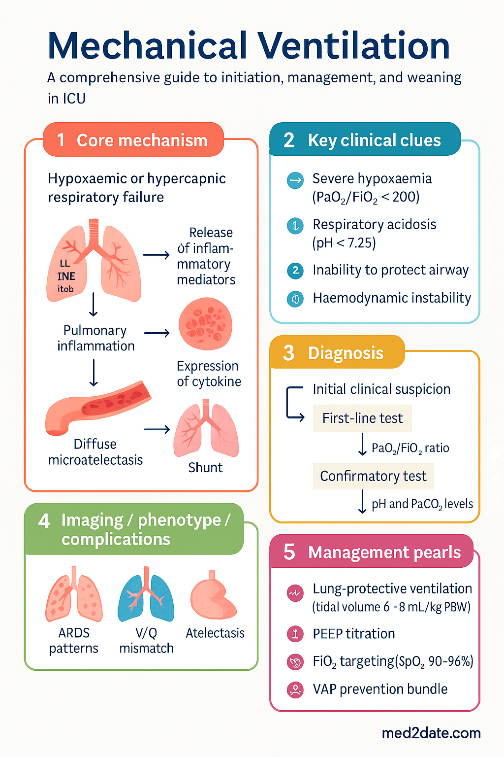
- Indications for invasive mechanical ventilation include severe hypoxaemic respiratory failure (PaO₂/FiO₂ <200), hypercapnic respiratory failure with respiratory acidosis (pH <7.25), inability to protect the airway, and haemodynamic instability requiring positive-pressure support.
- Respiratory failure types: Hypoxaemic (Type I) — impaired oxygenation from shunt, V/Q mismatch, or diffusion impairment; Hypercapnic (Type II) — alveolar hypoventilation from neuromuscular disease, chest wall restriction, or severe airflow obstruction.
- Initial ventilator settings for lung-protective ventilation: tidal volume 6–8 mL/kg predicted body weight (PBW), plateau pressure ≤30 cmH₂O, driving pressure ≤15 cmH₂O, and respiratory rate 20–30 breaths/min titrated to pH >7.25.
- Volume-controlled (VCV) delivers set tidal volume at variable pressure; pressure-controlled (PCV) delivers set pressure at variable volume. Both are acceptable; choose based on clinical context and patient comfort.
- PEEP selection should balance alveolar recruitment against haemodynamic compromise. Start at 5 cmH₂O and titrate using FiO₂/PEEP tables, driving pressure response, or ARDSNet protocol.
- FiO₂ titration: target SpO₂ 90–96% (88–92% in COPD). Wean FiO₂ first once PEEP is stabilised to minimise oxygen toxicity.
- Ventilator-induced lung injury (VILI) occurs via barotrauma, volutrauma, atelectrauma, and biotrauma. Lung-protective ventilation reduces mortality in ARDS (NNT ~10 per ARDSNetwork trial).
- VAP prevention bundle includes head-of-bed elevation 30–45°, daily sedation interruption, daily spontaneous breathing trial (SBT), oral care with chlorhexidine 0.12%, peptic ulcer prophylaxis, and DVT prophylaxis.
- Weaning readiness requires: resolution of underlying cause, adequate oxygenation (PaO₂/FiO₂ ≥150–200 on PEEP ≤5–8 cmH₂O), haemodynamic stability, intact respiratory drive, and adequate mental status.
- Spontaneous breathing trial (SBT) using low-level pressure support (PS 5–8 cmH₂O) or T-piece for 30–120 minutes is the standard readiness test. SBT failure demands return to prior support and reassessment within 24 hours.
- Post-extubation high-flow nasal cannula (HFNC) reduces reintubation rates in at-risk patients. Prophylactic NIV is recommended for high-risk extubation (hypercapnia, cardiac failure, failed SBT).
- Patient–ventilator dyssynchrony — trigger, flow, cycle, or auto-PEEP dyssynchrony — increases work of breathing, sedation requirements, and ICU length of stay; managed by adjusting trigger sensitivity, flow rates, and mode selection.
Introduction & Australian Epidemiology
Mechanical ventilation is the cornerstone of life support in the intensive care unit (ICU). In Australia, approximately 80,000–100,000 patients receive invasive mechanical ventilation annually across ~230 ICUs coordinated through the Australian and New Zealand Intensive Care Society (ANZICS). Mechanical ventilation accounts for the majority of ICU bed-days and is a principal determinant of ICU mortality, particularly in acute respiratory distress syndrome (ARDS) and severe pneumonia.
The ANZICS Centre for Outcome and Resource Evaluation (CORE) Adult Patient Database reports that approximately 30–40% of ICU admissions require invasive ventilation for ≥24 hours. In-hospital mortality for mechanically ventilated patients ranges from 25–45% depending on severity of illness, with ARDS-associated mortality of 35–46% for moderate-to-severe disease. The COVID-19 pandemic (2020–2023) substantially increased mechanical ventilation demand, with ~15–20% of hospitalised COVID-19 patients requiring ICU admission and invasive ventilation, prompting expansion of ICU capacity across Australian tertiary centres.
This guideline provides a comprehensive, evidence-based approach to initiation, management, complication avoidance, and liberation from mechanical ventilation, tailored to Australian clinical practice and resources.
Indications & Initiation
Indications for Invasive Mechanical Ventilation
Invasive mechanical ventilation is indicated when non-invasive support is insufficient or contraindicated. The decision to intubate and ventilate is a clinical one integrating respiratory function, mental status, fatigue signs, and trajectory of illness.
| Indication Category | Clinical Examples | Key Thresholds |
|---|---|---|
| Hypoxaemic respiratory failure (Type I) | ARDS, severe pneumonia, pulmonary oedema, pulmonary embolism, pneumothorax | PaO₂ <60 mmHg on FiO₂ ≥0.6, or PaO₂/FiO₂ <200 despite NIV/HFNC |
| Hypercapnic respiratory failure (Type II) | COPD exacerbation, severe asthma, neuromuscular disease (GBS, MND), obesity hypoventilation, drug overdose | pH <7.25 with PaCO₂ >60 mmHg despite maximal NIV |
| Airway protection | Reduced GCS (≤8), massive aspiration, angioedema, post-cardiac arrest | GCS ≤8 or inability to clear secretions |
| Haemodynamic instability | Cardiogenic shock, septic shock with respiratory compromise, massive PE | Requiring positive-pressure ventilation to offload work of breathing |
| Anticipated deterioration | Major burns >40% TBSA with inhalation injury, major surgery (post-operative), progressive neuromuscular weakness | Clinical trajectory assessment |
Respiratory Failure Types
Type I (Hypoxaemic) respiratory failure results from impaired gas exchange with relative or absolute preservation of alveolar ventilation. The hallmark is PaO₂ <60 mmHg (8 kPa) with normal or low PaCO₂. Pathophysiology includes intrapulmonary shunt (e.g., lobar pneumonia, ARDS), V/Q mismatch (e.g., pulmonary embolism), and diffusion impairment (e.g., pulmonary fibrosis). Non-invasive approaches (HFNC, CPAP, NIV) may suffice in moderate hypoxaemia, but progression to PaO₂/FiO₂ <150 or failed trial of NIV mandates invasive ventilation.
Type II (Hypercapnic) respiratory failure results from alveolar hypoventilation with elevated PaCO₂ (>45 mmHg / 6 kPa) and secondary hypoxaemia. Causes include depressed central respiratory drive (opioid overdose, anaesthesia), neuromuscular weakness (Guillain–Barré syndrome, myasthenia gravis, motor neurone disease), chest wall restriction (kyphoscoliosis, flail chest), and severe airflow obstruction (COPD, acute severe asthma). NIV is first-line for acute COPD exacerbation with respiratory acidosis; failure (pH <7.25 after 1–4 hours) prompts intubation.
Ventilator Modes
Ventilator modes describe the combination of control variable (volume or pressure), phase variable (trigger, target, cycle), and whether mandatory or spontaneous breaths are delivered. The following are the modes most commonly used in Australian ICUs:
| Mode | Abbreviation | Description | Indications |
|---|---|---|---|
| Assist Control — Volume Control | AC/VC or A/C-VCV | Every breath (patient- or machine-triggered) delivers set tidal volume. Guaranteed minute ventilation. | Initial mode for most patients; ARDS with lung-protective strategy |
| Assist Control — Pressure Control | AC/PC or A/C-PCV | Every breath delivers set inspiratory pressure for set time. Tidal volume varies with compliance. | Severe ARDS (with decelerating flow), patients with bronchopleural fistula |
| Synchronised Intermittent Mandatory Ventilation | SIMV | Set number of mandatory breaths (volume or pressure) + spontaneous breaths at patient effort. Spontaneous breaths may have PS support. | Weaning (largely supplanted by PS); some centres use for initial support |
| Pressure Support Ventilation | PSV | All breaths are patient-triggered, pressure-limited, flow-cycled. Patient controls rate and tidal volume. | SBT, weaning, NIV (BiPAP), post-extubation support |
| Pressure-Regulated Volume Control | PRVC | Pressure-controlled breaths with automatic adjustment of pressure to achieve target tidal volume. Decelerating flow pattern. | Hybrid mode; delivers volume guarantee with pressure waveform benefits |
| Neurally Adjusted Ventilatory Assist | NAVA | Uses diaphragmatic electrical activity (Edi) to trigger and cycle breaths. Proportional assist based on neural drive. | Improved synchrony; research tool and specialist centres |
| Airway Pressure Release Ventilation | APRV | Continuous positive airway pressure with brief, intermittent pressure releases for ventilation. Allows spontaneous breathing throughout. | Severe ARDS (evidence limited); used in some Australian centres |
Initial Ventilator Settings — Lung-Protective Strategy
All mechanically ventilated patients should receive lung-protective ventilation as the default strategy, not only those with ARDS. This is based on the landmark ARDSNetwork (2000) trial demonstrating 22% relative mortality reduction with low tidal volume ventilation.
Ventilator Management
Volume-Controlled vs Pressure-Controlled Ventilation
The choice between volume-controlled (VCV) and pressure-controlled (PCV) ventilation has been extensively debated. Current evidence demonstrates no significant difference in clinical outcomes (mortality, duration of ventilation) between the two modes when lung-protective targets are achieved. The decision should be guided by clinical context:
| Parameter | Volume-Controlled (VCV) | Pressure-Controlled (PCV) |
|---|---|---|
| Guaranteed parameter | Tidal volume (constant) | Inspiratory pressure (constant) |
| Varying parameter | Pressure varies with compliance and resistance | Tidal volume varies with compliance and resistance |
| Flow pattern | Square (constant) or decelerating | Decelerating (inherent) |
| Pplat monitoring | Essential — mandatory end-inspiratory hold | Approximate — peak pressure ≈ Pplat (brief pause) |
| Advantages | Guaranteed minute ventilation; precise volume control; easy titration of VT | Lower peak pressures; better patient comfort; improved distribution in heterogeneous lungs |
| Disadvantages | Higher peak pressures if compliance deteriorates; less comfortable if flow demand not met | VT not guaranteed — may rise or fall with changing compliance; requires close monitoring |
| Preferred when | Strict VT control needed (ARDS); precise CO₂ targeting | Severe ARDS with high PEEP; bronchopleural fistula; patient comfort priority |
PEEP Selection
Positive end-expiratory pressure (PEEP) prevents alveolar derecruitment at end-expiration, improves oxygenation, and reduces intrapulmonary shunt. However, excessive PEEP increases intrathoracic pressure, reduces venous return, and may cause overdistension of non-dependent lung regions.
ARDSNet FiO₂/PEEP Table (low PEEP strategy):
| FiO₂ | 0.3 | 0.4 | 0.4 | 0.5 | 0.5 | 0.6 | 0.7 | 0.7 | 0.8 | 0.9 | 1.0 |
|---|---|---|---|---|---|---|---|---|---|---|---|
| PEEP (cmH₂O) | 5 | 5 | 8 | 8 | 10 | 10 | 10 | 12 | 14 | 14 | 14–18 |
High PEEP strategy (ART trial, Alvarado et al. 2017): Higher PEEP levels (using open-lung approach with PEEP titration based on best compliance, recruitment manoeuvres, or decremental PEEP trial) have not shown consistent mortality benefit over low PEEP strategy. However, the EPVent-2 trial (2019) using oesophageal manometry to titrate transpulmonary pressure showed improved oxygenation with physiological PEEP titration. In practice, consider higher PEEP (12–18 cmH₂O) in moderate–severe ARDS (P/F ratio <200) if plateau pressure remains ≤30 cmH₂O.
FiO₂ Titration
The goal of oxygen therapy in mechanically ventilated patients is to maintain adequate tissue oxygenation while minimising the risks of hyperoxia (absorption atelectasis, oxidative lung injury, coronary and cerebral vasoconstriction) and hypoxaemia.
- Target SpO₂ 90–96% for most mechanically ventilated patients
- Target SpO₂ 88–92% in patients with COPD, chronic CO₂ retention, or those at risk of hypercapnic respiratory failure
- Initial FiO₂ 1.0 during intubation — rapidly wean to ≤0.6 within the first 30–60 minutes once PEEP is established
- Wean FiO₂ in decrements of 0.05–0.10 guided by continuous pulse oximetry and arterial blood gas (ABG) analysis
- Wean FiO₂ before PEEP — reduce FiO₂ to ≤0.60 before reducing PEEP below the target level
- Limit FiO₂ to ≤0.60 long-term to reduce oxygen toxicity; if higher FiO₂ is needed, consider recruitment manoeuvres or prone positioning
Sedation Protocols
Sedation is a critical component of mechanical ventilation management, balancing patient comfort, safety, ventilator synchrony, and the adverse effects of excessive sedation (prolonged ventilation, ICU-acquired weakness, delirium).
Analgosedation approach: Current best practice favours an analgesia-first (analgosedation) strategy. Assess and treat pain first using the Critical-Care Pain Observation Tool (CPOT) or Behavioural Pain Scale (BPS). Commence IV paracetamol (1 g QID) and opioid (fentanyl or morphine) for analgesia. Add propofol or dexmedetomidine only if agitation persists despite adequate analgesia. Avoid benzodiazepine-based sedation where possible due to association with delirium (MIDEX/PRODEX trials).
Delirium Management
ICU delirium affects 45–87% of mechanically ventilated patients and is independently associated with increased 6-month mortality. Routine screening with the Confusion Assessment Method for ICU (CAM-ICU) or Intensive Care Delirium Screening Checklist (ICDSC) every 8–12 hours is mandatory per ACSQHC guidelines. Non-pharmacological interventions include reorientation, early mobilisation, sleep hygiene, hearing aids, and family presence. Pharmacological management with antipsychotics (haloperidol or quetiapine) is reserved for distressing delirium or safety concerns.
Complications of Mechanical Ventilation
Ventilator-Induced Lung Injury (VILI)
VILI is the acute lung injury caused or exacerbated by mechanical ventilation. It is mediated by four primary mechanisms, collectively termed the "four traumas of VILI":
Barotrauma
Barotrauma occurs in 5–15% of mechanically ventilated patients and up to 20% of those with severe ARDS. Manifestations include pneumothorax (most common), pneumomediastinum, subcutaneous emphysema, pneumoperitoneum, and tension physiology. Risk factors include high PEEP (>15 cmH₂O), high Pplat (>30 cmH₂O), ARDS (especially with cystic changes on CT), and COPD with bullous disease.
Management of pneumothorax: Tension pneumothorax requires immediate needle decompression (2nd intercostal space, mid-clavicular line) followed by intercostal catheter (ICC) insertion. Non-tension pneumothorax in a ventilated patient always warrants ICC insertion due to positive-pressure ventilation risk of tension conversion. Use a 20–24 Fr ICC connected to underwater seal with −20 cmH₂O suction.
Ventilator-Associated Pneumonia (VAP)
VAP is pneumonia occurring >48 hours after endotracheal intubation. It is the most common ICU-acquired infection, affecting 10–20% of mechanically ventilated patients, with attributable mortality of 9–13%. In Australia, the VAP rate is approximately 8–12 episodes per 1000 ventilator-days (ANZICS infection monitoring).
Diagnostic criteria (ATS/IDSA 2016): New or progressive infiltrate on chest imaging + ≥2 of: fever >38°C or hypothermia <36°C, leucocytosis (>12 × 10⁹/L) or leucopenia (<4 × 10⁹/L), purulent secretions. Confirm with quantitative BAL (≥10⁴ CFU/mL) or endotracheal aspirate culture (≥10⁶ CFU/mL).
VAP prevention bundle components:
VAP Empirical Antibiotic Therapy
Empirical antibiotic therapy for suspected VAP should be guided by local antibiograms (which vary across Australian hospitals) and risk factors for multidrug-resistant organisms (MDROs). Use eTG Antibiotic guidelines and local protocols. Duration of therapy: 7 days for uncomplicated VAP (per IDSA/ATS 2016; Pugh et al. 2016).
Patient–Ventilator Dyssynchrony
Dyssynchrony occurs when the ventilator and patient are not properly coordinated. It increases work of breathing, oxygen consumption, sedation requirements, and may worsen VILI. The four major types:
| Type | Description | Cause | Management |
|---|---|---|---|
| Trigger dyssynchrony | Patient efforts fail to trigger a breath (ineffective triggering) or trigger extra breaths (auto-triggering) | Weak respiratory effort, auto-PEEP, trigger sensitivity too low, cardiac oscillations | Reduce trigger threshold; treat auto-PEEP (bronchodilators, reduce RR); consider PSV mode |
| Flow dyssynchrony | Patient inspiratory flow demand exceeds or falls below ventilator flow delivery | Fixed inspiratory flow too low (VCV); excessive drive from pain, anxiety, or acidosis | Increase peak flow (VCV) or use decelerating waveform; switch to PCV or PSV; treat underlying cause |
| Cycle dyssynchrony | Ventilator ends inspiration before (short cycling) or after (prolonged cycling) the patient's neural inspiration | Inspiratory time mismatch; short cycling in obstructive physiology; prolonged cycling in weak patients | Adjust inspiratory time; reduce flow termination criterion (PSV cycle-off); adjust I:E ratio |
| Auto-PEEP dyssynchrony | Intrinsic PEEP prevents triggering because patient must generate enough effort to overcome auto-PEEP before triggering threshold | Dynamic hyperinflation in COPD/asthma; high minute ventilation; inadequate expiratory time | Reduce respiratory rate; increase inspiratory flow; shorten I:E ratio; administer bronchodilators; set external PEEP to 80% of auto-PEEP |
Additional complications:
- ICU-acquired weakness (ICU-AW): Occurs in 25–50% of patients ventilated >7 days. Risk factors include corticosteroid use, neuromuscular blocking agents, sepsis, and immobility. Diagnosed by MRC sum score <48 or grip strength <11 kg (males) / <7 kg (females). Prevention through early mobilisation and minimising sedation is key.
- Endotracheal tube complications: Mucosal injury, subglottic stenosis, vocal cord granuloma. Use appropriately sized ETT (7.0–8.0 for adult females, 7.5–8.5 for adult males). Subglottic secretion drainage ETTs reduce VAP risk.
- Haemodynamic compromise: Positive-pressure ventilation reduces preload (venous return) and increases right ventricular afterload. Monitor for hypotension after PEEP increases. Fluid responsiveness may be assessed by pulse pressure variation (PPV) or passive leg raise test.
Weaning & Liberation from Mechanical Ventilation
Weaning Classification
Weaning accounts for approximately 40% of total ventilation duration. Patients are classified into three categories based on difficulty and duration of weaning:
Readiness to Wean — Screening Criteria
Daily screening for weaning readiness should be performed every morning by the bedside nurse or physiotherapist. All of the following criteria must be met:
- Resolution or improvement of cause of respiratory failure
- Adequate oxygenation: PaO₂/FiO₂ ≥150 (or SpO₂ ≥90% on FiO₂ ≤0.40 and PEEP ≤8 cmH₂O)
- Haemodynamic stability: No active myocardial ischaemia; no clinically significant hypotension (noradrenaline <0.1 mcg/kg/min or no vasopressor requirement)
- Adequate respiratory drive: Capable of initiating spontaneous breaths
- No planned procedures or surgery requiring heavy sedation/paralysis
- Adequate mental status: GCS ≥13, following commands (modified criteria acceptable in patients with baseline cognitive impairment)
- Adequate cough reflex and manageable secretions
- Core temperature 36–38.5°C
- Corrected electrolytes: K⁺ ≥3.5 mmol/L, Mg²⁺ ≥0.8 mmol/L, PO₄³⁻ ≥0.65 mmol/L, Ca²⁺ ≥1.1 mmol/L (ionised)
Spontaneous Breathing Trial (SBT)
The SBT is the gold-standard test of readiness for extubation. It assesses the patient's ability to breathe spontaneously with minimal support. Two principal methods are used in Australian ICUs:
| SBT Method | Settings | Duration | Evidence |
|---|---|---|---|
| Low-level Pressure Support | PS 5–8 cmH₂O, PEEP 5 cmH₂O, FiO₂ 0.40 | 30–120 minutes (30 min is sufficient per Perren et al. 2002) | Preferred in most Australian ICUs; provides unloading of ETT resistance |
| T-Piece Trial | T-piece connected to ETT; supplemental O₂ via flow-by | 30–120 minutes | More physiological; slightly more demanding; equivalent outcomes |
| CPAP | CPAP 5 cmH₂O, FiO₂ 0.40 | 30–120 minutes | Acceptable alternative; similar to PS 0 but eliminates ETT resistance compensation |
SBT failure criteria: Any of the following within 30–120 minutes warrants return to prior ventilator support:
- Respiratory rate >35 breaths/min sustained for >5 minutes
- SpO₂ <88% on FiO₂ ≤0.50
- Heart rate >140 or <50 bpm, or sustained change of >20% from baseline
- Systolic BP >180 mmHg or <90 mmHg
- Agitation, diaphoresis, anxiety, or altered mental status
- Evidence of increased work of breathing: accessory muscle use, paradoxical breathing, nasal flaring
Extubation Criteria & Procedure
Successful SBT is necessary but not sufficient for extubation. Additional extubation readiness criteria include:
- Adequate cough: Semi-quantitative cough strength score ≥3 (can cough against resistance)
- Secretion burden: Manageable with suctioning ≤ every 2 hours
- Upper airway patency: Cuff leak test positive (leak volume >110 mL or >15% of expired VT). Negative cuff leak suggests laryngeal oedema — consider corticosteroid prophylaxis (see below).
- No active upper airway obstruction or stridor on deflation of ETT cuff
Corticosteroids for laryngeal oedema prevention: For patients with a failed cuff leak test, administer methylprednisolone 20 mg IV 4 hours before planned extubation, then every 4 hours for 24 hours total (4 doses). This reduces stridor and reintubation for post-extubation laryngeal oedema (François et al. 2007, Chest; FACCLE trial).
Post-Extubation Care
Post-extubation respiratory failure occurs in 10–25% of critically ill patients and is associated with significantly increased ICU and hospital mortality.
Reintubation
Reintubation rates of 10–20% are expected. Risk factors for reintubation include: age >65, APACHE II >12, heart failure, COPD, duration of ventilation >7 days, secretions copious or unmanageable, failed SBT, and post-extubation stridor. Reintubation within 48–72 hours is associated with significantly higher ICU mortality (up to 40%). A structured post-extubation protocol reduces reintubation by 40–50%.
Tracheostomy for Prolonged Ventilation
Tracheostomy should be considered when prolonged ventilation (>10–14 days) is anticipated. Early tracheostomy (within 7–10 days) may reduce ICU length of stay, sedation requirements, and laryngeal injury but has not been shown to reduce mortality (TracMan trial, 2013). Percutaneous dilatational tracheostomy (PDT) is the preferred technique in Australian ICUs, performed at the bedside under bronchoscopic guidance. Typically placed at the 2nd–3rd tracheal ring. Key considerations include anticoagulation management, anatomical assessment (thyromental distance, neck mobility), and timing relative to decannulation planning.
Special Populations
Pregnancy
Paediatrics
Elderly (≥65 years)
Renal Impairment
Hepatic Impairment
Immunocompromised
Obesity
Aboriginal and Torres Strait Islander Health Considerations
Aboriginal and Torres Strait Islander Australians experience disproportionately higher rates of respiratory disease and critical illness. The AIHW reports that Indigenous Australians are hospitalised for respiratory conditions at 2.3 times the rate of non-Indigenous Australians. Chronic suppurative lung disease, bronchiectasis, and chronic obstructive pulmonary disease (COPD) are more prevalent, often presenting at younger ages with greater severity. The burden of rheumatic heart disease, obesity, diabetes, and chronic kidney disease contributes to increased complexity when invasive mechanical ventilation is required.
Critically ill Indigenous patients more frequently present from remote and very remote areas, where access to tertiary ICUs requires aeromedical retrieval (Royal Flying Doctor Service, CareFlight). Delays in retrieval and the physiological stress of transport may result in more advanced illness at the time of ICU admission. ANZICS data indicate that Indigenous Australians admitted to ICU have higher severity of illness scores (APACHE III) at admission but comparable or improved mortality when adjusting for severity, suggesting effective ICU care when patients reach tertiary centres.
📚 References
- 1. Acute Respiratory Distress Syndrome Network, Brower RG, Matthay MA, et al. Ventilation with lower tidal volumes as compared with traditional tidal volumes for acute lung injury and the acute respiratory distress syndrome. N Engl J Med. 2000;342(18):1301–1308.
- 2. Amato MBP, Meade MO, Slutsky AS, et al. Driving pressure and survival in the acute respiratory distress syndrome. N Engl J Med. 2015;372(8):747–755.
- 3. Girard TD, Kress JP, Fuchs BD, et al. Efficacy and safety of a paired sedation and ventilator weaning protocol for mechanically ventilated patients in intensive care (Awakening and Breathing Controlled trial): a randomised controlled trial. Lancet. 2008;371(9607):126–134.
- 4. Kalil AC, Metersky ML, Klompas M, et al. Management of adults with hospital-acquired and ventilator-associated pneumonia: 2016 clinical practice guidelines by the Infectious Diseases Society of America and the American Thoracic Society. Clin Infect Dis. 2016;63(5):e61–e111.
- 5. Hernández G, Vaquero C, González P, et al. Effect of postextubation high-flow nasal cannula vs conventional oxygen therapy on reintubation in low-risk patients: a randomized clinical trial. JAMA. 2016;315(13):1354–1361.
- 6. Thille AW, Muller G, Gacouin A, et al. Effect of postextubation high-flow nasal oxygen with noninvasive ventilation vs high-flow nasal oxygen alone on reintubation among patients at high risk of extubation failure: a randomized clinical trial. JAMA. 2019;322(15):1465–1475.
- 7. Young D, Harrison DA, Cuthbertson BH, Rowan K; TracMan Collaborators. Effect of early vs late tracheostomy on duration of mechanical ventilation and other outcomes: the TracMan randomized trial. JAMA. 2013;309(20):2121–2129.
- 8. François B, Bellissant E, Gissot V, et al. 12-h pretreatment with methylprednisolone versus placebo for prevention of postextubation laryngeal oedema: a randomised double-blind trial. Lancet. 2007;369(9567):1083–1089.
- 9. National Health and Medical Research Council (NHMRC). Australian guidelines for the prevention and control of infection in healthcare. Canberra: NHMRC; 2019 (updated 2024).
- 10. Australian Commission on Safety and Quality in Health Care (ACSQHC). National Safety and Quality Health Service Standards. 2nd ed. Sydney: ACSQHC; 2021.
- 11. Australian and New Zealand Intensive Care Society (ANZICS). Centre for Outcome and Resource Evaluation (CORE) Annual Report 2023. Melbourne: ANZICS; 2023.
- 12. Australian Institute of Health and Welfare (AIHW). Aboriginal and Torres Strait Islander Health Performance Framework 2023 summary report. Canberra: AIHW; 2023.
- 13. Goligher EC, Tomlinson G, Hajage D, et al. PEEP, driving pressure, and lung-protective ventilation in ARDS: a systematic review and meta-analysis of individual patient data. Am J Respir Crit Care Med. 2017;196(6):730–739.
- 14. Needham DM, Colantuoni E, Mendez-Tellez PA, et al. Lung protective mechanical ventilation and two year survival in patients with acute lung injury: prospective cohort study. BMJ. 2012;344:e2124.
- 15. RHDAustralia (Rheumatic Heart Disease Australia). RHDAustralia clinical guidelines: Acute rheumatic fever and rheumatic heart disease. 3rd ed. Darwin: RHDAustralia; 2020.