📋 Key Information Summary
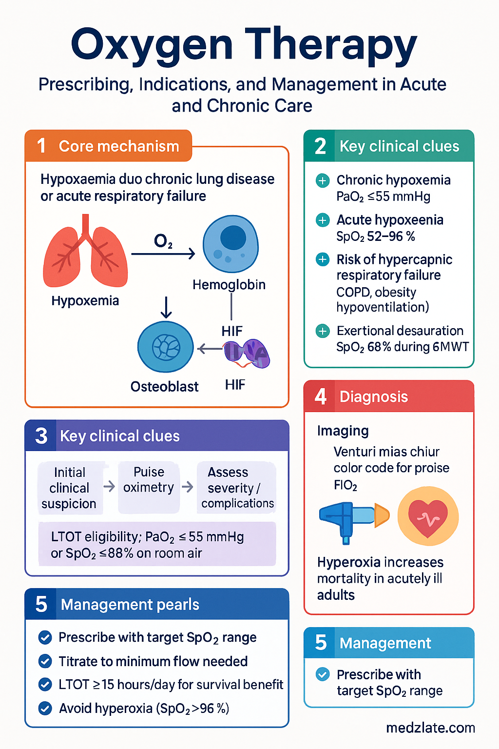
- Oxygen is a medication — it must be prescribed with a target SpO₂ range, flow rate, delivery device, and duration.
- In acute care, target SpO₂ 92–96% for most patients; 88–92% for those at risk of hypercapnic respiratory failure (COPD, obesity hypoventilation, bronchiectasis, neuromuscular disease).
- Long-term oxygen therapy (LTOT) is indicated for chronic resting hypoxaemia: PaO₂ ≤55 mmHg (≤7.3 kPa) or SpO₂ ≤88%, or PaO₂ 56–59 mmHg with cor pulmonale, polycythaemia (Hct >55%), or pulmonary hypertension.
- LTOT must be prescribed for ≥15 hours/day (ideally continuous) to improve survival; the BTS and TGA criteria require documentation of persistent hypoxaemia on two occasions ≥3 weeks apart.
- Nasal cannulae deliver 1–6 L/min (≈24–44% FiO₂); simple masks 5–10 L/min (≈35–55%); Venturi masks deliver precise fixed FiO₂ (24%, 28%, 31%, 35%, 40%, 60%).
- High-flow nasal cannula (HFNC) delivers heated, humidified oxygen at 20–80 L/min with precise FiO₂ 21–100%; indicated in acute hypoxaemic respiratory failure, post-extubation, and pre-oxygenation.
- Ambulatory oxygen is indicated when patients on LTOT demonstrate exertional desaturation (SpO₂ <88% during a 6-minute walk test) despite supplemental oxygen during rest.
- Pulse-dose (demand) delivery conserves oxygen and suits mobile patients; continuous flow is preferred during sleep, exercise, and for patients with high minute ventilation.
- Always titrate oxygen to the minimum flow/FiO₂ needed to achieve target SpO₂; hyperoxia (SpO₂ >96%) increases mortality in acutely ill adults and should be avoided.
- Oxygen therapy equipment includes concentrators (electrically powered, stationary), compressed gas cylinders, and liquid oxygen systems; selection depends on flow needs, mobility, and home environment.
- Aboriginal and Torres Strait Islander peoples experience higher rates of chronic lung disease and respiratory hospitalisations; access to LTOT and pulmonary rehabilitation must be culturally safe and geographically supported.
Introduction & Australian Epidemiology
Oxygen therapy is one of the most commonly prescribed treatments in acute and chronic medicine. Despite its ubiquity, inappropriate use — both under-treatment of hypoxaemia and injudicious hyperoxia — is associated with increased morbidity and mortality. The Australian Commission on Safety and Quality in Health Care (ACSQHC) and the Thoracic Society of Australia and New Zealand (TSANZ) recommend that oxygen be treated as a drug: prescribed, titrated, monitored, and reviewed.
In Australia, chronic obstructive pulmonary disease (COPD) affects approximately 7.5% of adults aged ≥40 years (AIHW, 2023), and is the fifth leading cause of death. Long-term oxygen therapy (LTOT) is used by an estimated 30,000–40,000 Australians at any given time, predominantly for COPD-related chronic hypoxaemia. Idiopathic pulmonary fibrosis (IPF), bronchiectasis, cystic fibrosis, pulmonary hypertension, and neuromuscular diseases account for the remaining indications.
The Ambulance Victoria and NSW clinical frameworks mandate target SpO₂-directed oxygen therapy, and the National Safety and Quality Health Service (NSQHS) Standards include oxygen safety as a core clinical governance item (Standard 5: Comprehensive Care).
Indications & Prescribing
Core Indications for Supplemental Oxygen
| Category | Indication | Target SpO₂ |
|---|---|---|
| Acute hypoxaemia | SpO₂ <92% (or <88% in known CO₂ retainers) | 92–96% (88–92% if hypercapnic risk) |
| Critical illness | Sepsis, shock, major trauma, cardiac arrest | 92–96% (after initial stabilisation) |
| Exertional desaturation | SpO₂ <88% during 6-minute walk test (6MWT) | ≥88–90% with ambulatory O₂ |
| Nocturnal hypoxaemia | SpO₂ <88% for >30% of sleep time (oximetry/polygraphy) | ≥88–90% overnight |
| Chronic resting hypoxaemia (LTOT) | PaO₂ ≤55 mmHg (≤7.3 kPa) or SpO₂ ≤88% on room air, documented twice ≥3 weeks apart | PaO₂ >60 mmHg / SpO₂ >90% |
| Near-threshold with complications | PaO₂ 56–59 mmHg with cor pulmonale, Hct >55%, or pulmonary hypertension | Same as LTOT |
| Carbon monoxide poisoning | Any confirmed or suspected COHb elevation | 100% FiO₂ until COHb <3% |
| Palliative / symptom relief | Refractory dyspnoea in advanced disease | Symptom-directed; comfort SpO₂ |
Resting Hypoxaemia Criteria (LTOT Eligibility)
The Australian Therapeutic Goods Administration (TGA) and TSANZ align with international BTS/ERS criteria. LTOT eligibility requires measurement of arterial blood gas (ABG) on room air (after ≥20 minutes off supplemental oxygen) on two separate occasions at least 3 weeks apart, during a period of clinical stability (no acute exacerbation for ≥4 weeks). Either arterial or arterialised capillary samples are acceptable.
Prescribing Template (Australian Standard)
- Medication: Oxygen (O₂)
- Device: e.g., nasal cannula, Venturi mask, HFNC
- Flow rate / FiO₂: e.g., 2 L/min nasal cannula or 28% Venturi
- Target SpO₂: e.g., 92–96% or 88–92%
- Duration: e.g., continuous, PRN for SpO₂ <90%, nocturnal, during exertion
- Review date: e.g., within 24 hours / at next ward round
When NOT to Prescribe Oxygen
Oxygen is not indicated for breathlessness in the absence of hypoxaemia. Routine use in acute myocardial infarction with SpO₂ ≥94% is harmful (DETO2X-AMI trial, NEJM 2017). Hyperoxia (SpO₂ >96%) in acutely ill patients increases free radical injury, coronary and cerebral vasoconstriction, and is associated with higher in-hospital mortality in ICU populations (OXYGEN-ICU trial, ICU 2016).
Delivery Systems
The choice of delivery system depends on the severity of hypoxaemia, the required FiO₂ precision, patient tolerance, and whether humidification is needed. Devices range from low-flow systems (where the patient supplements inspired gas with room air) to high-flow systems that can meet or exceed inspiratory demand.
Low-Flow Devices
| Device | Flow Rate | Approximate FiO₂ | Key Considerations |
|---|---|---|---|
| Nasal cannula | 1–6 L/min | 24–44% | Most common in acute and home settings; comfortable; flows >4 L/min dry nasal mucosa — use humidification |
| Simple face mask | 5–10 L/min | 35–55% | Minimum 5 L/min to prevent CO₂ rebreathing; not for eating/drinking; useful for short-term moderate hypoxaemia |
| Reservoir (non-rebreather) mask | 10–15 L/min | 60–90% | Reservoir bag must remain >½ full; one-way valve prevents rebreathing; critical/emergency use |
| Reservoir (partial rebreather) mask | 8–12 L/min | 50–75% | No one-way valve; some rebreathing permitted; less common |
High-Flow / Precise-FiO₂ Devices
| Device | Flow Rate | Approximate FiO₂ | Key Considerations |
|---|---|---|---|
| Venturi mask | Variable (4–15 L/min depending on adapter) | 24%, 28%, 31%, 35%, 40%, 60% (fixed by colour-coded adapter) | Delivers precise FiO₂; preferred in COPD and hypercapnic respiratory failure; each adapter requires a specified minimum flow |
| High-flow nasal cannula (HFNC) | 20–80 L/min | 21–100% | Heated, humidified; reduces anatomical dead space; generates low-level CPAP (~2–5 cmH₂O); see dedicated section below |
| T-piece / tracheostomy collar | Variable (typically 10–40 L/min) | Up to 100% | Used for spontaneously breathing tracheostomy patients; heated humidification recommended |
Venturi Mask Colour Codes & Flow Requirements
| Colour | FiO₂ (%) | Minimum Flow (L/min) | Typical Use |
|---|---|---|---|
| Blue | 24 | 4 | COPD — mild hypoxaemia, target SpO₂ 88–92% |
| White | 28 | 4 | COPD — moderate hypoxaemia |
| Orange | 31 | 6 | COPD — moderate-severe hypoxaemia |
| Yellow | 35 | 8 | Non-hypercapnic moderate hypoxaemia |
| Red | 40 | 10 | Non-hypercapnic moderate-severe hypoxaemia |
| Green | 60 | 15 | Severe hypoxaemia pending escalation |
Home Oxygen Therapy
Long-Term Oxygen Therapy (LTOT)
LTOT is one of the few interventions in COPD that improves survival when prescribed ≥15 hours/day. The landmark NOTT (Nocturnal Oxygen Therapy Trial, 1980) and MRC (Medical Research Council, 1981) trials established that continuous LTOT reduces mortality, polycythaemia, and pulmonary vascular resistance in patients with severe chronic hypoxaemia.
LTOT Eligibility Criteria (Australian)
Ambulatory Oxygen Therapy
Ambulatory oxygen is indicated for patients who desaturate during physical activity (SpO₂ <88% on a 6-minute walk test) and who demonstrate improvement in exercise capacity and/or dyspnoea during a supervised trial of supplemental oxygen during exercise. It may be prescribed independently of LTOT or in addition to it.
- Assessment: standardised 6MWT on room air, then repeated with supplemental oxygen at titrated flow.
- Minimum meaningful improvement: ≥10% increase in 6MWT distance or significant reduction in dyspnoea (Borg scale ≥2-point improvement).
- Typical prescription: 2–6 L/min via nasal cannula during exertion.
- Equipment: lightweight portable cylinder (D, E size), portable oxygen concentrator (POC), or liquid oxygen portable unit (LOX).
Pulse-Dose vs Continuous Flow
| Feature | Continuous Flow | Pulse-Dose (Demand) |
|---|---|---|
| Mechanism | Constant flow regardless of respiratory cycle | Delivers a bolus at the start of each detected inspiratory effort |
| Oxygen conservation | Higher consumption | 3:1 to 5:1 conservation ratio |
| Battery life (POC) | Shorter | Substantially longer |
| Preferred during sleep | Yes — pulse may not detect shallow sleep breathing | No — unreliable triggering in sleep or very shallow breaths |
| Preferred during exercise | Yes — reliable at high respiratory rates | Variable — may not keep up at high rates >25/min |
| PBS funding | Concentrator or cylinders via LTOT approval | POC or cylinders; additional PBS authority required for ambulatory component |
Equipment Selection for Home Oxygen
Titration of Home Oxygen
- Initial titration: Performed by a respiratory scientist or specialist nurse; ABG or SpO₂-guided increase in flow until PaO₂ >60 mmHg or SpO₂ >90% at rest while awake.
- Nocturnal titration: Overnight oximetry to confirm SpO₂ ≥88–90% throughout sleep; increase nocturnal flow by 0.5–1 L/min above daytime if nocturnal desaturation persists.
- Exertional titration: 6MWT-guided; increase flow by 1–2 L/min above resting flow to maintain SpO₂ ≥88% during standardised exercise.
- Review schedule: Clinical review at 1, 3, and 6 months after initiation, then at least 6-monthly. ABG and functional assessment should be repeated at each review.
- Weaning/discontinuation: Reassess at 60–90 days of stability; if PaO₂ >59 mmHg on repeat ABG, consider trial off oxygen with close monitoring.
High-Flow Nasal Cannula (HFNC)
Overview & Mechanism
High-flow nasal cannula (HFNC) therapy delivers heated (37°C), fully humidified oxygen/air mixtures at flows of 20–80 L/min with precise FiO₂ control from 0.21 to 1.0. HFNC has become a cornerstone of respiratory support in Australian emergency departments and ICUs.
HFNC provides several physiological benefits beyond simple oxygen delivery:
- Washout of nasopharyngeal dead space: High flows flush CO₂ from the anatomical dead space, improving alveolar ventilation and reducing the work of breathing.
- Low-level positive end-expiratory pressure (PEEP): Generates approximately 2–5 cmH₂O of PEEP at flow rates of 30–60 L/min (mouth-closed); recruitment of alveoli, improved oxygenation.
- Reduced inspiratory resistance: Matches or exceeds patient peak inspiratory flow demand, reducing entrainment of room air and providing more consistent FiO₂.
- Mucociliary clearance: Humidification at 37°C preserves ciliary function and reduces inspissated secretions.
Clinical Indications
Additional HFNC Indications
- Immunocompromised patients: Early HFNC in febrile neutropenia and post-HSCT respiratory failure reduces intubation and ICU mortality (HIGH trial, Lancet Resp Med 2018).
- Acute heart failure (cardiogenic pulmonary oedema): HFNC improves dyspnoea and reduces work of breathing; may be used as a bridge to NIV or in mild-moderate cases (B UIStoryboardSegue trial).
- Bronchiolitis (paediatric): HFNC at 1–2 L/kg/min is first-line respiratory support in infants with moderate-severe bronchiolitis (PREDICT/ANZICS guidelines, 2019).
- Palliative care: HFNC for refractory dyspnoea in end-stage respiratory disease when conventional oxygen fails to provide comfort.
Flow & FiO₂ Settings
| Parameter | Adult Starting | Adult Escalation | Paediatric Starting | Notes |
|---|---|---|---|---|
| Flow rate | 30–40 L/min | Titrate up to 50–60 L/min (max 80 L/min) | 1–2 L/kg/min (max 8 L/kg in bronchiolitis) | Higher flow = better dead-space washout and PEEP; comfort is a limiting factor |
| FiO₂ | Set to match current delivery (e.g., 0.50) | Titrate in 0.05–0.10 increments every 15–30 min to target SpO₂ | 0.30–0.50 initially | Wean FiO₂ first once SpO₂ stabilised; wean flow last |
| Temperature | 37°C | 37°C (34°C if patient febrile or uncomfortable) | 37°C | Full humidification at 37°C maximises mucociliary clearance |
Initiation & Escalation Protocol
Contraindications to HFNC
- Cardiac or respiratory arrest (immediate intubation/airway management)
- Severe haemodynamic instability requiring vasopressors (relative)
- Inability to protect airway (GCS ≤8, absent gag/cough reflex)
- Upper airway obstruction (e.g., epiglottitis, tumour) — will not bypass obstruction
- Severe facial trauma preventing nasal cannula application
- Untreated pneumothorax (relative — low-level PEEP risk)
Pathophysiology of Hypoxaemia
Understanding the mechanisms of hypoxaemia guides the appropriate use of supplemental oxygen. There are five principal mechanisms, which often coexist:
| Mechanism | A–a Gradient | Response to O₂ | Common Causes |
|---|---|---|---|
| V/Q mismatch | Elevated | Good — easily corrected | COPD, asthma, pneumonia, PE, pulmonary oedema |
| Shunt (true R→L) | Elevated | Poor — does not respond to 100% O₂ | Intrapulmonary (ARDS, large consolidation); intracardiac (ASD, VSD) |
| Diffusion impairment | Elevated (worse with exercise) | Good at rest; moderate with exercise | IPF, asbestosis, pulmonary fibrosis, emphysema (reduced surface area) |
| Hypoventilation | Normal | Corrects hypoxaemia but may worsen hypercapnia | Obesity hypoventilation, OHS, neuromuscular disease, opioid overdose |
| Low PiO₂ | Normal | Excellent | High altitude (not relevant to Australian practice at sea level) |
The alveolar–arterial (A–a) oxygen gradient is calculated as: A–a gradient = [FiO₂ × (Patm – PH₂O) – (PaCO₂ / R)] – PaO₂, where Patm ≈ 760 mmHg, PH₂O = 47 mmHg, R = 0.8. A normal A–a gradient is approximately (Age/4) + 4 mmHg.
Investigations
Monitoring
Acute Inpatient Monitoring
Home Oxygen Monitoring
- Patient self-monitoring: Spot-check SpO₂ with personal oximeter (≥2× daily); report SpO₂ <88% to GP or respiratory nurse.
- GP review: At 1, 3, and 6 months post-initiation, then 6-monthly. Assess compliance (hours/day), functional status, oxygen flow requirements, and side effects (nasal dryness, epistaxis, skin breakdown).
- Repeat ABG: At 60–90 days to assess response; annually or if clinically deteriorating.
- Equipment servicing: Concentrators: serviced every 12 months by supplier. Cylinders: checked for valve integrity. Portable units: battery health check at each supplier visit.
- Re-assessment for discontinuation: If stable and non-smoking with improved PaO₂ on repeat ABG, a supervised trial off oxygen may be considered at 6–12 months.
Special Populations
Pregnancy
Paediatrics
Elderly
Renal Impairment
Hepatic Impairment
Immunocompromised
Aboriginal and Torres Strait Islander Health Considerations
Aboriginal and Torres Strait Islander peoples experience a disproportionate burden of chronic respiratory disease, including COPD, bronchiectasis, and rheumatic heart disease–associated pulmonary hypertension. COPD prevalence in Indigenous Australians is approximately 2–3 times that of non-Indigenous Australians, with onset a decade earlier and higher rates of hospitalisation and death (AIHW, 2023). These conditions drive substantial demand for long-term and acute oxygen therapy.
📚 References
- 1. British Thoracic Society (BTS). Guideline for oxygen use in adults in healthcare and emergency settings. BMJ Open Resp Res. 2017;4:e000170.
- 2. Beasley R, Chien J, Douglas J, et al. Thoracic Society of Australia and New Zealand oxygen guidelines for acute oxygen use in adults. Respirology. 2016;21(8):1434–1448.
- 3. Stoller JK, Panos RJ, Krachman S, et al. Oxygen therapy for patients with COPD: current evidence and the long-term oxygen treatment trial. Chest. 2010;138(1):179–187.
- 4. Frat JP, Thille AW, Mercat A, et al. High-flow oxygen through nasal cannula in acute hypoxemic respiratory failure. N Engl J Med. 2015;372(23):2185–2196. (FLORALI trial)
- 5. Hernandez G, Vaquero C, Gonzalez P, et al. Effect of postextubation high-flow nasal cannula vs conventional oxygen therapy on reintubation in low-risk patients: a randomized clinical trial. JAMA. 2016;315(13):1354–1361.
- 6. Hofmann R, James SK, Jernberg T, et al. Oxygen therapy in suspected acute myocardial infarction. N Engl J Med. 2017;377(13):1240–1249. (DETO2X-AMI trial)
- 7. Chu DK, Kim LH, Young PJ, et al. Mortality and morbidity in acutely ill adults treated with liberal versus conservative oxygen therapy (IOTA): a systematic review and meta-analysis. Lancet. 2018;391(10131):1693–1705.
- 8. Australian Institute of Health and Welfare (AIHW). Chronic obstructive pulmonary disease (COPD). AIHW; 2023.
- 9. Huang DT, Angus DC, Moss M, et al. Design and rationale of the reevaluation of systemic early neuromuscular blockade trial for acute respiratory distress syndrome. Ann Am Thorac Soc. 2017;14(1):124–133. (Relevant: OXYGEN-ICU trial, Girardis et al. JAMA 2016;316:1583–1589)
- 10. Renda T, Corrado A, Iskandar G, et al. High-flow nasal oxygen therapy in intensive care and anaesthesia. Br J Anaesth. 2018;120(1):18–27.
- 11. Frank LH, Greenough A, et al. British Thoracic Society guideline for home oxygen use in adults. Thorax. 2015;70(Suppl 1):i1–i43.
- 12. Franklin D, Babl FE, Schlapbach LJ, et al. A randomized trial of high-flow oxygen therapy in infants with bronchiolitis. N Engl J Med. 2018;378(12):1121–1131. (PREDICT/ANZICS)
- 13. Australian Commission on Safety and Quality in Health Care (ACSQHC). National Safety and Quality Health Service Standards. 2nd ed. Sydney: ACSQHC; 2021.
- 14. Chang AB, Bell SC, Torzillo PJ, et al. Chronic suppurative lung disease and bronchiectasis in children and adults in Australia and New Zealand. Thoracic Society of Australia and New Zealand (TSANZ) Bronchiectasis Guidelines. Med J Aust. 2015;202(1):21–23.
- 15. Yan S, Chai X, et al. ROX index as a predictor of high-flow nasal cannula outcome: a systematic review and meta-analysis. Crit Care. 2022;26:258.