📋 Key Information Summary
- Pulmonary embolism (PE) affects approximately 8,000–10,000 Australians annually; incidence increases with age and is higher in Aboriginal and Torres Strait Islander populations.
- Use the Wells score to stratify pre-test probability; apply the PERC rule to safely exclude PE in low-risk emergency presentations without requiring D-dimer testing.
- D-dimer has high sensitivity but low specificity; age-adjusted cutoffs (age × 10 µg/L for patients >50 years) improve diagnostic utility and reduce unnecessary imaging.
- CT pulmonary angiography (CTPA) is the first-line imaging modality; V/Q scanning is the preferred alternative when CTPA is contraindicated (contrast allergy, severe renal impairment, pregnancy).
- Hemodynamic classification into massive (with shock), submassive (RV dysfunction without shock), and low-risk PE guides treatment intensity and escalation pathways.
- Systemic anticoagulation with LMWH (enoxaparin 1 mg/kg SC BD) or UFH IV infusion is the immediate treatment for non-massive PE; DOACs (apixaban, rivaroxaban) can be initiated as first-line oral therapy.
- Systemic thrombolysis (alteplase 100 mg IV over 2 hours) is indicated for massive PE with haemodynamic instability; catheter-directed therapy is an alternative for patients with contraindications to systemic lysis.
- Submassive PE with signs of clinical deterioration warrants multidisciplinary discussion regarding escalation to thrombolysis or catheter-directed therapy.
- Anticoagulation duration: minimum 3 months for provoked PE; ≥6 months (often extended/indefinite) for unprovoked PE guided by bleeding risk assessment using HAS-BLED or VTE-BLEED scores.
- All patients with PE should be screened for chronic thromboembolic pulmonary hypertension (CTEPH) at 3–6 months if dyspnoea persists; echocardiography is the initial screening tool.
- DOACs are generally preferred over warfarin for long-term anticoagulation due to convenience, predictable pharmacokinetics, and comparable or superior safety profiles (Hokusai-VTE, EINSTEIN trials).
- Beware of PE in Aboriginal and Torres Strait Islander patients presenting with acute breathlessness; address geographic barriers to CTPA access, consider point-of-care D-dimer, and facilitate telemedicine-guided management in remote settings.
Introduction & Australian Epidemiology
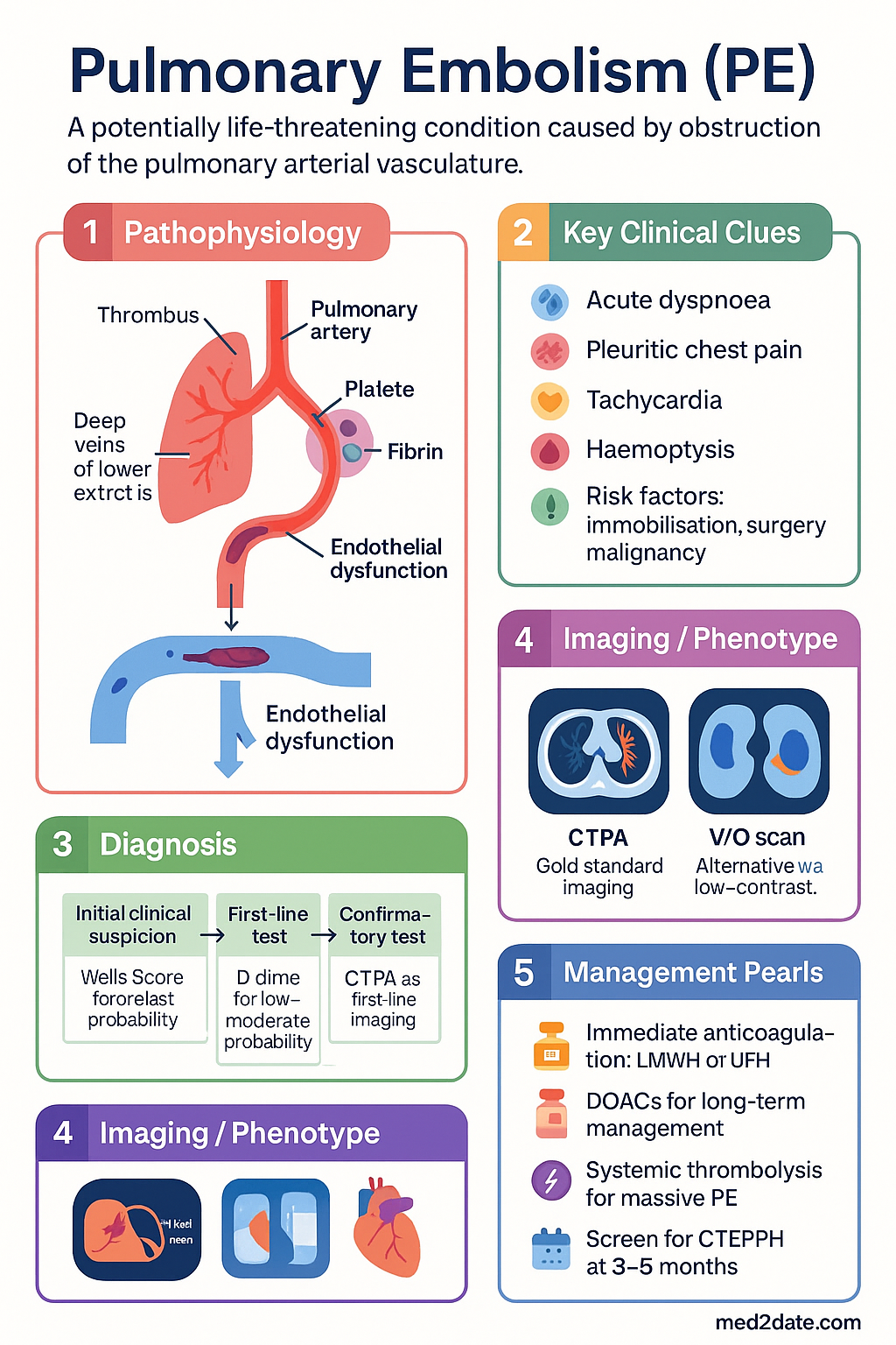
Pulmonary embolism (PE) is a potentially life-threatening condition caused by obstruction of the pulmonary arterial vasculature, most commonly by thrombus originating from the deep veins of the lower extremities or pelvis. PE and deep vein thrombosis (DVT) together constitute venous thromboembolism (VTE), which represents a significant cause of morbidity and mortality in Australia.
In Australia, VTE accounts for an estimated 8,000–10,000 hospital admissions per year, with PE responsible for approximately 3,000 deaths annually. The incidence of VTE in Australia is estimated at 0.8–1.6 per 1,000 person-years, rising sharply with age to exceed 5 per 1,000 person-years in those over 80 years. The Australian Institute of Health and Welfare (AIHW) data indicate that PE-related hospital separations have increased over the past decade, partly reflecting improved detection with modern imaging.
Key Australian epidemiological considerations include:
- Age distribution: Median age at diagnosis is approximately 63 years; rarely seen in children except in the context of central venous catheters, malignancy, or inherited thrombophilia.
- Sex differences: Slightly higher incidence in males overall, though females have additional risk factors including combined oral contraceptive use, hormone replacement therapy, and pregnancy.
- Indigenous health disparities: Aboriginal and Torres Strait Islander Australians experience higher rates of VTE, particularly in remote communities, driven by higher prevalence of comorbidities including rheumatic heart disease, obesity, diabetes, and limited access to thromboprophylaxis.
- Rural and remote access: Geographic isolation poses challenges for timely diagnosis (limited CTPA availability) and specialist input; transfer to tertiary centres may be required for massive PE management.
- Hospital-acquired VTE: Approximately 50–60% of hospital-associated VTE events are considered preventable with appropriate thromboprophylaxis, as emphasised by the National Safety and Quality Health Service (NSQHS) Standards.
Diagnosis & Risk Stratification
Clinical Decision Rules: Wells Score
The Wells score for PE is the most widely validated clinical prediction rule and should be applied to all patients presenting with suspected PE. It stratifies patients into low, moderate, or high pre-test probability categories, guiding subsequent diagnostic testing.
| Criterion | Points |
|---|---|
| Clinical signs/symptoms of DVT (leg swelling, palpable deep vein tenderness) | 3 |
| PE is the #1 diagnosis, or equally likely | 3 |
| Heart rate > 100 bpm | 1.5 |
| Immobilisation (≥3 days) or surgery within the past 4 weeks | 1.5 |
| Previous DVT/PE | 1.5 |
| Haemoptysis | 1 |
| Active malignancy (treatment within 6 months or palliative) | 1 |
| Score | Probability | Prevalence of PE | Next Step |
|---|---|---|---|
| 0–4 (or <5) | Low / PE-unlikely | ~3–8% | Apply PERC rule or D-dimer |
| ≥5 (or ≥6 if two-level) | High / PE-likely | ~35–50% | Proceed directly to CTPA |
PERC Rule (Pulmonary Embolism Rule-out Criteria)
The PERC rule is applied only to patients classified as low risk by Wells score. If all 8 PERC criteria are negative, PE can be safely excluded without D-dimer testing, avoiding unnecessary imaging.
D-Dimer Interpretation
D-dimer (fibrin degradation product) testing is highly sensitive (~95%) but poorly specific (~40–50%) for PE. It is most useful for excluding PE in low-to-moderate probability patients.
- Standard cutoff: 500 µg/L (FEU) — a negative result in a low-risk patient effectively excludes PE (NPV >99%).
- Age-adjusted cutoff (patients >50 years): Age × 10 µg/L (e.g., cutoff for a 65-year-old = 650 µg/L). This strategy reduces false positives and unnecessary CTPA in older adults. Validated in the ADJUST-PE study.
- Limitations: D-dimer is elevated in malignancy, infection, pregnancy, recent surgery, inflammation, liver disease, and advanced age — it should not be used to exclude PE in high-risk patients regardless of the result.
- Availability: Available at all Australian public hospital laboratories; point-of-care D-dimer assays are increasingly available in rural EDs but vary in sensitivity.
CT Pulmonary Angiography (CTPA)
CTPA is the gold-standard first-line imaging modality for PE diagnosis in the Australian setting. Modern multidetector CT scanners achieve sensitivity of ~97–100% and specificity of ~97% for PE in the main, lobar, and segmental pulmonary arteries.
- MBS item: MBS item 57355 (CT angiography, chest). Requires a valid clinical request from a medical practitioner.
- Contrast considerations: IV iodinated contrast is required. Assess renal function (eGFR) prior; avoid if eGFR <30 mL/min/1.73 m² unless benefits outweigh risks. Premedicate patients with prior contrast reactions.
- Radiation: Estimated effective dose 3–5 mSv. Use judiciously in young women (mammary tissue irradiation) and during pregnancy (low-dose protocols available).
- Availability: Available 24/7 in major metropolitan hospitals; limited after-hours availability in some regional centres — may require patient transfer.
V/Q (Ventilation-Perfusion) Scanning
V/Q scanning is the preferred alternative to CTPA when iodinated contrast is contraindicated (severe contrast allergy, significant renal impairment) or in selected pregnancy cases. A normal V/Q scan effectively excludes PE; a high-probability scan is diagnostic. However, the majority of V/Q scans yield intermediate or low-probability results, which are non-diagnostic.
- MBS item: MBS item 61358 (nuclear medicine, pulmonary perfusion study).
- Technetium-99m macroaggregated albumin (⁹⁹ᵐTc-MAA) is used for perfusion; ventilation agents include ⁹⁹ᵐTc-DTPA aerosol or ⁸¹ᵐKr gas.
- SPECT V/Q: Single-photon emission CT V/Q has improved sensitivity over planar imaging and is increasingly available in Australian nuclear medicine departments.
- Limitations: Less specific than CTPA; inferior diagnostic performance in patients with pre-existing lung disease (COPD, pneumonia). Requires nuclear medicine facilities — not universally available in smaller regional hospitals.
Diagnostic Algorithm
Additional Investigations
Acute PE Management
Hemodynamic Classification
The European Society of Cardiology (ESC) haemodynamic classification, widely adopted in Australian practice, stratifies acute PE into three categories that directly guide management intensity:
Initial Anticoagulation
Anticoagulation should be initiated as soon as PE is diagnosed (or strongly suspected) in the absence of absolute contraindications. The goal is to prevent thrombus propagation and recurrent embolisation while endogenous fibrinolytic mechanisms resolve the existing burden.
Anticoagulation Pathway: DOACs vs LMWH → Warfarin
In Australian practice, the preferred approach for acute PE anticoagulation depends on clinical stability, renal function, and patient factors:
- Haemodynamically stable, low-risk PE: Initiate a DOAC directly (apixaban 10 mg BD × 7 days then 5 mg BD; or rivaroxaban 15 mg BD × 21 days then 20 mg OD). No need for initial LMWH/UFH bridge. Supported by AMPLIFY and EINSTEIN-PE trials. This is the preferred pathway for most patients and allows early discharge.
- LMWH → warfarin pathway: Use when DOACs are contraindicated (antiphospholipid syndrome with triple positivity, mechanical heart valve, severe renal impairment eGFR <15, drug interactions). Start warfarin on day 1 with LMWH; continue LMWH for ≥5 days and until INR ≥2.0 for ≥24 hours.
- Massive/submassive PE: Initiate UFH IV bolus + infusion. Transition to DOAC or LMWH → warfarin once stable. UFH is preferred as it is rapidly reversible with protamine and has a short half-life.
- Cancer-associated PE: LMWH (dalteparin 200 IU/kg SC OD × 1 month, then 150 IU/kg SC OD) or DOACs (edoxaban, apixaban per CARAVAGGIO, ADAM-VTE trials). Caution with GI malignancy due to increased GI bleeding with DOACs.
Thrombolysis Indications in Acute PE
Systemic thrombolysis is indicated for the following:
- Absolute indication: Massive PE with haemodynamic instability (SBP <90 mmHg ≥15 min, vasopressor requirement, cardiac arrest).
- Relative indication: Submassive PE with clinical deterioration — worsening RV function, rising biomarkers, haemodynamic trajectory toward shock, severe hypoxaemia despite supplemental oxygen.
- Cardiac arrest with suspected PE: Empirical thrombolysis should be considered during CPR. Alteplase 50 mg IV bolus during cardiac arrest (repeat once if no ROSC).
Massive & Submassive PE
Massive PE (High-Risk)
Massive PE is defined by haemodynamic instability: sustained hypotension (SBP <90 mmHg for ≥15 minutes or requiring inotropic support), pulselessness, or persistent profound bradycardia (HR <40 bpm with signs of shock). In-hospital mortality ranges from 25–65% if untreated.
Catheter-Directed Therapy (CDT)
Catheter-directed therapy is an emerging option for patients with massive or submassive PE who have contraindications to systemic thrombolysis or who have failed systemic lysis. CDT includes catheter-directed thrombolysis (low-dose alteplase via pulmonary artery catheter), mechanical thrombectomy, and aspiration thrombectomy devices (FlowTriever, Indigo, EKOS ultrasound-assisted thrombolysis).
- Advantages: Lower systemic thrombolytic dose (reduced bleeding risk), direct clot delivery, mechanical disruption of thrombus.
- Evidence: SEATTLE-II, FLARE, and OPTALYSE trials demonstrated improvement in RV/LV ratio and pulmonary artery pressure with acceptable safety profiles.
- Availability in Australia: Performed at select tertiary centres (Royal Melbourne, Royal Prince Alfred, Royal Adelaide, Princess Alexandra, Fiona Stanley). Requires interventional radiology or cardiology expertise. Available on a case-by-case basis in 2024.
- MBS: No dedicated MBS item for catheter-directed PE therapy; costs may be covered under hospital casemix funding or compassionate access programs.
Surgical Pulmonary Embolectomy
Surgical embolectomy is reserved for patients with massive PE when thrombolysis is contraindicated or has failed, and catheter-directed therapy is not available or appropriate.
- Procedure: Median sternotomy, cardiopulmonary bypass (CPB), pulmonary arteriotomy with direct clot extraction.
- Outcomes: Operative mortality 20–30% in contemporary series (improved from historical rates). Best outcomes when performed before the onset of multiorgan failure.
- Centres: Cardiac surgical centres with cardiothoracic surgery capability (most major Australian tertiary hospitals).
Extracorporeal Membrane Oxygenation (ECMO)
Veno-arterial (VA) ECMO may be used as a bridging strategy in patients with massive PE and refractory cardiogenic shock or cardiac arrest, to provide circulatory and respiratory support while definitive reperfusion (thrombolysis, CDT, or embolectomy) is arranged.
- Indications: Refractory cardiogenic shock despite maximal medical therapy · Cardiac arrest with suspected PE · Bridge to surgical embolectomy in centres with cardiac surgery.
- Complications: Bleeding (systemic anticoagulation required), limb ischaemia, stroke, circuit thrombosis, sepsis.
- Australian ECMO centres: Alfred Hospital (Melbourne), Royal Melbourne, Royal Prince Alfred (Sydney), St Vincent's (Sydney), Princess Alexandra (Brisbane), Royal Adelaide, Fiona Stanley (Perth).
Submassive PE Risk Stratification Scores
| Score | Components | Use |
|---|---|---|
| sPESI (Simplified PE Severity Index) | Age >80, cancer, chronic cardiopulmonary disease, HR ≥110, SBP <100, SpO₂ <90% | Identifies low-risk patients safe for outpatient management (sPESI = 0) |
| Bova Score | SBP 90–100 (2 pts), troponin elevated (2 pts), RV dysfunction (2 pts), HR ≥110 (1 pt) | Substratifies intermediate-risk PE; Stage III (≥5 pts) identifies highest-risk patients |
| HESTIA Criteria | 11 clinical criteria for outpatient PE management eligibility | Safe selection of patients for home treatment of low-risk PE |
Long-term Anticoagulation
Duration Decisions: Provoked vs Unprovoked PE
The duration of anticoagulation following acute PE is determined by the balance between the risk of VTE recurrence and the risk of major bleeding. The key determinant is whether a transient, reversible provoking factor was identified.
| Category | Examples | Recommended Duration | Recurrence Risk (off anticoagulation) |
|---|---|---|---|
| Major provoked (transient risk factor) | Surgery, plaster cast, hospitalisation ≥3 days, major trauma within 3 months | 3 months (stop) | ~3% at 1 year |
| Minor provoked | OCP/HRT, long-haul travel (>8 hours), minor surgery, pregnancy | 3–6 months (individualise) | ~5% at 1 year |
| Unprovoked (no identifiable trigger) | Idiopathic, or associated with minor/non-persistent risk factors only | ≥6 months, consider extended/indefinite therapy | ~10–15% at 1 year; ~30% at 5 years |
| Cancer-associated | Active malignancy (diagnosis, treatment, or palliative) | Extended (indefinite) until cancer resolved or treatment complete | ~20% at 1 year |
| Recurrent VTE | Second or subsequent unprovoked VTE | Extended/indefinite (strongly recommended) | ~30–50% at 5 years |
Extended Therapy: DOAC Selection
For patients requiring extended (indefinite) anticoagulation, DOACs are generally preferred over warfarin due to their convenience (no INR monitoring), predictable pharmacokinetics, favourable food/drug interaction profiles, and comparable or superior safety data.
- Apixaban 5 mg BD → 2.5 mg BD for extended prevention: The AMPLIFY-EXT trial demonstrated that apixaban 2.5 mg BD reduced recurrent VTE by 80% compared with placebo, with no significant increase in major bleeding. This is the preferred extended regimen in many Australian centres.
- Rivaroxaban 20 mg OD → 10 mg OD for extended prevention: EINSTEIN-CHOICE showed rivaroxaban 10 mg OD was superior to placebo and comparable to 20 mg OD for preventing recurrence, with lower bleeding risk.
- Warfarin (INR 2.0–3.0): Remains appropriate for patients with antiphospholipid syndrome, mechanical heart valves, or those who prefer it. Requires regular INR monitoring (at least monthly at steady state); target TTR (time in therapeutic range) >65%.
Bleeding Risk Assessment
Before committing to extended anticoagulation, bleeding risk must be formally assessed. Two validated scores are commonly used:
Hypertension (uncontrolled, SBP >160) · Abnormal renal/liver function (1 pt each) · Stroke · Bleeding history · Labile INR (TTR <60%) · Elderly (>65) · Drugs/alcohol (1 pt each). Score ≥3 = high bleeding risk. Requires careful review of modifiable risk factors.
Specifically validated for VTE patients on extended anticoagulation. Components: active cancer (2 pts), male with uncontrolled hypertension (1 pt), anaemia (1.5 pts), bleeding history (1.5 pts), age ≥60 (1.5 pts), renal insufficiency (1.5 pts). Score ≥2 = high risk. More discriminatory than HAS-BLED for VTE-specific bleeding.
Thrombophilia Testing — When and For Whom
Routine thrombophilia testing is not recommended for all PE patients. Testing is considered in specific circumstances:
- Young patients (<50 years) with unprovoked PE, especially if the result would change management (e.g., decision regarding extended anticoagulation).
- Recurrent VTE in unusual sites (cerebral, splanchnic, portal).
- Strong family history of VTE (multiple first-degree relatives).
- Testing should be performed ≥2 weeks after completing acute anticoagulation (ideally at 4–6 weeks off therapy) to avoid false negatives.
- Do NOT test during acute VTE or while on anticoagulation (LMWH and DOACs affect antithrombin levels, lupus anticoagulant, and protein C/S assays).
Chronic Thromboembolic Disease
CTEPH Screening Post-PE
Chronic thromboembolic pulmonary hypertension (CTEPH) is a serious long-term complication of acute PE, affecting an estimated 2–4% of PE survivors. It results from incomplete resolution of pulmonary thromboemboli, leading to organised fibrotic obstruction of the pulmonary vasculature and progressive pulmonary hypertension. CTEPH is the only potentially curable form of pulmonary hypertension through pulmonary endarterectomy (PEA).
Screening Algorithm
CTEPH Treatment Options
- Pulmonary endarterectomy (PEA): Treatment of choice for operable CTEPH. Median sternotomy, deep hypothermic circulatory arrest. Performed at select centres in Australia (Royal Prince Alfred Hospital, Sydney, is the primary PHSANZ-designated CTEPH centre). Perioperative mortality 2–5% at experienced centres. Can be curative.
- Balloon pulmonary angioplasty (BPA): Catheter-based treatment for patients with inoperable CTEPH or residual PH after PEA. Performed in a staged fashion (multiple sessions). Increasingly available in Australian PH centres. Improves haemodynamics and functional capacity in the CHEST-UK and Japanese registries.
- Medical therapy: Riociguat (soluble guanylate cyclase stimulator) is PBS-listed (Authority Required) for inoperable CTEPH. WHO functional class improvement demonstrated in the CHEST-1 trial. Other PH-specific therapies (macitentan, sildenafil, epoprostenol) may be used off-label in specialist centres.
- Lifelong anticoagulation: All CTEPH patients require indefinite anticoagulation (typically warfarin; DOACs increasingly used but less evidence in CTEPH specifically).
Residual Symptoms Post-PE
Up to 50% of PE survivors report persistent symptoms at 6 months, including exertional dyspnoea, exercise intolerance, and reduced quality of life. This entity is increasingly recognised as "post-PE syndrome" and may occur in the absence of CTEPH.
- Pathophysiology: May include residual pulmonary vascular obstruction, deconditioning, cardiac remodelling (RV dysfunction), microvascular disease, and chronic thromboembolic disease without meeting haemodynamic criteria for CTEPH.
- Investigations: Echocardiography, 6-minute walk test (6MWT), cardiopulmonary exercise testing (CPET), and pulmonary function tests. CT pulmonary angiography may demonstrate residual thrombus.
- Management: Supervised exercise-based cardiac rehabilitation has shown benefit (PEITHO-2, EINSTEIN CHOICE sub-studies). Pulmonary rehabilitation programs available through Australian public hospitals and community services.
- Dyspnoea evaluation: Comprehensive assessment should include cardiac (echocardiography, BNP), pulmonary (PFTs, CT chest), deconditioning (CPET), and psychological factors (anxiety, PTSD post-acute PE are common).
Specialist Referral Pathways
Special Populations
Pregnancy
Paediatrics
Elderly (≥65 years)
Renal Impairment
Hepatic Impairment
Immunocompromised
Aboriginal and Torres Strait Islander Health Considerations
Aboriginal and Torres Strait Islander Australians experience a higher burden of venous thromboembolism than the non-Indigenous population, with contributing factors including higher prevalence of cardiovascular comorbidities, rheumatic heart disease, obesity, diabetes, and chronic kidney disease. Delays in diagnosis and challenges in accessing specialist care contribute to poorer outcomes. The following considerations apply to PE management in this population.
📚 References
- 1. Konstantinides SV, Meyer G, Becattini C, et al. 2019 ESC Guidelines for the diagnosis and management of acute pulmonary embolism. Eur Heart J. 2020;41(4):543-603. doi:10.1093/eurheartj/ehz405.
- 2. Wells PS, Anderson DR, Rodger M, et al. Derivation of a simple clinical model to categorize patients probability of pulmonary embolism: increasing the models utility with the SimpliRED D-dimer. Thromb Haemost. 2000;83(3):416-420.
- 3. Kline JA, Courtney DM, Kabrhel C, et al. Prospective multicenter evaluation of the pulmonary embolism rule-out criteria. J Thromb Haemost. 2008;6(5):772-780. doi:10.1111/j.1538-7836.2008.02944.x.
- 4. Righini M, Van Es J, Den Exter PL, et al. Age-adjusted D-dimer cutoff levels to rule out pulmonary embolism: the ADJUST-PE study. JAMA. 2014;311(11):1117-1124. doi:10.1001/jama.2014.2135.
- 5. Agnelli G, Buller HR, Cohen A, et al. Oral apixaban for the treatment of acute venous thromboembolism (AMPLIFY). N Engl J Med. 2013;369(9):799-808. doi:10.1056/NEJMoa1302507.
- 6. Bauersachs R, Berkowitz SD, Brenner B, et al. Oral rivaroxaban for symptomatic venous thromboembolism (EINSTEIN-PE). N Engl J Med. 2010;363(26):2499-2510. doi:10.1056/NEJMoa1007903.
- 7. Agnelli G, Buller HR, Cohen A, et al. Apixaban for extended treatment of venous thromboembolism (AMPLIFY-EXT). N Engl J Med. 2013;368(8):699-708. doi:10.1056/NEJMoa1207541.
- 8. Weitz JI, Lensing AWA, Prins MH, et al. Rivaroxaban or aspirin for extended treatment of venous thromboembolism (EINSTEIN-CHOICE). N Engl J Med. 2017;376(13):1211-1222. doi:10.1056/NEJMoa1700518.
- 9. Ghofrani HA, D'Armini AM, Grimminger F, et al. Riociguat for the treatment of chronic thromboembolic pulmonary hypertension (CHEST-1). N Engl J Med. 2013;369(4):319-329. doi:10.1056/NEJMoa1209657.
- 10. Goldhaber SZ, Visani L, De Rosa M. Acute pulmonary embolism: clinical outcomes in the International Cooperative Pulmonary Embolism Registry (ICOPER). Lancet. 1999;353(9162):1386-1389. doi:10.1016/S0140-6736(98)07534-5.
- 11. Australian Institute of Health and Welfare. Cardiovascular disease in Australia 2024. AIHW, Canberra. https://www.aihw.gov.au/reports/cardiovascular/cardiovascular-disease-in-australia.
- 12. RHDAustralia (RHD Australia), a program of Menzies School of Health Research. The 2020 Australian guideline for prevention, diagnosis and management of acute rheumatic fever and rheumatic heart disease. 3rd ed. Darwin: RHDAustralia; 2020.
- 13. Klok FA, Dzikowska-Diduch O, Kostrubiec M, et al. Derivation of a clinical prediction score for chronic thromboembolic pulmonary hypertension after acute pulmonary embolism. J Thromb Haemost. 2016;14(1):124-131. doi:10.1111/jth.13175.
- 14. National Safety and Quality Health Service Standards. 2nd ed. Australian Commission on Safety and Quality in Health Care (ACSQHC), Sydney; 2021.
- 15. Stein PD, Fowler SE, Goodman LR, et al. Multidetector computed tomography for acute pulmonary embolism (PIOPED II). N Engl J Med. 2006;354(22):2317-2327. doi:10.1056/NEJMoa052367.
- 16. Bova C, Sanchez O, Prandoni P, et al. Identification of intermediate-risk patients with acute symptomatic pulmonary embolism. Eur Respir J. 2014;44(3):694-703. doi:10.1183/09031936.00006114.
- 17. Jiménez D, Aujesky D, Moores L, et al. Simplification of the pulmonary embolism severity index for prognostic assessment of patients with acute symptomatic pulmonary embolism (sPESI). Arch Intern Med. 2010;170(15):1383-1389. doi:10.1001/archinternmed.2010.199.