📋 Key Information Summary
- Bronchiectasis is defined by permanent abnormal bronchial dilatation on HRCT (broncho-arterial ratio >1, or lack of tapering within 2 cm of pleural surface), with a rising prevalence in Australia, particularly among Aboriginal and Torres Strait Islander peoples.
- Key aetiologies include post-infectious (childhood adenovirus, pertussis, measles, TB), idiopathic (≈30–40%), COPD overlap, cystic fibrosis (CF), primary ciliary dyskinesia (PCD), and immunodeficiency (IgG subclass deficiency, CVID).
- All patients require sputum microscopy, culture and sensitivity (MC&S) at baseline and during every exacerbation; Pseudomonas aeruginosa colonisation is the single most important prognostic marker.
- Airway clearance is the cornerstone of chronic management — active cycle of breathing technique (ACBT), oscillating PEP devices, and autogenic drainage; chest physiotherapy referral is essential.
- Mucoactive agents: hypertonic saline (7%) nebulised, mannitol dry powder (Bronchitol® PBS-listed), and mucolytics (N-acetylcysteine) improve mucus clearance and quality of life.
- Long-term macrolide prophylaxis (azithromycin 500 mg Mon/Wed/Fri or 250 mg daily) reduces exacerbation frequency by ~50% — monitor ECG for QTc, liver function, and audiometry; confirm sputum NTM-negative before initiation.
- Acute exacerbations: treat with 14 days of antibiotics guided by prior sputum culture; first-line oral amoxicillin 500 mg–1 g TDS or doxycycline 200 mg then 100 mg daily if Haemophilus influenzae/atypical suspected.
- Pseudomonal exacerbations require ciprofloxacin 750 mg PO BD or IV anti-pseudomonal β-lactam (piperacillin-tazobactam, ceftazidime, or meropenem) for 14 days; consider inhaled tobramycin (300 mg BD) or aztreonam lysine for chronic colonisation.
- Hemoptysis — minor: oral tranexamic acid 1 g TDS; major/life-threatening: interventional radiology for bronchial artery embolisation (BAE); surgical resection reserved for localised disease refractory to embolisation.
- Complications include respiratory failure (home oxygen assessment), pulmonary hypertension/cor pulmonale (echocardiography screening), and rarely amyloidosis; pneumococcal and influenza vaccination are mandatory.
- Aboriginal and Torres Strait Islander peoples have the highest prevalence of bronchiectasis globally (up to 1,470/100,000 children in Top End communities); early childhood infection, overcrowding, and limited access to specialist care drive disease burden.
- Refer to respiratory specialist for all newly diagnosed cases, suspected CF/PCD/immunodeficiency, frequent exacerbations (≥3/year), chronic Pseudomonas colonisation, hemoptysis, or consideration of surgical resection.
Introduction & Australian Epidemiology
Bronchiectasis is a chronic respiratory condition characterised by permanent, pathological dilatation of the bronchi resulting from cycles of airway inflammation, infection, and impaired mucociliary clearance. Once considered an "orphan disease," bronchiectasis is increasingly recognised as a significant cause of chronic respiratory morbidity in Australia, with rising hospitalisation rates and healthcare utilisation over the past two decades.
The Australian Bronchiectasis Registry (ABR), established in 2015 through the Lung Foundation Australia, has provided crucial data on disease burden, aetiology, and outcomes in the Australian population. Current estimates suggest a prevalence of approximately 500–700 per 100,000 adults in the general Australian population, though this is likely an underestimate given under-diagnosis in primary care.
The disease exhibits a bimodal age distribution: a paediatric peak (often post-infectious, frequently resolving with growth) and an adult peak (predominantly idiopathic, post-infectious, or associated with COPD). Female sex predominates in adult-onset idiopathic bronchiectasis. The economic burden is substantial, with direct healthcare costs estimated at >$10,000 per patient per annum, driven largely by hospitalisations for acute exacerbations.
Key Australian Statistics
- Hospital separations for bronchiectasis (non-CF) have increased by ~30% over the past decade (AIHW data).
- Median age at diagnosis in adults: 60–65 years; in children: 4–6 years.
- ~40% of adult cases are classified as idiopathic after thorough investigation.
- Chronic Pseudomonas aeruginosa colonisation is present in 15–30% of Australian adult patients and is associated with faster FEV₁ decline and increased mortality.
- Indigenous Australians with bronchiectasis present earlier, have more severe disease, higher rates of Pseudomonas colonisation, and worse outcomes than non-Indigenous Australians.
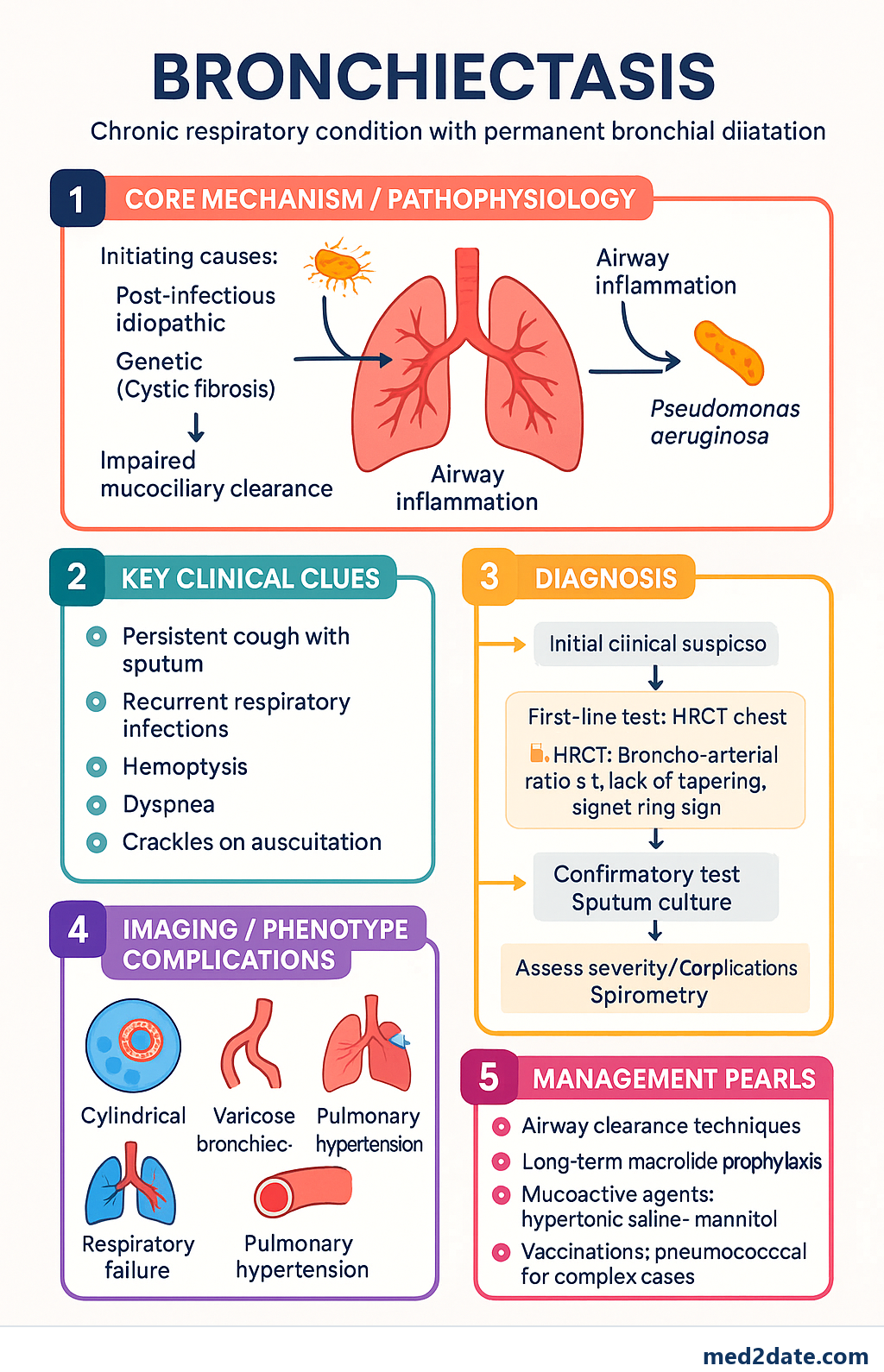
Diagnosis & Aetiology
HRCT Diagnostic Criteria
High-resolution computed tomography (HRCT) of the chest is the gold standard for diagnosing bronchiectasis. Scans should be performed at full inspiration (1 mm collimation, 10 mm intervals) and, where available, expiratory images should be obtained to identify air trapping and small airway disease.
| HRCT Sign | Description | Sensitivity |
|---|---|---|
| Broncho-arterial ratio >1 | Internal bronchial diameter exceeds accompanying pulmonary artery diameter — the hallmark sign | High (most specific) |
| Lack of tapering | Bronchi fail to taper normally within 2 cm of the pleural surface or visualised bronchus extends to the pleural margin | High |
| Signet ring sign | Dilated bronchus seen in cross-section adjacent to a smaller pulmonary artery — resembles a signet ring | Moderate |
| Tram-track sign | Parallel thickened bronchial walls seen in longitudinal section (cylindrical bronchiectasis) | Moderate |
| Mucus plugging | Bronchial filling with mucus, often finger-in-glove pattern | Variable |
HRCT Morphological Classification (Reid Classification)
| Type | Morphology | Characteristics |
|---|---|---|
| Cylindrical (varicose) | Smooth, uniform dilatation | Most common type; parallel walls, mild disease |
| Varicose | Beaded, irregular dilatation | Intermediate severity; areas of narrowing and dilation |
| Cystic | Grape-like clusters of dilated airways | Most severe; often associated with Pseudomonas colonisation |
Aetiology — Systematic Workup
An identifiable cause can be found in 60–70% of cases. A structured aetiological workup is recommended for all newly diagnosed patients, guided by clinical phenotype and severity.
| Aetiology | Proportion | Key Features & Investigations |
|---|---|---|
| Idiopathic | 30–40% | Diagnosis of exclusion; female predominance; often lower lobe predominant |
| Post-infectious | 25–30% | Childhood adenovirus, pertussis, measles, RSV, TB, non-tuberculous mycobacteria (NTM); history of severe childhood pneumonia |
| COPD overlap | 10–15% | Smoking history, concurrent emphysema, COPD phenotype; CT bronchiectasis + emphysema |
| Allergic bronchopulmonary aspergillosis (ABPA) | 2–5% | Central bronchiectasis, eosinophilia, elevated total IgE >1,000 IU/mL, positive Aspergillus-specific IgE/IgG, serum precipitins |
| Cystic fibrosis (CF) | Separate entity | Sweat chloride test (MBS 11900), CFTR genotyping; upper lobe predominant; sinus disease; pancreatic insufficiency |
| Primary ciliary dyskinesia (PCD) | 2–5% | Situs inversus (50%), neonatal respiratory distress, chronic rhinositis, infertility; nasal NO screening, ciliary biopsy (electron microscopy), genetic testing |
| Immunodeficiency | 5–10% | CVID, IgG subclass deficiency; measure serum IgG, IgA, IgM, IgG subclasses, functional antibody responses (pre/post pneumococcal vaccine titres) |
| NTM infection | 5–10% | M. avium complex (MAC), M. abscessus; Lady Windermere syndrome (right middle lobe/lingula); requires sputum AFB cultures ×3, NTM PCR |
| Rheumatoid arthritis | 2–3% | Pre- or post-RA onset; may be worsened by methotrexate or anti-TNF therapy |
Recommended Aetiological Investigations
Chronic Management
Chronic management of bronchiectasis is centred on four pillars: airway clearance, mucoactive therapy, infection prevention/treatment, and management of underlying causes. A multidisciplinary approach involving respiratory physicians, physiotherapists, pharmacists, and dietitians is recommended.
Airway Clearance Techniques
Airway clearance is the single most important intervention in bronchiectasis management. All patients should be referred to a respiratory physiotherapist for individualised assessment and technique prescription.
Mucoactive Agents
Bronchodilators
Bronchodilators are not disease-modifying in bronchiectasis but are indicated when there is coexistent airflow obstruction (reversible or fixed) or significant dyspnoea. Always assess response objectively with spirometry.
Anti-Inflammatory Therapy
Chronic airway inflammation is a hallmark of bronchiectasis. While inhaled corticosteroids (ICS) are not routinely recommended for bronchiectasis in the absence of asthma or COPD overlap, macrolide antibiotics have anti-inflammatory and immunomodulatory properties and are used as long-term prophylaxis in frequent exacerbators (see Exacerbation Management section).
Inhaled corticosteroids may be considered in the following scenarios:
- Coexistent asthma with documented eosinophilic inflammation (FeNO >40 ppb or blood eosinophils >300 cells/μL)
- COPD overlap with frequent exacerbations despite LABA/LAMA
- ABPA: systemic corticosteroids (prednisolone 0.5 mg/kg/day for 2 weeks, then taper over 6–8 weeks) + itraconazole 200 mg BD (PBS Authority Required)
Vaccination & Preventive Care
- Influenza vaccine: Annual (free under NIP for all Australians with chronic respiratory disease)
- Pneumococcal vaccine: Pneumococcal conjugate (PCV13 or PCV20) followed by pneumococcal polysaccharide (PPSV23) at ≥8-week interval — funded under NIP for at-risk groups
- COVID-19: Annual booster recommended (chronic respiratory condition — priority group)
- Pertussis booster: dTpa every 10 years if not recently received
- Smoking cessation: Absolute requirement; offer NRT, varenicline (Champix®), or bupropion (Zyban®); referral to Quitline (13 7848)
Exacerbation Management
A bronchiectasis exacerbation is defined as a sustained deterioration (≥48 hours) in at least three of the following symptoms beyond normal day-to-day variation: cough, sputum volume, sputum purulence, breathlessness, exercise tolerance, fatigue, or hemoptysis. Exacerbations drive disease progression, accelerate lung function decline, and reduce quality of life.
Sputum Culture — Essential in Every Exacerbation
Sputum MC&S should be collected before initiating antibiotics whenever feasible. If sputum is difficult to obtain, consider hypertonic saline nebulisation induction. Patients with known chronic colonisation may be started on antibiotics based on prior culture results, but a fresh specimen should still be sent.
Antibiotic Selection — Exacerbations
Antibiotic duration for bronchiectasis exacerbations is typically 14 days — longer than for acute COPD exacerbations (5–7 days) due to the higher bacterial burden and biofilm formation within dilated airways. Antibiotic choice should be guided by the most recent sputum culture result.
| Scenario | First-Line Antibiotic | Duration | PBS Status |
|---|---|---|---|
| No prior isolate / H. influenzae | Amoxicillin 500 mg–1 g PO TDS | 14 days | PBS General Benefit |
| Penicillin allergy / atypical organisms | Doxycycline 200 mg day 1 then 100 mg PO daily | 14 days | PBS General Benefit |
| Moraxella catarrhalis | Amoxicillin-clavulanate 875/125 mg PO BD | 14 days | PBS General Benefit |
| Pseudomonas aeruginosa (mild–moderate) | Ciprofloxacin 750 mg PO BD | 14 days | PBS General Benefit |
| Pseudomonas (severe / resistant / failing oral) | Piperacillin-tazobactam 4.5 g IV TDS or Ceftazidime 2 g IV TDS or Meropenem 1 g IV TDS | 14 days (IV then consider oral step-down) | Hospital PBS |
| MRSA | Vancomycin IV (target trough 15–20 mg/L) or linezolid 600 mg PO/IV BD | 14 days | Hospital PBS |
Inhaled Antibiotics — Chronic Pseudomonas Colonisation
For patients with chronic Pseudomonas aeruginosa colonisation (≥3 positive cultures in 12 months) and frequent exacerbations (≥3/year), long-term inhaled antibiotic therapy should be considered. These agents achieve high intrapulmonary concentrations with minimal systemic absorption.
Macrolide Prophylaxis
Long-term macrolide therapy is the most evidence-based intervention for reducing exacerbation frequency in bronchiectasis. The landmark BAT (Bronchiectasis and Long-Term Azithromycin Treatment), EMBRACE, and BAT-2 trials demonstrated a ~50% reduction in exacerbations. The mechanism involves both anti-inflammatory (neutrophilic) and anti-biofilm effects, in addition to antimicrobial activity against H. influenzae and atypical organisms.
Complications
Hemoptysis Management
Hemoptysis occurs in 20–50% of bronchiectasis patients and ranges from blood-streaked sputum to life-threatening massive hemoptysis. Bronchiectasis is one of the most common causes of non-massive hemoptysis in Australia.
| Severity | Definition | Initial Management | Escalation |
|---|---|---|---|
| Blood-streaked sputum | Streaks of blood in sputum; <20 mL/day | Reassure; review and optimise airway clearance; rule out coagulopathy; ensure not on excessive anticoagulation | If recurrent, refer to respiratory physician for HRCT review |
| Mild–moderate hemoptysis | 20–200 mL/24 hr | Tranexamic acid 1 g PO/IV TDS (antifibrinolytic); correct coagulopathy; monitor haemoglobin; respiratory physician review | If not settling in 48 hr, consider CT pulmonary angiography and referral for bronchial artery embolisation |
| Major / life-threatening hemoptysis | >200 mL/24 hr or haemodynamic instability | Emergency: Secure airway (affected side down, intubation if needed), IV access, cross-match blood, IV tranexamic acid 1 g stat, resuscitation | Urgent bronchial artery embolisation (BAE) by interventional radiology — first-line definitive treatment; success rate 85–95% |
Respiratory Failure
Progressive bronchiectasis can lead to chronic respiratory failure, typically Type II (hypoxic + hypercapnic), particularly in advanced disease with extensive bilateral involvement. Assessment follows standard COPD/respiratory failure pathways.
- Pulse oximetry: SpO₂ target 88–92% in patients at risk of hypercapnia; arrange arterial blood gas (ABG) if SpO₂ <92% or clinically suspected Type II failure.
- Long-term oxygen therapy (LTOT): Indicated if PaO₂ ≤55 mmHg (or ≤59 mmHg with cor pulmonale/polycythaemia) — assess via ABG on room air, confirmed on two occasions ≥3 weeks apart. Funded under state-based Home Oxygen Programs.
- Non-invasive ventilation (NIV): Consider for chronic hypercapnic respiratory failure (PaCO₂ >50 mmHg) with recurrent admissions, particularly if comorbid obstructive sleep apnoea (overlap syndrome). Refer for sleep study and NIV titration.
- Pulmonary rehabilitation: Evidence-based improvement in exercise capacity and quality of life; available at major hospitals and community health centres. Funded under Medicare for chronic respiratory disease.
Cor Pulmonale & Pulmonary Hypertension
Pulmonary hypertension secondary to chronic hypoxia occurs in advanced bronchiectasis. Clinical features include progressive dyspnoea, peripheral oedema, raised JVP, loud P₂, and right ventricular heave.
Management of cor pulmonale in bronchiectasis focuses on optimising the underlying lung disease (airway clearance, infection control, oxygen therapy), salt/fluid restriction, and diuretics (furosemide 20–40 mg PO daily, spironolactone 25–50 mg PO daily). Specific pulmonary vasodilator therapy is generally not indicated and requires specialist PH centre management.
Surgical Resection — Indications
Surgical resection for bronchiectasis has become less common with improvements in medical management but remains an important option in selected patients with localised disease.
Special Populations
Paediatrics
Pregnancy
Elderly (>65 years)
Renal Impairment
Hepatic Impairment
Immunocompromised
Aboriginal and Torres Strait Islander Health Considerations
Bronchiectasis is a disease of profound health inequity for Aboriginal and Torres Strait Islander Australians. Indigenous children in remote communities of the Northern Territory, Far North Queensland, and Western Australia have among the highest prevalence rates of non-CF bronchiectasis documented anywhere in the world. The burden of disease reflects the cumulative impact of early childhood respiratory infections, environmental factors, socioeconomic disadvantage, and healthcare access barriers.
Key Risk Factors in Indigenous Communities
- Early childhood infections: High rates of severe lower respiratory tract infections in infancy (RSV, adenovirus, Streptococcus pneumoniae, Haemophilus influenzae) — often inadequately treated due to access delays.
- Environmental tobacco smoke: Smoking rates among Aboriginal and Torres Strait Islander adults remain ~40% (vs ~10% non-Indigenous); passive smoke exposure significantly increases childhood respiratory infection severity.
- Overcrowded housing: Persistent overcrowding in remote communities facilitates respiratory pathogen transmission; the Australian Government's National Partnership on Remote Housing aims to address this but progress remains slow.
- Malnutrition: Protein-energy malnutrition and micronutrient deficiencies (vitamin D, zinc) impair immune function and are associated with more severe childhood respiratory infections.
- Healthcare access: Remote communities rely on Aboriginal Community Controlled Health Organisations (ACCHOs) and visiting specialist services; specialist respiratory review may require medical evacuation to regional centres (Alice Springs, Darwin, Cairns, Townsville).
Culturally Responsive Care
Key Organisations & Resources
- Lung Foundation Australia — Indigenous Lung Health: Culturally tailored resources for bronchiectasis education and management.
- RHDAustralia (Menzies School of Health Research): Evidence-based guidelines for chronic lung disease in remote Indigenous communities.
- Australian Indigenous HealthInfoNet: Summaries and resources on respiratory health for Aboriginal and Torres Strait Islander peoples.
- NACCHO (National Aboriginal Community Controlled Health Organisation): Advocacy and support for ACCHOs delivering respiratory care.
- CARPA Clinical Procedures Manual: Primary care guidelines for remote Aboriginal health services, including bronchiectasis management.
Quick Reference — Exacerbation Antibiotic Guide
Monitoring & Follow-Up
Regular monitoring is essential to detect disease progression, colonising organisms, treatment complications, and the impact of management interventions. The following schedule is recommended for stable bronchiectasis patients managed in primary care with respiratory physician co-management.
| Assessment | Frequency | Details |
|---|---|---|
| Respiratory physician review | 3–6-monthly (stable); sooner if frequent exacerbations | Symptom review, management optimisation, vaccination status |
| Spirometry (MBS 11505) | 6–12-monthly | FEV₁, FVC, bronchodilator response; track trajectory |
| Sputum MC&S | 3-monthly during stability + every exacerbation | Track colonisation status; detect Pseudomonas acquisition |
| HRCT chest | Not routinely repeated; clinical deterioration or new symptoms | Baseline HRCT sufficient for most; repeat if concern for progression, NTM, or new focal abnormality |
| Quality of life (QoL-B or Leicester Cough Questionnaire) | 6–12-monthly | Validated disease-specific tools; monitor treatment response |
| Exercise capacity (6MWT) | Annually or as clinically indicated | Functional assessment; guide pulmonary rehabilitation referral |
| Echocardiography | If FEV₁ <50% predicted or clinical features of cor pulmonale | Screen for pulmonary hypertension and RV dysfunction |
| ECG (if on macrolide) | Baseline, 1 month, then annually | QTc monitoring; discontinue macrolide if QTc >500 ms |
| Audiometry (if on macrolide/tobramycin) | Baseline, 6-monthly for inhaled tobramycin | Detect ototoxicity; prompt dose adjustment or cessation |
| Renal function (if on aminoglycosides) | Monthly during active treatment | eGFR, creatinine; electrolytes if on diuretics |
📚 References
- 1. Chang AB, Bell SC, Byrnes CA, et al. Thoracic Society of Australia and New Zealand (TSANZ) position statement: chronic suppurative lung disease and bronchiectasis in children and adults in Australia and New Zealand. Respirology. 2010;15(5):739–753.
- 2. Pasteur MC, Bilton D, Hill AT; British Thoracic Society Bronchiectasis non-CF Guideline Group. British Thoracic Society guideline for non-CF bronchiectasis. Thorax. 2010;65(Suppl 1):i1–i58.
- 3. Polverino E, Goeminne PC, McDonnell MJ, et al. European Respiratory Society guidelines for the management of adult bronchiectasis. Eur Respir J. 2017;50(3):1700629.
- 4. Goyal V, Grimwood K, Marchant J, et al. Does failed chronic wet cough response to antibiotics predict bronchiectasis? Arch Dis Child. 2014;99(6):522–525.
- 5. Wong C, Jayaram L, Karalus N, et al. Azithromycin for prevention of exacerbations in non-cystic fibrosis bronchiectasis (EMBRACE): a randomised, double-blind, placebo-controlled trial. Lancet. 2012;380(9842):660–667.
- 6. Altenburg J, de Graaff CS, Stienstra Y, et al. Effect of azithromycin maintenance treatment on infectious exacerbations among patients with non-cystic fibrosis bronchiectasis: the BAT randomized controlled trial. JAMA. 2013;309(12):1251–1259.
- 7. Serisier DJ, Martin ML, McGuckin MA, et al. Effect of long-term, low-dose erythromycin on pulmonary exacerbations among patients with non-cystic fibrosis bronchiectasis: the BLESS randomized controlled trial. JAMA. 2013;309(12):1260–1267.
- 8. Chang AB, Upham JW, Masters IB, et al. Protracted bacterial bronchitis: the last decade and the road ahead. Pediatr Pulmonol. 2016;51(3):225–242.
- 9. Singleton RJ, Valery PC, Morris P, et al. Indigenous children from three countries with non-cystic fibrosis chronic suppurative lung disease/bronchiectasis. Pediatr Pulmonol. 2014;49(2):189–200.
- 10. Australian Institute of Health and Welfare (AIHW). Aboriginal and Torres Strait Islander Health Performance Framework: Respiratory disease. Canberra: AIHW; 2023.
- 11. Lung Foundation Australia. Australian Bronchiectasis Registry: Annual Report 2022–2023. Milton, QLD: Lung Foundation Australia; 2023.
- 12. Chalmers JD, McDonnell MJ, Rutherford R, et al. The generalised bronchiectasis severity index and responsiveness to treatment: international multicentre cohort study. Eur Respir J. 2016;48(3):683–693.
- 13. Murray MP, Turnbull K, MacQuarrie S, Hill AT. Assessing response to treatment of exacerbations of bronchiectasis in adults. Eur Respir J. 2009;33(2):312–318.
- 14. Bilton D, Tino G, Barker AF, et al. Inhaled mannitol for non-cystic fibrosis bronchiectasis: a randomised, controlled trial. Thorax. 2014;69(12):1073–1079.
- 15. McDonnell MJ, Aliberti S, Goeminne PC, et al. Multidimensional severity assessment in bronchiectasis: an analysis of seven European cohorts. Thorax. 2016;71(12):1110–1118.
- 16. National Aboriginal Community Controlled Health Organisation (NACCHO). National Guide to a Preventive Health Assessment for Aboriginal and Torres Strait Islander People. 3rd ed. Melbourne: RACGP/NACCHO; 2018.