📋 Key Information Summary
- Lung cancer is the leading cause of cancer death in Australia, with approximately 13,000 new diagnoses annually; five-year survival remains below 20% due to late-stage presentation.
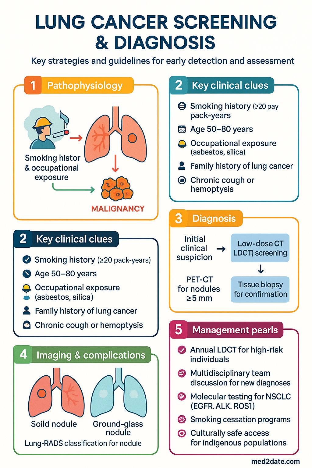
- Low-dose CT (LDCT) screening reduces lung cancer mortality by 20% in high-risk populations; recommended annually for adults aged 50–80 years with ≥20 pack-year smoking history and current smoking or cessation within 15 years (USPSTF 2021 criteria).
- Australia has not yet implemented a national lung cancer screening programme, though the Department of Health has announced LungCheck — a planned national programme expected to commence in 2025.
- Pulmonary nodule evaluation follows Fleischner Society 2017 guidelines for incidental nodules and Lung-RADS for screening-detected nodules; solid nodules <6 mm in low-risk patients typically require no further follow-up.
- PET-CT is indicated for solid nodules ≥8 mm with intermediate-to-high pre-test probability of malignancy; it has limited utility for ground-glass nodules and nodules <8 mm.
- Tissue diagnosis via bronchoscopy (EBUS, navigational bronchoscopy) or CT-guided transthoracic needle biopsy is required before treatment; the approach depends on lesion location, size, and accessibility.
- Endobronchial ultrasound-guided transbronchial needle aspiration (EBUS-TBNA) is the first-line modality for mediastinal lymph node staging, with sensitivity of 89–93% and specificity approaching 100%.
- Molecular testing (EGFR, ALK, ROS1, BRAF, KRAS G12C, PD-L1, NTRK, MET, RET) is mandatory for all newly diagnosed non-small cell lung cancer (NSCLC) specimens, regardless of histological subtype.
- TNM 8th edition staging guides treatment planning; staging is clinical (cTNM) at diagnosis and pathological (pTNM) after surgical resection.
- Multidisciplinary team (MDT) discussion is mandatory for every new lung cancer diagnosis in Australia, as per Optimal Care Pathway for Lung Cancer (Cancer Council Victoria / ACSQHC).
- Aboriginal and Torres Strait Islander Australians have 1.8× higher lung cancer incidence and significantly lower five-year survival than non-Indigenous Australians; culturally safe screening access and smoking cessation programmes are critical.
- Performance status (ECOG/Karnofsky) is a key determinant of treatment eligibility; ECOG 0–2 generally qualifies for curative-intent therapy.
Introduction & Australian Epidemiology
Lung cancer remains the most lethal malignancy in Australia and worldwide, responsible for more deaths than colorectal, breast, and prostate cancers combined. In 2024, an estimated 13,800 new cases were diagnosed in Australia, with a five-year survival rate of approximately 18–22% — a figure that has improved only modestly over the past two decades, largely because most patients present with advanced-stage disease.
The majority (approximately 85%) of lung cancers are non-small cell lung cancer (NSCLC), comprising adenocarcinoma (~40%), squamous cell carcinoma (~25%), and large cell carcinoma (~10–15%). Small cell lung cancer (SCLC) accounts for approximately 13–15% and is strongly associated with smoking. Rarer subtypes include large cell neuroendocrine carcinoma and pulmonary carcinoid tumours.
Australian Burden of Disease
- Incidence: Lung cancer is the fifth most commonly diagnosed cancer in Australia (age-standardised rate ~35 per 100,000).
- Mortality: Approximately 9,400 deaths per year; the age-standardised mortality rate is ~25 per 100,000.
- Trends: Incidence in males has declined since the 1980s reflecting reduced smoking rates; female incidence has plateaued and may increase due to later adoption of smoking by women.
- Aboriginal and Torres Strait Islander peoples: Age-standardised incidence is 1.8 times higher, with mortality rates 1.9 times higher than non-Indigenous Australians (AIHW 2023).
- Socioeconomic disparity: Lung cancer incidence is approximately 2.5 times higher in the most disadvantaged quintile compared to the most advantaged.
- Geographic variation: Rural and remote populations experience later-stage diagnosis and reduced access to specialist services, particularly PET-CT and bronchoscopy.
Risk Factors
| Risk Factor | Relative Risk | Australian Relevance |
|---|---|---|
| Active smoking | 15–30× | 11% of Australian adults smoke daily (2022–23 NDSHS) |
| Former smoking (quit <15 years) | 5–10× | Large eligible screening population |
| Second-hand smoke | 1.2–1.3× | Relevant in household and workplace settings |
| Occupational carcinogens (asbestos, silica, diesel exhaust) | 2–5× | Mining and construction industries; legacy asbestos in older buildings |
| Radon exposure | 1.5–3× | Lower prevalence than Northern Hemisphere; relevant in some geological regions |
| Family history (first-degree relative) | 1.5–2× | Independent of smoking status |
| COPD / pulmonary fibrosis | 2–5× | High prevalence in smoking population and Aboriginal communities |
| Prior chest radiotherapy | 2–10× | E.g., for Hodgkin lymphoma or breast cancer |
Screening Guidelines
Lung cancer screening with low-dose computed tomography (LDCT) is the only evidence-based modality shown to reduce lung cancer mortality. Two landmark randomised controlled trials — the National Lung Screening Trial (NLST, 2011) and the NELSON trial (2020) — demonstrated 20% and 24% relative reductions in lung cancer mortality, respectively, in high-risk populations screened with LDCT compared to chest radiography or no screening.
USPSTF 2021 Screening Recommendations
The United States Preventive Services Task Force (USPSTF) provides the most widely adopted eligibility criteria:
- Age 50 to 80 years
- ≥20 pack-year smoking history (1 pack/day × 20 years, or equivalent)
- Currently smoking OR quit within the past 15 years
| Criterion | USPSTF 2021 | NELSON Trial Criteria | Proposed Australian (LungCheck) |
|---|---|---|---|
| Age | 50–80 years | 50–75 years | 50–80 years |
| Smoking history | ≥20 pack-years | >15 cigs/day for >25 yrs OR >10 cigs/day for >30 yrs | ≥20 pack-years (anticipated) |
| Smoking status | Current or quit ≤15 years | Current or quit ≤10 years | Current or quit ≤15 years (anticipated) |
| Screening interval | Annual | Baseline, 1 yr, 3 yrs, 5.5 yrs | Annual (anticipated) |
| Mortality reduction | 20% (NLST) | 24% (NELSON) | Expected comparable |
Australian Screening Context
Low-Dose CT Protocol
- Technique: Non-contrast, volumetric acquisition through the entire chest; slice thickness ≤1.25 mm (ideally ≤1 mm); low radiation dose ≤2 mGy (CTDIvol); no IV contrast required.
- Reporting: Should use Lung-RADS (Lung CT Screening Reporting and Data System) classification.
- Nodule volumetry: Automated volumetric measurement with volumetric doubling time (VDT) calculation is preferred over manual diameter measurement (NELSON protocol).
- Incidental findings: Coronary artery calcification, emphysema, osteoporosis, thyroid nodules, adrenal lesions, and hepatic lesions are frequently identified and must be reported.
Lung-RADS Classification for Screening Nodules
| Lung-RADS | Category | Findings | Management |
|---|---|---|---|
| 1 | Negative | No pulmonary nodules; clearly benign nodules | Continue annual LDCT |
| 2 | Benign | Solid nodule <6 mm; new solid <4 mm; part-solid <6 mm total; GGN <30 mm stable | Continue annual LDCT |
| 3 | Probably Benign | Solid 6–<8 mm; new solid 4–<6 mm; part-solid ≥6 mm with solid <6 mm; new GGN ≥30 mm | 6-month LDCT follow-up |
| 4A | Suspicious | Solid 8–<15 mm; new/growing 6–<8 mm; part-solid with solid component 6–<8 mm | 3-month LDCT; PET-CT; tissue sampling |
| 4B | Very Suspicious | Solid ≥15 mm; new/growing ≥8 mm; part-solid with solid ≥8 mm | PET-CT; tissue sampling; consider surgical biopsy |
| 4X | Suspicious + Extra-thoracic | Category 3/4 with additional suspicious features (e.g., lymphadenopathy, pleural effusion) | Urgent tissue sampling; specialist referral |
| S | Other | Clinically significant non-pulmonary findings | Appropriate workup per finding |
Harms and Limitations of Screening
- False-positive rate: Approximately 23–27% of screening rounds in the NLST yielded a false-positive result; the positive predictive value of a positive screen is approximately 3–4%.
- Overdiagnosis: Estimated at 10–20% of screen-detected cancers, particularly slow-growing adenocarcinomas and ground-glass nodules.
- Radiation exposure: Cumulative dose over multiple rounds is low (~1.5 mSv per scan) but must be considered in the context of repeated screening.
- Psychological impact: False-positive results cause significant anxiety; shared decision-making is essential before initiating screening.
- Invasive procedures: Approximately 1–3% of screened individuals undergo an invasive procedure for a benign nodule.
Shared Decision-Making
Current guidelines (USPSTF, NCCN, ACS) recommend a shared decision-making conversation before ordering screening LDCT. This discussion should cover:
- The individual's lung cancer risk (pack-years, age, comorbidities)
- Benefits of screening (20–24% mortality reduction)
- Potential harms (false positives, invasive procedures, overdiagnosis)
- Importance of continued smoking cessation
- Willingness to undergo further investigation and potential treatment
Pulmonary Nodule Evaluation
Pulmonary nodules are increasingly detected as incidental findings on CT imaging performed for other indications (cardiac CT, CT pulmonary angiography, low-dose CT for calcium scoring, COVID-19 pneumonia follow-up). Incidental pulmonary nodules are identified in approximately 20–30% of chest CT scans. The Fleischner Society guidelines provide evidence-based recommendations for managing incidental pulmonary nodules based on size, morphology, and patient risk factors.
Nodule Morphology and Terminology
| Type | Definition | Differential Diagnosis | Malignancy Risk |
|---|---|---|---|
| Solid nodule | Homogeneous soft tissue attenuation that completely obscures parenchyma | Granuloma, hamartoma, intrapulmonary lymph node, lung cancer, metastasis | Varies by size and risk factors |
| Ground-glass nodule (GGN) | Hazy increased attenuation without obscuring bronchial/vascular markings | Atypical adenomatous hyperplasia (AAH), adenocarcinoma in situ (AIS), inflammation | Generally lower but may represent early adenocarcinoma |
| Part-solid (mixed) nodule | Both ground-glass and solid components | Minimally invasive adenocarcinoma (MIA), invasive adenocarcinoma | Highest malignancy risk (63–80% if ≥8 mm solid component) |
| Subsolid nodule | Umbrella term for GGN + part-solid | See above | Intermediate |
| Cavitary nodule | Nodule with central lucency; wall thickness ≥4 mm suggests malignancy | Squamous cell carcinoma, infection (TB, fungal), granulomatosis with polyangiitis | Increased with thick irregular walls |
| Calcified nodule | Central, laminated, popcorn, or diffuse calcification pattern | Granuloma, hamartoma (popcorn calcification) | Very low (typically benign) |
Fleischner Society 2017 Guidelines — Incidental Solid Nodules
Low-Risk Patients
(Minimal or absent smoking history; no other risk factors)
| Nodule Size | Recommendation | Follow-up |
|---|---|---|
| <6 mm | No routine follow-up | None required |
| 6–8 mm | CT at 6–12 months, then consider CT at 18–24 months | 6–12 months, then 18–24 months |
| >8 mm | CT at 3 months; PET-CT; or tissue sampling | 3 months (CT), or consider PET-CT/biopsy at presentation |
High-Risk Patients
(Smoking, family history, upper-lobe emphysema, occupational exposures)
| Nodule Size | Recommendation | Follow-up |
|---|---|---|
| <6 mm | Optional CT at 12 months | 12 months (optional) |
| 6–8 mm | CT at 6–12 months, then CT at 18–24 months | 6–12 months, then 18–24 months |
| >8 mm | CT at 3 months; PET-CT; or tissue sampling | 3 months (CT), or consider PET-CT/biopsy at presentation |
Fleischner Society 2017 — Incidental Subsolid Nodules
| Nodule Type | Size | Recommendation | Follow-up |
|---|---|---|---|
| Solitary GGN | <6 mm | No routine follow-up | None required |
| Solitary GGN | ≥6 mm | CT at 6–12 months, then every 2 years for 5 years | 6–12 mo, then 2-yearly × 5 yrs |
| Solitary part-solid | ≥6 mm | CT at 3–6 months; if solid component ≥6 mm and persists → PET-CT or biopsy | 3–6 months; further based on evolution |
| Multiple subsolid | Variable | CT at 3–6 months; if stable/dominant lesion ≥6 mm → CT at 2 and 4 years | 3–6 months, then 2 and 4 years |
PET-CT Indications
FDG PET-CT (MBS item 61338 — requires Medicare-eligible indication) plays a critical role in nodule evaluation and staging but has specific strengths and limitations:
Biopsy Techniques for Nodule Sampling
Nodule Growth Assessment
- Solid nodule growth: An increase in mean diameter of ≥2 mm on CT is considered significant growth.
- Volumetric doubling time (VDT): Malignant solid nodules typically have VDT of 100–400 days; VDT <100 days suggests infection or rapidly growing malignancy (SCLC, metastasis); VDT >400 days suggests benignity.
- Subsolid nodule growth: Increase in size of the ground-glass component or new/increasing solid component is concerning for progression to invasive adenocarcinoma.
- Stability: A solid nodule that remains unchanged in size on CT over ≥2 years is very likely benign (exception: pure GGN may require longer surveillance, up to 5 years).
Tissue Diagnosis
Obtaining an adequate tissue sample is a prerequisite for accurate histological classification, molecular testing, and treatment planning. The choice of biopsy technique depends on tumour location, size, proximity to airways, patient comorbidities, and local expertise. In Australia, access to advanced bronchoscopy techniques (EBUS, navigational bronchoscopy) has expanded significantly, with major tertiary centres offering these procedures routinely.
Bronchoscopy Techniques
Conventional Flexible Bronchoscopy
Endobronchial Ultrasound–Guided Transbronchial Needle Aspiration (EBUS-TBNA)
Navigational Bronchoscopy
CT-Guided Transthoracic Needle Biopsy (TTNB)
Surgical Biopsy
- Video-assisted thoracoscopic surgery (VATS) wedge resection: Minimally invasive approach; gold standard for indeterminate nodules when less invasive methods are non-diagnostic; diagnostic accuracy ~100%.
- VATS is preferred over open thoracotomy for diagnostic wedge resection due to reduced pain, shorter hospital stay, and lower morbidity.
- Open thoracotomy: Reserved for cases where VATS is technically not feasible, or when diagnostic resection is combined with definitive lobectomy.
- Mediastinoscopy: Historically the gold standard for mediastinal staging; now largely replaced by EBUS-TBNA; still indicated for station 5/6 sampling or EBUS non-diagnostic cases. MBS item 38504.
Molecular Testing (Comprehensive Genomic Profiling)
Molecular testing is mandatory for all newly diagnosed advanced NSCLC (stage IIIB–IV) and recommended for earlier stages where neoadjuvant/perioperative targeted therapy is being considered. Adequate tissue collection is critical — core biopsy specimens are strongly preferred over cytology alone.
| Biomarker | Test Method | Prevalence in NSCLC | Approved Targeted Therapy (Australia) |
|---|---|---|---|
| EGFR mutations (ex19del, L858R, exon 20 ins, others) | PCR / NGS | 15–20% (higher in never-smokers, Asian descent) | Osimertinib (Tagrisso®), erlotinib, gefitinib, afatinib, amivantamab, lazertinib |
| ALK rearrangement | FISH / IHC / NGS | 3–5% | Alectinib (Alecensa®), lorlatinib, crizotinib, brigatinib, ceritinib |
| ROS1 rearrangement | FISH / NGS | 1–2% | Crizotinib (Xalkori®), entrectinib (Rozlytrek®) |
| BRAF V600E | PCR / NGS / IHC | 2–4% | Dabrafenib + trametinib (Tafinlar® + Mekinist®) |
| KRAS G12C | NGS | 13% | Sotorasib (Lumakras®) |
| PD-L1 expression (TPS) | IHC (22C3, 28-8, SP263) | 50–60% ≥1%; 20–30% ≥50% | Pembrolizumab, atezolizumab, nivolumab, durvalumab |
| NTRK fusions | NGS / FISH / IHC | <1% | Larotrectinib (Vitrakvi®), entrectinib (Rozlytrek®) |
| MET exon 14 skipping | NGS / RNA-based | 3–4% | Capmatinib (Tabrecta®), tepotinib |
| RET fusions | NGS / FISH | 1–2% | Selpercatinib (Retevmo®), pralsetinib (Gavreto®) |
| HER2 mutations | NGS | 2–3% | Trastuzumab deruxtecan (Enhertu®) |
Liquid Biopsy (Circulating Tumour DNA)
- Role: When tissue biopsy is insufficient, technically not feasible, or as complement to tissue testing. Guardant360®, FoundationOne® Liquid CDx available in Australia through private pathology.
- Sensitivity: 60–80% (lower than tissue for EGFR exon 20, ALK, and fusions); specificity >95%.
- If liquid biopsy is negative: Tissue biopsy should still be pursued if clinically indicated (negative liquid biopsy does not exclude actionable mutations).
- MBS status: Not currently funded under Medicare; out-of-pocket cost $2,000–5,000 through private pathology laboratories.
Staging & Treatment Planning
Accurate staging is the cornerstone of lung cancer management. The TNM 8th edition classification (IASLC, 2017) is the current standard, applied in both clinical (cTNM) and pathological (pTNM) forms. Staging determines operability, guides adjuvant therapy decisions, and identifies patients who may benefit from neoadjuvant or definitive chemoradiation approaches.
TNM 8th Edition — Key Changes and Definitions
T (Primary Tumour)
| T Category | Size / Extent | Key Details |
|---|---|---|
| Tis | Carcinoma in situ | AIS or squamous carcinoma in situ |
| T1a(mi) | Minimally invasive adenocarcinoma | ≤3 cm lepidic predominant, invasion ≤0.5 cm |
| T1a | ≤1 cm | |
| T1b | >1 cm, ≤2 cm | |
| T1c | >2 cm, ≤3 cm | |
| T2a | >3 cm, ≤4 cm | Or involves main bronchus (not carina); visceral pleura; atelectasis to hilum |
| T2b | >4 cm, ≤5 cm | |
| T3 | >5 cm, ≤7 cm | Or separate tumour nodule in same lobe; invasion of chest wall, pericardium, phrenic nerve |
| T4 | >7 cm | Or invasion of mediastinum, heart, great vessels, trachea, recurrent laryngeal nerve, oesophagus, vertebral body, carina; separate nodule in different ipsilateral lobe |
N (Regional Lymph Nodes)
| N Category | Description | Stations Involved |
|---|---|---|
| N0 | No regional lymph node metastasis | — |
| N1 | Ipsilateral peribronchial/hilar nodes | Stations 10, 11, 12, 13 |
| N2 | Ipsilateral mediastinal/subcarinal nodes | Stations 2, 3, 4, 7, 8, 9 |
| N3 | Contralateral mediastinal/hilar; scalene/supraclavicular | Contralateral stations 2, 3, 4, 10; station 1 |
M (Distant Metastasis)
| M Category | Description |
|---|---|
| M0 | No distant metastasis |
| M1a | Contrateral lung nodules; pleural/pericardial nodules or effusion |
| M1b | Single extrathoracic metastasis in a single organ |
| M1c | Multiple extrathoracic metastases (one or multiple organs) |
Stage Grouping (AJCC 8th Edition)
Staging Investigations
Mediastinal Staging Algorithm
Accurate mediastinal staging is critical because occult mediastinal nodal disease changes management from primary surgery to neoadjuvant therapy or definitive chemoradiation.
Performance Status Assessment
Performance status (PS) is a critical determinant of treatment eligibility and prognosis. Australian lung cancer MDTs commonly use the ECOG (Eastern Cooperative Oncology Group) scale:
| ECOG Grade | Description | Treatment Implications |
|---|---|---|
| 0 | Fully active; no restrictions | All treatment options (surgery, chemotherapy, immunotherapy, targeted therapy, chemoradiation) |
| 1 | Restricted in strenuous activity; ambulatory; light work possible | All treatment options with close monitoring |
| 2 | Ambulatory; capable of self-care; unable to work; up and about >50% of waking hours | May tolerate combination chemotherapy; immunotherapy; reduced-dose regimens. Surgery case-by-case. |
| 3 | Limited self-care; confined to bed/chair >50% of waking hours | Single-agent or immunotherapy preferred; best supportive care; palliative intent |
| 4 | Completely disabled; no self-care; confined to bed/chair | Best supportive care; palliative radiation if symptomatic |
| 5 | Dead | — |
Multidisciplinary Team (MDT) Discussion
Key elements discussed at MDT:
- Histological subtype and molecular results
- Clinical and pathological TNM stage
- Fitness for surgery (PFTs, cardiopulmonary exercise testing if borderline)
- Fitness for systemic therapy (ECOG PS, comorbidities, geriatric assessment if ≥70 years)
- Patient preferences and goals of care
- Access to clinical trials
- Palliative care integration (recommended from diagnosis for all patients, not just advanced disease)
Investigations Summary — Complete Workup Before Treatment
Special Populations
Elderly Patients (≥70 years)
Renal Impairment
Young Adults (<40 years)
Immunocompromised Patients
Patients with Severe COPD / Pulmonary Fibrosis
Aboriginal and Torres Strait Islander Health Considerations
Aboriginal and Torres Strait Islander Australians experience a disproportionate burden of lung cancer — the most commonly diagnosed cancer and leading cause of cancer death in Indigenous Australians. The age-standardised incidence rate is 1.8 times that of non-Indigenous Australians, and mortality rates are 1.9 times higher. Five-year survival is significantly lower (11% vs 19%), reflecting later-stage presentation, higher comorbidity burden, and reduced access to specialist services, particularly in remote communities.
Key Disparities
Culturally Safe Practice Recommendations
- Yarning-based communication: Use culturally appropriate communication approaches. Allow time for yarning and relationship building before discussing diagnosis. Understand that a cancer diagnosis carries significant cultural and spiritual weight in many Aboriginal and Torres Strait Islander communities.
- Aboriginal and Torres Strait Islander health workers/practitioners: Involve Aboriginal health workers in screening, education, navigation, and follow-up. They provide cultural brokerage and build trust within communities.
- Smoking cessation: Tackling smoking is the single most impactful intervention. The Tackling Indigenous Smoking programme provides community-based support. Pharmacotherapy (varenicline, NRT) should be offered with culturally appropriate counselling. Varenicline is PBS-listed as a general benefit.
- Telehealth and outreach: Use telehealth for MDT discussions, pre-biopsy counselling, and follow-up. Outreach bronchoscopy and specialist clinics (e.g., through RHDAustralia, Cancer Council programmes) improve access in remote communities.
- Family and community: Recognise the importance of family and community in decision-making. Treatment decisions may involve extended family and Elders. Ensure appropriate consent processes that respect collective decision-making preferences.
- Palliative care: Culturally safe palliative care must be available, including support for dying on country where possible. Community-controlled palliative care services are limited but growing.
- Data sovereignty: Engage with Aboriginal Community Controlled Health Organisations (ACCHOs) in programme design and evaluation. Respect data sovereignty principles in screening programme governance.
📚 References
- 1. US Preventive Services Task Force. Screening for lung cancer: US Preventive Services Task Force recommendation statement. JAMA. 2021;325(10):962–970. doi:10.1001/jama.2021.1117
- 2. National Lung Screening Trial Research Team. Reduced lung-cancer mortality with low-dose computed tomographic screening. N Engl J Med. 2011;365(5):395–409. doi:10.1056/NEJMoa1102873
- 3. de Koning HJ, van der Aalst CM, de Jong PA, et al. Reduced lung-cancer mortality with volume CT screening in a randomized trial. N Engl J Med. 2020;382(6):503–513. doi:10.1056/NEJMoa1911793
- 4. MacMahon H, Naidich DP, Goo JM, et al. Guidelines for management of incidental pulmonary nodules detected on CT images: from the Fleischner Society 2017. Radiology. 2017;284(1):228–243. doi:10.1148/radiol.2017161659
- 5. American College of Radiology. Lung-RADS Version 1.1: Lung CT Screening Reporting and Data System. Reston, VA: ACR; 2019.
- 6. Detterbeck FC, Boffa DJ, Kim AW, Tanoue LT. The eighth edition lung cancer stage classification. Chest. 2017;151(1):193–203. doi:10.1016/j.chest.2016.10.010
- 7. Navani N, Nankivell M, Lawrence DR, et al. Lung cancer diagnosis and staging with endobronchial ultrasound-guided transbronchial needle aspiration compared with conventional approaches: an open-label, pragmatic, randomised controlled trial. Lancet Respir Med. 2015;3(4):282–289. doi:10.1016/S2213-2600(15)00029-6
- 8. Cancer Council Victoria. Optimal Care Pathway for People with Lung Cancer. 2nd ed. Melbourne: Cancer Council Victoria; 2022.
- 9. Australian Institute of Health and Welfare. Cancer in Australia 2024. AIHW Cancer Series No. 142. Canberra: AIHW; 2024.
- 10. Conroy M, Pinsky PF. Indolent lung cancer—the overdiagnosis problem. JAMA Intern Med. 2023;183(3):283–284. doi:10.1001/jamainternmed.2022.6434
- 11. Ren S, Wang J, Ying J, et al. Consensus for molecular biomarker testing in non-small cell lung cancer. J Thorac Oncol. 2022;17(5):624–639. doi:10.1016/j.jtho.2021.12.013
- 12. Australian Government Department of Health and Aged Care. National Lung Cancer Screening Programme — Discussion Paper. Canberra: Commonwealth of Australia; 2023.
- 13. Lim E, Baldwin D, Beckles M, et al. Guidelines on the radical management of patients with lung cancer. Thorax. 2010;65(Suppl 3):iii1–iii27. doi:10.1136/thx.2010.145938
- 14. Aberle DR, Adams AM, Berg CD, et al. Reduced lung-cancer mortality with low-dose computed tomographic screening. N Engl J Med. 2011;365(5):395–409. doi:10.1056/NEJMoa1102873
- 15. Aiolfi A, Nosotti M, Bertoglio P, et al. Navigational bronchoscopy for peripheral pulmonary nodules: a systematic review and meta-analysis. Respiration. 2023;102(11):967–979. doi:10.1159/000534518