📋 Key Information Summary
- Pulmonary aspergillosis encompasses a spectrum from invasive disease in immunocompromised hosts to chronic colonisation in structurally abnormal lungs.
- Invasive pulmonary aspergillosis (IPA) carries mortality of 30–90% depending on host factors; early diagnosis and prompt antifungal therapy are critical.
- Key risk factors for IPA include prolonged neutropenia (>10 days), haematological malignancy on intensive chemotherapy, haematopoietic stem cell transplant (HSCT), solid organ transplant (SOT), high-dose corticosteroids, and advanced HIV (CD4 <50).
- CT chest showing the halo sign (ground-glass opacity surrounding a nodule) in an immunocompromised patient is highly suggestive of IPA and should prompt urgent workup.
- Serum galactomannan (GM) and (1→3)-β-D-glucan (BDG) are essential non-invasive biomarkers; bronchoalveolar lavage (BAL) GM has higher sensitivity for pulmonary disease.
- Voriconazole (Vfend®) is the first-line antifungal for IPA; therapeutic drug monitoring (TDM) of trough levels (1–5.5 mg/L) is mandatory to optimise efficacy and reduce toxicity.
- Chronic pulmonary aspergillosis (CPA) affects patients with pre-existing structural lung disease (tuberculosis, COPD, bronchiectasis); Aspergillus IgG serology is the principal diagnostic marker.
- Aspergilloma may present with life-threatening haemoptysis; bronchial artery embolisation is the first-line intervention for massive haemoptysis, with surgical resection reserved for localised disease.
- Itraconazole is the first-line oral antifungal for CPA; voriconazole is an alternative for refractory or azole-resistant disease.
- Azole-resistant Aspergillus fumigatus is emerging in Australia; susceptibility testing should be performed on culture isolates from patients failing first-line therapy.
- Antifungal toxicity monitoring includes LFTs, FBC, electrolytes (voriconazole: hepatotoxicity, visual disturbance, photosensitivity; amphotericin B: nephrotoxicity, electrolyte wasting).
- Aboriginal and Torres Strait Islander Australians may have higher rates of underlying structural lung disease and reduced access to specialist antifungal care in remote settings.
Introduction & Australian Epidemiology
Pulmonary aspergillosis is a spectrum of lung diseases caused by Aspergillus species, most commonly A. fumigatus, A. niger, and A. flavus. The clinical manifestations range from life-threatening invasive disease in severely immunocompromised patients to chronic localised infection and simple colonisation in those with structural lung abnormalities.
In Australia, invasive aspergillosis (IA) is an increasingly recognised complication of intensive immunosuppressive therapies. The Australian Mycoses Registry reports an annual incidence of approximately 3–5 cases per 100,000 hospital admissions, with peaks during winter months correlating with construction activity and environmental spore counts. Chronic pulmonary aspergillosis is estimated to affect 1,500–2,500 Australians, many undiagnosed, particularly among those with prior tuberculosis, chronic obstructive pulmonary disease (COPD), or bronchiectasis.
The incidence of azole-resistant A. fumigatus has risen globally and has been detected in Australian agricultural regions, with resistance rates of 3–8% reported in surveillance studies. This has implications for empirical antifungal selection and mandates susceptibility testing where culture is available.
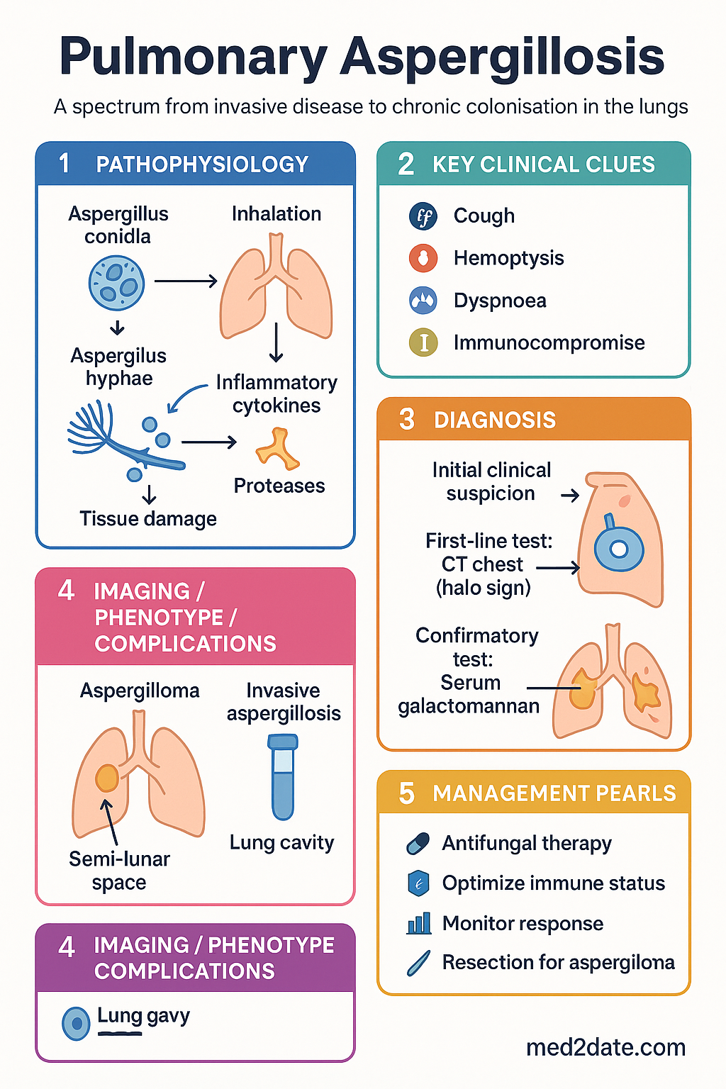
Pathophysiology
Aspergillus conidia (2–3 µm) are inhaled and deposited in the terminal airways. In immunocompetent hosts, alveolar macrophages and neutrophilic oxidative killing clear conidia effectively. Disease occurs when host defences are impaired:
- Invasive pulmonary aspergillosis (IPA): Conidia germinate into hyphae that invade pulmonary vasculature, causing thrombosis, haemorrhagic infarction, and tissue necrosis. Angioinvasion leads to dissemination to brain, liver, kidneys, and skin.
- Chronic pulmonary aspergillosis (CPA): In the setting of pre-existing cavitary or fibrotic lung disease, Aspergillus colonises cavities, forming fungal balls (aspergilloma) or driving progressive cavitary or fibrosing disease. Host immune response is partially intact, resulting in chronic granulomatous inflammation rather than angioinvasion.
- Allergic bronchopulmonary aspergillosis (ABPA): Hypersensitivity reaction in atopic individuals (especially asthma and cystic fibrosis) — characterised by eosinophilic airway inflammation, mucoid impaction, and proximal bronchiectasis. Distinct from IPA and CPA but part of the broader Aspergillus disease spectrum.
Virulence factors of A. fumigatus include thermotolerance (growth at 37–50°C), melanin production (protects against reactive oxygen species), gliotoxin secretion (immunosuppressive mycotoxin), and protease enzymes facilitating tissue invasion.
Invasive Aspergillosis
Invasive pulmonary aspergillosis (IPA) is the most severe manifestation of Aspergillus disease, occurring almost exclusively in immunocompromised hosts. It requires rapid diagnosis and immediate initiation of antifungal therapy.
Risk Factors
Host risk factors are categorised by degree of immunosuppression:
Diagnosis of Invasive Aspergillosis
Diagnosis follows the revised EORTC/MSG criteria, integrating host factors, clinical features, mycological evidence, and radiology. A classification of proven, probable, or possible IA guides management urgency.
CT Halo Sign
The CT halo sign — a zone of ground-glass attenuation surrounding a pulmonary nodule or mass — represents haemorrhage around angioinvasive hyphae. It is seen in 30–60% of early IPA cases in neutropenic patients and is highly suggestive in the appropriate host context. The air crescent sign (crescent of air separating necrotic centre from surrounding lung) appears later during neutrophil recovery and indicates cavity formation.
Galactomannan and Biomarkers
| Biomarker | Specimen | Sensitivity | Specificity | Notes |
|---|---|---|---|---|
| Serum galactomannan (GM) | Blood | 60–70% | 85–95% | Index ≥0.5 positive; reduced by mould-active azole prophylaxis; twice-weekly monitoring recommended in neutropenic patients |
| BAL galactomannan | Bronchoalveolar lavage | 80–90% | 85–95% | Higher sensitivity than serum for pulmonary IPA; index ≥0.8 recommended cutoff; perform BAL early when IPA suspected |
| (1→3)-β-D-glucan (BDG) | Blood | 55–75% | 70–85% | Non-specific (also elevated in Candida, Pneumocystis); cutoff ≥80 pg/mL; useful combined with GM |
| Aspergillus PCR (blood/BAL) | Blood or BAL | 70–85% | 80–95% | Not yet standardised; increasingly available at reference laboratories; positive predictive value increases in high-risk hosts |
| Histopathology / culture | Tissue biopsy | 30–50% | >99% | Required for proven IA; shows septate hyphae with acute-angle branching; culture identifies species and allows susceptibility testing |
Voriconazole Therapy for IPA
Voriconazole remains the first-line treatment for IPA as established by the landmark Herbrecht et al. (2002) trial demonstrating superior efficacy and survival compared with conventional amphotericin B.
Chronic Pulmonary Aspergillosis
Chronic pulmonary aspergillosis (CPA) is a slowly progressive Aspergillus infection of the lung occurring in patients with pre-existing structural lung disease and mildly impaired immunity (e.g., malnutrition, diabetes, COPD, prior TB). Unlike IPA, CPA is not immediately life-threatening but causes significant morbidity and progressive lung destruction if untreated.
Clinical Subtypes
| Subtype | Description | Key Features | Imaging |
|---|---|---|---|
| Simple aspergilloma | Fungal ball within a pre-existing pulmonary cavity | Often asymptomatic or mild haemoptysis; stable over years | Mobile intracavitary mass with air crescent sign; upper lobe predilection |
| Chronic cavitary pulmonary aspergillosis (CCPA) | One or more expanding pulmonary cavities with progressive fibrosis | Chronic cough, weight loss, haemoptysis, fatigue; progressive over months–years | Increasing cavity size, new cavities, pericavitary infiltrates, pleural thickening |
| Chronic fibrosing pulmonary aspergillosis (CFPA) | End-stage CCPA with extensive fibrosis | Severe breathlessness, respiratory failure; often treatment-refractory | Dense upper lobe fibrosis with volume loss; may mimic fibrotic TB |
| Aspergillus nodule | Single or multiple parenchymal nodules | Usually incidental; may mimic malignancy | Solitary or multiple nodules; central necrosis possible |
Diagnosis of CPA
Diagnostic criteria for CPA (modified from Denning et al., 2016):
- Chronic (>3 months) pulmonary or systemic symptoms (cough, sputum, haemoptysis, weight loss, fatigue)
- Radiological evidence of progressive pulmonary cavity, infiltrate, or fibrosis on CT chest
- Aspergillus IgG antibody positive (or other serological/mycological evidence)
- Exclusion of alternative diagnoses (TB, malignancy, other fungal infection)
- Mild or no immunocompromise (i.e., not meeting criteria for IPA)
Antifungal Indications in CPA
- Aspergilloma: Treat only if symptomatic (haemoptysis, cough) or enlarging on imaging. Many simple aspergillomata are stable and require surveillance only.
- CCPA / CFPA: Antifungal therapy is indicated for all symptomatic patients and those with radiological progression. Early treatment may prevent progression to fibrosing disease.
- Aspergillus nodule: Resection may be diagnostic (to exclude malignancy) and curative; antifungal therapy is considered if resection is incomplete or the patient is unfit for surgery.
Clinical Presentation & Diagnostic Criteria
Invasive Pulmonary Aspergillosis
- Persistent fever unresponsive to broad-spectrum antibiotics in neutropenic patients (classic presentation)
- Cough, pleuritic chest pain, dyspnoea, haemoptysis (may be absent early)
- Tachypnoea, hypoxia, pleural rub on auscultation
- Disseminated disease: cutaneous lesions (necrotic papules), mental status changes (cerebral aspergillosis), hepatosplenic lesions
Chronic Pulmonary Aspergillosis
- Insidious onset over months to years
- Chronic productive cough, haemoptysis (minor to massive), weight loss, fatigue, exertional dyspnoea
- May be initially misattributed to underlying COPD, bronchiectasis, or TB
- Physical signs: clubbing (late), crackles, upper lobe signs of fibrosis/cavitation
EORTC/MSG Criteria for Invasive Aspergillosis (2020 Revision)
| Classification | Host Criteria | Clinical Criteria | Mycological Criteria |
|---|---|---|---|
| Proven | Not required | Not required | Histological evidence of septate, acutely branching hyphae with tissue invasion; or positive sterile-site culture for Aspergillus |
| Probable | ≥1 host factor (immunosuppression as above) | ≥1 clinical feature (CT halo sign, air crescent, cavity, new infiltrate not responding to antibiotics) | ≥1 mycological criterion (positive GM, positive culture from BAL, positive Aspergillus PCR from BAL, positive BDG) |
| Possible | ≥1 host factor | ≥1 clinical feature | No mycological evidence |
Investigations
Risk Stratification
Risk stratification guides intensity of surveillance and urgency of empirical therapy:
Prophylaxis Agents
Treatment Strategies
First-Line Therapy for IPA
Antifungal Selection
Treatment Algorithm
Surgical Resection
Surgical resection is considered in the following scenarios:
- Aspergilloma with life-threatening haemoptysis not controlled by bronchial artery embolisation — lobectomy or segmentectomy if disease is localised and patient is fit for surgery.
- Localised IPA failing medical therapy (e.g., pulmonary lesion invading great vessels or chest wall).
- Aspergillus nodule where malignancy cannot be excluded — diagnostic and potentially curative wedge resection.
Duration of Therapy
| Condition | Minimum Duration | End-of-Treatment Criteria |
|---|---|---|
| IPA (haematological) | 6–12 weeks | Radiological resolution/stabilisation; clinical improvement; immune reconstitution (ANC recovery, reduction of immunosuppression); GM normalisation |
| IPA (SOT recipient) | 12+ weeks; often lifelong | May require lifelong suppression if ongoing immunosuppression; specialist transplant ID guidance essential |
| CPA (CCPA) | 6 months minimum | Symptom improvement; stable/improving imaging; declining Aspergillus IgG; many patients require lifelong therapy |
| Simple aspergilloma | 3–6 months if symptomatic | Resolution of haemoptysis; stable size on imaging |
Monitoring & Complications
Antifungal Therapy Monitoring
| Parameter | Frequency | Purpose / Target |
|---|---|---|
| Voriconazole trough level | Day 3–5, then weekly for first month; monthly thereafter | Target 1–5.5 mg/L; levels <1 associated with treatment failure; >5.5 with neurotoxicity and hepatotoxicity |
| LFTs (ALT, AST, ALP, bilirubin) | Baseline, twice weekly for 2 weeks, then weekly, then monthly | Voriconazole and itraconazole: hepatotoxicity; stop if ALT/AST >5× ULN or symptomatic hepatitis |
| FBC | Baseline, then weekly | Azole-related: rarely agranulocytosis; monitor for cytopaenias in combination with chemotherapy |
| Electrolytes (K⁺, Mg²⁺, Ca²⁺) | Daily during IV amphotericin B; weekly with azoles | Amphotericin B: hypokalaemia, hypomagnesaemia; aggressive supplementation often required |
| Creatinine / eGFR | Baseline, twice weekly during amphotericin B; weekly with azoles | Amphotericin B nephrotoxicity: reduce dose or switch if creatinine doubles from baseline |
| Serum galactomannan | Twice weekly during neutropenia for IPA; every 1–2 weeks during treatment | Declining GM correlates with response; rising GM suggests treatment failure or relapse |
| Aspergillus IgG (CPA) | Every 1–3 months during treatment | Declining titres indicate response; rising titres suggest progression or relapse |
| CT chest | 6–8 weeks post-treatment initiation; then 3–6 monthly for CPA | Assess cavity size, resolution of infiltrates, new lesions |
Voriconazole-Specific Adverse Effects
- Visual disturbance (30% of patients): blurred vision, photopsia, altered colour perception — typically transient and dose-dependent; usually resolves with continued therapy
- Photosensitivity and skin cancer risk: chronic use associated with squamous cell carcinoma and melanoma; mandatory sun protection (SPF 50+, protective clothing); regular dermatological review in long-term users
- Hepatotoxicity: jaundice, transaminitis; usually reversible on cessation
- Periosteal bone pain: rare but characteristically associated with prolonged voriconazole use (periostitis); consider switching antifungal
- Skin reactions: cheilosis, alopecia, erythema multiforme (rare)
Haemoptysis Management
Special Populations
Pregnancy
Paediatrics
Elderly
Renal Impairment
Hepatic Impairment
Immunocompromised
Aboriginal and Torres Strait Islander Health Considerations
Aboriginal and Torres Strait Islander Australians bear a disproportionate burden of structural lung disease, which increases the risk of chronic pulmonary aspergillosis. Historically high rates of tuberculosis (now largely controlled), chronic suppurative lung disease, bronchiectasis, and rheumatic heart disease contribute to a higher prevalence of underlying cavitation and fibrosis. Access to specialist antifungal care, advanced imaging, and mycology services is limited in many remote and regional communities.
📚 References
- 1. Herbrecht R, Denning DW, Patterson TF, et al. Voriconazole versus amphotericin B for primary therapy of invasive aspergillosis. N Engl J Med. 2002;347(6):408–415.
- 2. Ullmann AJ, Aguado JM, Arikan-Akdagli S, et al. Diagnosis and management of Aspergillus diseases: executive summary of the 2017 ESCMID-ECMM-ERS guideline. Clin Microbiol Infect. 2018;24(Suppl 1):e1–e38.
- 3. Denning DW, Cadranel J, Beigelman-Aubry C, et al. Chronic pulmonary aspergillosis: rationale and clinical guidelines for diagnosis and management. Eur Respir J. 2016;47(1):45–68.
- 4. Maertens JA, Raad II, Marr KA, et al. Isavuconazole versus voriconazole for primary treatment of invasive mould disease caused by Aspergillus and other filamentous fungi (SECURE): a phase 3, randomised-controlled, non-inferiority trial. Lancet. 2016;387(10017):760–769.
- 5. Donnelly JP, Chen SC, Kauffman CA, et al. Revision and update of the consensus definitions of invasive fungal disease from the European Organization for Research and Treatment of Cancer and the Mycoses Study Group Education and Research Consortium (EORTC/MSG). Clin Infect Dis. 2020;71(6):1367–1376.
- 6. Chang AB, Marsh RL, Upham JW, et al. Toward making inroads in reducing the disparity of lung health in Australian Indigenous and New Zealand Māori children. Front Pediatr. 2015;3:9.
- 7. Patterson TF, Thompson GR, Denning DW, et al. Practice guidelines for the diagnosis and management of aspergillosis: 2016 update by the Infectious Diseases Society of America. Clin Infect Dis. 2016;63(4):e1–e60.
- 8. Verweij PE, Ananda-Rajah M, Andes D, et al. International expert opinion on the management of infection caused by azole-resistant Aspergillus fumigatus. Drug Resist Updat. 2015;21–22:30–40.
- 9. Kolve H, Groll AH, Jürgens H, et al. Antifungal activity of posaconazole against clinical isolates of Aspergillus species and other moulds. Int J Antimicrob Agents. 2009;34(5):429–434.
- 10. Schwartz S, Ruhnke M, Ribaud P, et al. Improved outcome in central nervous system aspergillosis, using voriconazole treatment. Blood. 2005;106(8):2865–2870.
- 11. Felton T, Troke PF, Hope WW. Tissue penetration of antifungal agents. Clin Microbiol Rev. 2014;27(1):68–88.
- 12. Australian Institute of Health and Welfare (AIHW). Aboriginal and Torres Strait Islander Health Performance Framework: Summary report 2023. Canberra: AIHW; 2023.
- 13. Moran C, O'Brien S, Doffman S, et al. Voriconazole-associated squamous cell carcinoma in lung transplant recipients. J Heart Lung Transplant. 2018;37(8):1014–1021.
- 14. Agarwal R, Chakrabarti A, Shah A, et al. Allergic bronchopulmonary aspergillosis: review of literature and proposal of new diagnostic and classification criteria. Clin Exp Allergy. 2013;43(8):850–873.