📋 Key Information Summary
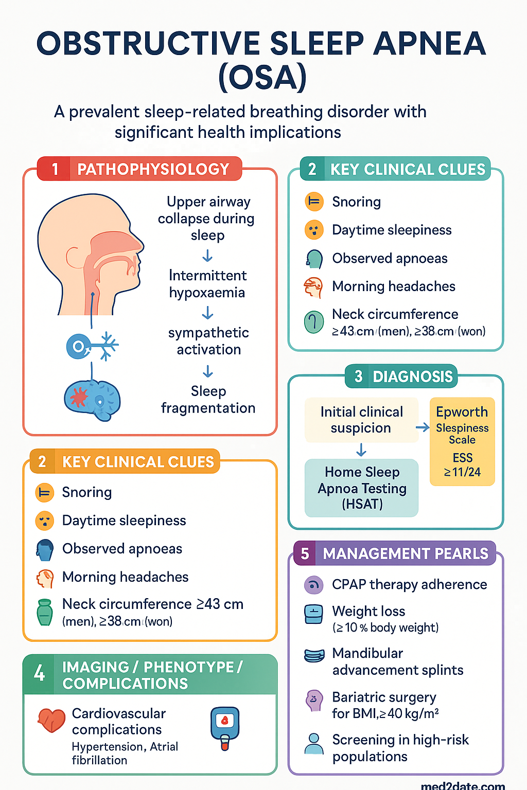
- Obstructive sleep apnoea (OSA) affects an estimated 20–30% of Australian adults, with prevalence rising in parallel with obesity rates; moderate-to-severe disease is present in approximately 10% of men and 5% of women aged 30–70 years.
- The Epworth Sleepiness Scale (ESS ≥ 11/24) supports referral but is insensitive — many patients with significant OSA report normal daytime alertness, so a low score does not exclude disease.
- In-laboratory polysomnography (PSG) remains the gold standard; however, Medicare-rebatable home sleep apnoea testing (HSAT, MBS item 12250) is an acceptable first-line alternative for uncomplicated adult patients with high pre-test probability.
- AHI classification: mild 5–14, moderate 15–29, severe ≥30 events/hour; treatment is recommended for symptomatic patients at any severity and for asymptomatic patients with moderate-to-severe disease due to cardiovascular risk.
- Continuous positive airway pressure (CPAP) is first-line therapy; adherence is the primary barrier, with ≥4 hours/night on ≥70% of nights considered adequate; mask fitting, humidification, and telemonitoring improve long-term use.
- Auto-titrating CPAP (APAP) is effective for uncomplicated OSA; bilevel positive airway pressure (BiPAP) is reserved for CPAP-intolerant patients, obesity hypoventilation syndrome, or elevated CO₂.
- Mandibular advancement splints (MAS) fitted by a dentist are a valid alternative for mild-to-moderate OSA or CPAP-intolerant patients; effectiveness depends on adequate mandibular advancement (≥50% maximum).
- Weight loss of ≥10% body weight can reduce AHI by 26–50%; bariatric surgery should be considered for patients with BMI ≥40 kg/m² (or ≥35 with comorbidities) who have failed conservative measures.
- OSA is an independent risk factor for resistant hypertension, atrial fibrillation, coronary artery disease, stroke, type 2 diabetes, and perioperative respiratory complications — screening is mandatory in these populations.
- Aboriginal and Torres Strait Islander Australians have a disproportionately higher burden of OSA and comorbid cardiovascular disease; culturally safe screening, community-based sleep health programmes, and equitable access to CPAP are critical gaps requiring targeted action.
- Modafinil (PBS Authority Required) may be considered for residual excessive daytime sleepiness despite adequate CPAP adherence; routine pharmacotherapy is not a substitute for positive airway pressure therapy.
- Perioperative OSA screening using the STOP-BANG questionnaire is recommended for all patients undergoing general anaesthesia; unmanaged severe OSA increases the risk of difficult intubation, post-operative hypoxaemia, and cardiac events.
Introduction & Australian Epidemiology
Obstructive sleep apnoea (OSA) is a sleep-related breathing disorder characterised by repetitive episodes of partial (hypopnoea) or complete (apnoea) upper airway collapse during sleep, resulting in intermittent hypoxaemia, hypercapnia, intrathoracic pressure swings, sleep fragmentation, and sympathetic nervous system activation. It is the most common sleep-disordered breathing condition and is a significant contributor to cardiovascular morbidity, metabolic dysfunction, neurocognitive impairment, occupational accidents, and reduced quality of life.
In Australia, the 2016 Sleep Health Foundation National Survey estimated that approximately 8% of adults have been diagnosed with sleep apnoea, with many more undiagnosed. Population-based studies using objective sleep monitoring suggest that the true prevalence of moderate-to-severe OSA (AHI ≥ 15) is around 10–14% in men and 5–8% in women aged 30–70 years. Prevalence increases sharply with age, male sex, obesity (BMI ≥ 30 kg/m²), neck circumference >43 cm (men) or >38 cm (women), craniofacial factors, and menopausal status in women.
The economic burden is substantial: the Sleep Health Foundation estimated that sleep disorders cost the Australian economy over $26 billion annually in healthcare expenditure, lost productivity, and accident-related costs. OSA contributes to increased road traffic accidents (two- to seven-fold risk), workplace injuries, reduced work productivity, and increased utilisation of primary care and emergency department services.
Key Australian Risk Factors
| Risk Factor | Details | Population Prevalence |
|---|---|---|
| Obesity (BMI ≥ 30) | Central adiposity, neck fat deposition, reduced lung volumes | 31% of Australian adults (AIHW 2023) |
| Male sex | 2–3× higher prevalence than pre-menopausal women | Ratio narrows post-menopause |
| Age >50 years | Pharyngeal muscle tone decline, central respiratory control changes | Prevalence peaks 50–70 years |
| Family history | First-degree relative with OSA; craniofacial genetics | 2–4× increased risk |
| Craniofacial features | Retrognathia, micrognathia, macroglossia, tonsillar hypertrophy | Higher in some Asian and ATSI populations |
| Alcohol and sedative use | Pharyngeal muscle relaxation, arousal threshold elevation | Common contributing factor |
| Nasal obstruction | Deviated septum, allergic rhinitis, nasal polyps | Contributes in up to 30% of cases |
Pathophysiology
OSA results from the interplay of three principal mechanisms operating during sleep when pharyngeal dilator muscle tone naturally declines:
Mechanisms of Upper Airway Collapse
- Anatomical narrowing (critical closing pressure, Pcrit): Bony and soft-tissue craniofacial structure, tongue volume, lateral pharyngeal wall fat, and tonsillar/adenoid hypertrophy determine the baseline calibre of the upper airway. Obesity increases Pcrit by depositing fat in the parapharyngeal space and reducing lung volumes (which normally provide caudal traction on the trachea, splinting the pharynx open).
- Compensatory neuromuscular drive failure: Pharyngeal dilator muscles (particularly genioglossus) respond to negative intraluminal pressure and hypercapnia during wakefulness to maintain airway patency. During sleep, this reflexive activation diminishes, and in susceptible individuals is insufficient to counterbalance the collapsing forces.
- Loop instability (low arousal threshold, high loop gain): Some patients have unstable ventilatory control — small perturbations in ventilation cause large corrective responses (high controller gain), leading to over-breathing followed by central apnoea, which destabilises the airway. Patients with a low arousal threshold wake before oxygen normalises, perpetuating repetitive cycling.
Pathophysiological Consequences
- Intermittent hypoxaemia → sympathetic activation: Cyclical desaturation and reoxygenation generates reactive oxygen species (ROS), activates the sympathetic nervous system, and increases catecholamine release, driving systemic hypertension.
- Intrathoracic pressure swings (Müller manoeuvre): Vigorous inspiratory efforts against a closed airway generate intrathoracic pressures as low as −60 cmH₂O, increasing transmural cardiac wall stress, favouring atrial fibrillation and impairing cardiac output.
- Sleep fragmentation: Repeated cortical arousals suppress slow-wave and REM sleep, impairing memory consolidation, executive function, and daytime alertness.
- Inflammatory cascade: Intermittent hypoxia activates NF-κB, increases CRP, IL-6, TNF-α, and endothelin-1, promoting endothelial dysfunction, atherosclerosis, and insulin resistance.
- Metabolic dysregulation: Intermittent hypoxaemia impairs glucose tolerance independently of obesity, increases hepatic gluconeogenesis, and promotes visceral adiposity through HPA axis activation.
Diagnosis & Testing
Clinical Screening Tools
| Tool | Components | Interpretation | Australian Context |
|---|---|---|---|
| Epworth Sleepiness Scale (ESS) | 8 questions; probability of dozing in various situations (0–3 each) | Normal: 0–10; Mild EDS: 11–14; Moderate: 15–17; Severe: 18–24 | Validated in Australian populations; available in multiple languages. Sensitivity ~50–60% for OSA — many patients score normally. |
| STOP-BANG Questionnaire | Snoring, Tiredness, Observed apnoeas, Pressure (hypertension), BMI >35, Age >50, Neck circumference >40 cm, male Gender | 0–2: Low risk; 3–4: Intermediate risk; 5–8: High risk | Recommended by ANZCA for pre-anaesthetic screening; high sensitivity for moderate-to-severe OSA. |
| Berlin Questionnaire | 10 items across 3 categories: snoring/daytime somnolence, obesity, hypertension | ≥2 positive categories = high risk | Less commonly used in Australian practice; lower specificity than STOP-BANG. |
| NoSAS Score | Neck circumference, Obesity, Snoring, Age, Sex | 0–7: Low; 8–12: Intermediate; ≥13: High risk | Useful in primary care as a rapid triage tool. |
Polysomnography (PSG)
Full, attended, in-laboratory polysomnography remains the gold standard for diagnosis of sleep-disordered breathing. PSG records electroencephalography (EEG), electrooculography (EOG), electromyography (EMG), nasal/oral airflow, thoracoabdominal effort, oximetry (SpO₂), ECG, body position, and leg movements. It is performed in accredited sleep laboratories under the supervision of qualified sleep scientists.
| PSG Indication | MBS Item | Details |
|---|---|---|
| Diagnostic PSG | 12203 | Full overnight attended polysomnography; referral from sleep/respiratory physician required. |
| Split-night PSG (diagnostic + CPAP titration) | 12204 | If AHI ≥ 20 in first half, proceeds to CPAP titration; saves one night of study. |
| CPAP titration PSG | 12206 | Dedicated titration study when split-night not performed or was suboptimal. |
Home Sleep Apnoea Testing (HSAT)
Home sleep apnoea testing (HSAT), also known as portable monitoring or home sleep studies, uses simplified devices (typically Level 3 or Level 4) to record airflow, respiratory effort, oximetry, and heart rate in the patient's own home over 1–3 nights.
| Parameter | HSAT | In-Lab PSG |
|---|---|---|
| MBS item | 12250 | 12203 / 12204 / 12206 |
| Channels recorded | 4–7 (airflow, effort, SpO₂, HR; ± position) | 16+ (full EEG, EOG, EMG, airflow, effort, SpO₂, ECG, position, legs) |
| Measures RDI/AHI | Yes (RDI may underestimate due to limited EEG) | Yes — gold standard AHI |
| Detects central/complex apnoea | Limited (no respiratory effort with some devices) | Yes |
| Detects sleep architecture | No | Yes |
| Suitable population | Adults with high pre-test probability, no significant comorbidities (COPD, CHF, neuromuscular disease) | All patients; mandatory for complex or inconclusive HSAT |
| Cost (approximate out-of-pocket) | $0–200 (Medicare-rebatable with referral) | $200–600 (Medicare-rebatable with referral) |
AHI Interpretation & Clinical Significance
Additional Sleep Parameters
- Respiratory Disturbance Index (RDI): Includes apnoeas, hypopnoeas, and respiratory effort-related arousals (RERAs). RDI ≥ 5 with symptoms defines Upper Airway Resistance Syndrome (UARS).
- ODI (Oxygen Desaturation Index): Number of ≥3% or ≥4% desaturation events per hour. ODI ≥ 5 correlates with clinically significant OSA. ODI ≥ 15 associated with increased cardiovascular risk.
- Mean and nadir SpO₂: Nadir SpO₂ <85% correlates with severe disease and increased cardiovascular risk. Mean SpO₂ <93% during sleep warrants evaluation for hypoventilation.
- T90 (time spent with SpO₂ <90%): T90 >30% of total sleep time indicates significant nocturnal hypoxaemia and may warrant supplemental oxygen in addition to CPAP.
- Central Apnoea Index (CAI): CAI ≥ 5/hour suggests central sleep apnoea component; CPAP alone may be insufficient and ASV or BiPAP may be required.
Investigations
CPAP Therapy
Continuous positive airway pressure (CPAP) is the gold-standard treatment for moderate-to-severe OSA and for mild OSA with significant symptoms. CPAP acts as a pneumatic splint, maintaining upper airway patency throughout the respiratory cycle, eliminating apnoeas, hypopnoeas, and snoring.
CPAP Modes
| Mode | Description | Indications |
|---|---|---|
| Fixed CPAP | Single pressure delivered continuously (e.g., 10 cmH₂O) | Established pressure from titration study; positional OSA; patient preference |
| Auto-titrating CPAP (APAP) | Algorithm adjusts pressure breath-by-breath within set range (e.g., 4–20 cmH₂O) | Uncomplicated OSA (first-line per ASA); initial therapy before PSG titration; positional and REM-related OSA |
| Bilevel PAP (BiPAP / BPAP) | Separate inspiratory (IPAP) and expiratory (EPAP) pressures | CPAP-intolerant patients (high fixed pressures); obesity hypoventilation syndrome (OHS); COPD-OSA overlap; neuromuscular disease |
| ASV (Adaptive Servo-Ventilation) | Adjusts pressure support to stabilise ventilation; targets a minute ventilation | Complex/central sleep apnoea; treatment-emergent central apnoea on CPAP; NOT for HFrEF with LVEF ≤ 45% (SERVE-HF trial) |
| EPAP valves (e.g., Provent®) | Nasal expiratory positive airway pressure via small disposable valves | Mild OSA; CPAP-intolerant patients; travel; limited evidence base |
CPAP Titration Protocols
The goal of titration is to identify the minimum effective pressure that eliminates obstructive apnoeas, hypopnoeas, flow limitation, snoring, and respiratory effort-related arousals in all sleep stages and body positions.
Mask Selection
| Mask Type | Best For | Considerations |
|---|---|---|
| Nasal mask | Most patients; first-line default | Requires nasal breathing; claustrophobia-friendly; least skin contact |
| Nasal pillows | Patients with claustrophobia; those with facial hair; side sleepers | May cause nasal dryness; not ideal for pressures >15 cmH₂O |
| Full-face (oronasal) mask | Mouth breathers; nasal obstruction; high pressures | Higher leak risk; more claustrophobic; may cause eye irritation; consider heated humidification |
| Total-face mask | Patients with facial deformities; pressure injuries from other masks | Higher dead space; used as a last resort |
Adherence Strategies
- Early follow-up (within 1–2 weeks): The first fortnight is critical. Troubleshoot mask fit, pressure comfort, and nasal symptoms early. Telehealth review is effective and increasingly used in Australian sleep services.
- Ramp and expiratory pressure relief (EPR/C-Flex): Ramp feature starts at a low pressure and gradually increases over 5–45 minutes to ease sleep onset. EPR reduces expiratory pressure by 1–3 cmH₂O, improving comfort.
- Heated humidification: Reduces nasal dryness, congestion, and epistaxis. Consider in all patients, especially those on high pressures, mouth breathers, or in dry climates (common in inland Australia).
- Mask refitting: Multiple mask trials may be necessary. Ensure adequate supply of replacement cushions (every 1–3 months) and headgear (every 6 months) — most Australian CPAP suppliers provide these on subscription.
- Patient education and support groups: Referral to sleep psychologist for CBT-based CPAP desensitisation for anxious patients. Support groups (e.g., Sleep Health Foundation resources, CPAP Australia online forums).
- Telemonitoring: ResMed AirView, Philips Care Orchestrator, and Löwenstein platforms allow real-time monitoring. Nursing staff can intervene when usage drops below thresholds. Widely adopted in Australian public and private sleep services.
- Ongoing follow-up: Review at 1 month, 3 months, and 6 months after initiation; annually thereafter. Download device data at each visit. Adjust pressure if residual AHI >5 or if significant leak persists.
Troubleshooting CPAP Problems
| Problem | Cause | Solution |
|---|---|---|
| Nasal congestion / rhinitis | Airflow drying nasal mucosa; allergic response to mask silicone | Heated humidification; intranasal corticosteroid (e.g., fluticasone); nasal saline irrigation; hypoallergenic mask cushions |
| Aerophagia (air swallowing) | Excessive pressure; open mouth during sleep | Reduce pressure or switch to APAP; chin strap or full-face mask; check for underlying gastroparesis |
| Mask leak | Poorly fitting mask; worn cushion; facial hair | Refit mask; replace cushion; consider nasal pillows if facial hair; mask liner pads |
| Claustrophobia | Sensation of confinement from mask | Switch to nasal pillows; desensitisation therapy; practice wearing mask during the day while reading; consider EPAP valves |
| Residual sleepiness despite good adherence | Inadequate pressure; other sleep disorder (narcolepsy, PLMS); insufficient sleep; depression | Review AHI from device data; repeat PSG on CPAP; screen for depression; consider MSLT; consider modafinil |
| Central apnoeas on CPAP | Treatment-emergent central apnoea; pre-existing central component | May resolve over 1–3 months; if persistent, switch to ASV (confirm LVEF >45%); sleep physician review |
CPAP Access & Funding in Australia
- Public hospital sleep services: Many state health services provide CPAP devices on loan (especially for concession card holders). Wait times vary (4–12 weeks).
- Private purchase: CPAP devices cost $800–2,500 AUD. Mask and supply kits $50–300. Most Australian suppliers offer rental-to-own and payment plans.
- Private health insurance: Many extras policies cover CPAP devices and supplies (partially or fully). Check individual fund limits.
- Department of Veterans' Affairs (DVA): CPAP devices and supplies are covered for eligible veterans with a Gold or White Card.
- NDIS: CPAP may be funded under NDIS for participants with disability-related sleep-disordered breathing.
Alternative Treatments
Alternative treatments are indicated when CPAP is refused, not tolerated despite adequate troubleshooting, or when the patient has mild-to-moderate OSA and prefers a non-CPAP approach. Treatment selection should be guided by disease severity, anatomical site of obstruction, patient preference, and availability of specialist services.
Oral Appliances (Mandibular Advancement Splints — MAS)
Mandibular advancement splints (MAS), also termed mandibular advancement devices (MAD), are custom-fitted oral appliances worn during sleep that protrude the mandible forward, increasing the anteroposterior dimension of the pharyngeal airway and reducing collapsibility.
Positional Therapy
Positional OSA (supine-predominant) is defined as an AHI at least twice as high in the supine position compared to the lateral position. Approximately 20–30% of OSA patients have a significant positional component.
- Positional devices: Vibrotactile devices (e.g., NightShift™, Philips NightBalance™) worn on the neck or chest deliver gentle vibrations when the patient adopts the supine position, prompting positional change without awakening. Available in Australia through sleep device suppliers.
- Modified sleep positioning: Elevated head of bed 30°; body positioning pillows; tennis ball technique (less tolerated long-term); side-sleeping pillows. Low cost but adherence is often poor.
- Evidence: Positional therapy alone may reduce AHI by 50% in positional OSA. Often used as adjunct to CPAP or MAS rather than standalone therapy for moderate-to-severe disease.
Upper Airway Surgery
Surgical options are considered when CPAP and oral appliances have failed or are refused, and when a surgically correctable anatomical abnormality is identified. Drug-induced sleep endoscopy (DISE) is increasingly used to guide surgical planning.
| Procedure | Indication | Efficacy | Availability |
|---|---|---|---|
| Tonsillectomy ± adenoidectomy | Tonsillar hypertrophy (especially paediatric OSA — first-line) | AHI cure rate ~75–80% in children with tonsillar hypertrophy; ~50% reduction in adults | Widely available; public and private ENT services |
| Uvulopalatopharyngoplasty (UPPP) | Palatal/retropalatal obstruction; CPAP-intolerant adults | AHI reduction ~40–60%; success (AHI <20 and 50% reduction) ~40–50% | Tertiary ENT services |
| Maxillomandibular advancement (MMA) | Multilevel obstruction with skeletal deficiency; severe CPAP-intolerant OSA | Most effective single surgical procedure; success rate 85–90%; AHI cure rate ~40–50% | Limited — major teaching hospitals with maxillofacial surgery (Melbourne, Sydney, Brisbane) |
| Septoplasty / turbinate reduction | Nasal obstruction contributing to OSA or CPAP intolerance | May improve CPAP tolerance; modest independent effect on AHI | Widely available ENT |
| Genioglossus advancement | Tongue base obstruction; adjunct to UPPP | Part of multilevel approach; insufficient as standalone | Select tertiary centres |
| Tracheostomy | Life-threatening OSA with acute respiratory failure; bariatric bridge; last resort | 100% effective but rarely performed in modern practice | Major hospitals only — emergency/life-saving indication |
Hypoglossal Nerve Stimulation (HGNS)
Hypoglossal nerve stimulation (e.g., Inspire® Upper Airway Stimulation) is an implantable device that delivers electrical stimulation to the hypoglossal nerve (specifically the genioglossus branch) during inspiration, synchronised with respiratory effort, to protrude the tongue and maintain airway patency.
- Candidacy criteria: Age ≥ 18; moderate-to-severe OSA (AHI 15–65); CPAP-intolerant (failed ≥ 3 months with optimised therapy); BMI <32 kg/m² (some protocols <35); absence of complete concentric collapse at the palatal level on DISE.
- Efficacy: STAR trial showed AHI reduction from 29 to 9 events/hour at 12 months; 66% of patients achieved AHI <15; ESS improved by ~6 points.
- Australian availability: The Inspire system received TGA approval. Implantation is available at select tertiary centres in Australia (e.g., Royal Melbourne Hospital, Westmead Hospital). Limited access and high cost (device + surgery ~$40,000–60,000 AUD) remain significant barriers. Not PBS-listed; private health fund coverage varies.
- Complications: Tongue abrasion, infection, device malfunction, nerve injury (rare); requires long-term follow-up and device programming.
Pharmacological Adjuncts
Comorbidity Management
OSA is independently associated with a range of cardiovascular, metabolic, and neurological comorbidities. Bidirectional relationships exist: OSA worsens comorbidity outcomes, and comorbidities may exacerbate OSA severity. Integrated management by the GP, sleep physician, and relevant specialists is essential.
Cardiovascular Disease
- OSA is an independent risk factor for coronary artery disease (adjusted OR 2.6), heart failure (OR 2.4), and stroke (OR 2–4). The Wisconsin Sleep Cohort study demonstrated a dose-response relationship between AHI severity and incident cardiovascular events.
- CPAP treatment reduces sympathetic activity, lowers nocturnal and daytime blood pressure (by 2–10 mmHg systolic), and improves endothelial function. The SAVE trial (2016) showed that CPAP did not significantly reduce major cardiovascular events in patients with moderate-to-severe OSA and established cardiovascular disease, but post-hoc analyses suggested benefit with adequate adherence (≥4 hours/night).
- Screen for OSA in all patients with heart failure (especially HFrEF), recent stroke, or recurrent atrial fibrillation — prevalence of OSA in these populations is 40–60%.
Resistant Hypertension
Atrial Fibrillation
- OSA increases the risk of atrial fibrillation (AF) 2–4 fold. Prevalence of OSA in AF populations is ~50–65%.
- Untreated OSA reduces the effectiveness of cardioversion, catheter ablation, and anti-arrhythmic therapy for AF. Recurrence rates after ablation are significantly higher in untreated OSA.
- Screen all AF patients for OSA (AHA/ASA recommendation Level B). Treat OSA concurrently with rhythm management. CPAP use in OSA patients with AF reduces AF recurrence post-ablation.
Metabolic Syndrome & Type 2 Diabetes
- OSA is independently associated with insulin resistance, impaired glucose tolerance, and type 2 diabetes mellitus, even after adjustment for BMI and waist circumference. Intermittent hypoxaemia directly impairs pancreatic β-cell function.
- Prevalence of OSA in type 2 diabetes is 50–70%. Conversely, the prevalence of impaired glucose tolerance in OSA is approximately 30–40%.
- CPAP treatment modestly improves insulin sensitivity and HbA1c (by 0.2–0.4% in some studies), though the effect is variable and less pronounced than with weight loss. CPAP and weight loss are complementary strategies.
- Screen for OSA in patients with metabolic syndrome (waist circumference ≥102 cm men / ≥88 cm women, triglycerides ≥1.7 mmol/L, HDL <1.0 men / <1.3 women, BP ≥130/85, fasting glucose ≥5.6 mmol/L).
Perioperative Risk
Weight Management
Obesity is the single most important modifiable risk factor for OSA. Weight gain exacerbates OSA through increased pharyngeal fat deposition, reduced lung volumes, and decreased upper airway calibre. Weight loss, conversely, reduces AHI, improves CPAP adherence, and may in some cases resolve OSA entirely.
Weight Loss Targets
| Weight Loss | Expected AHI Impact | Clinical Significance |
|---|---|---|
| 5% body weight | AHI reduction ~20% | Measurable improvement; may enable CPAP pressure reduction |
| 10% body weight | AHI reduction ~26–50% | Clinically significant; some patients may convert from severe to mild OSA |
| 15–20% body weight | AHI reduction ~50–70% | May allow CPAP weaning in some patients; sustained weight maintenance essential |
| ≥20% body weight (bariatric surgery) | AHI reduction ~60–80%; remission in ~40–60% | Highest remission rate of any intervention; however, residual OSA often persists; post-operative sleep reassessment mandatory |
Weight Loss Interventions
- Lifestyle intervention: Structured diet and exercise programmes targeting 500–750 kcal/day deficit; Mediterranean or low-glycaemic-index diet preferred. Referral to accredited practising dietitian (APD) recommended. The Look AHEAD trial demonstrated that intensive lifestyle intervention improved AHI in overweight adults with type 2 diabetes.
- Pharmacotherapy for obesity: GLP-1 receptor agonists (liraglutide [Saxenda®], semaglutide [Wegovy®]) are TGA-approved for chronic weight management. The STEP trials showed mean weight loss of 10–17% with semaglutide 2.4 mg SC weekly. Subcutaneous tirzepatide (Mounjaro®) shows even greater weight loss (~20%). These agents may significantly improve OSA severity in parallel with weight loss. Access and cost remain barriers in Australia.
- Behavioural support: Multidisciplinary teams including dietitians, exercise physiologists, psychologists. Telehealth-delivered weight management programmes are increasingly used in rural and remote Australia.
Bariatric Surgery
Bariatric surgery is the most effective intervention for sustained weight loss in severe obesity and has the highest OSA remission rate of any treatment modality.
| Procedure | Mean Weight Loss | OSA Impact |
|---|---|---|
| Sleeve gastrectomy | 25–30% total body weight | AHI reduction ~60–70%; remission in ~40–50% |
| Roux-en-Y gastric bypass | 30–35% total body weight | AHI reduction ~70–80%; remission in ~50–60% |
| Adjustable gastric banding | 15–20% total body weight | AHI reduction ~50%; declining popularity due to lower efficacy and complications |
- Australian criteria: BMI ≥ 40 kg/m², or BMI ≥ 35 with obesity-related comorbidities (including OSA), who have failed ≥6 months of supervised conservative management. MBS items exist for bariatric surgery.
- Pre-operative requirement: Sleep study for all bariatric surgery candidates. CPAP initiation before surgery if OSA diagnosed — reduces perioperative anaesthetic risk.
- Post-operative requirement: Repeat sleep study 12–18 months post-surgery regardless of symptom improvement. Many patients with resolved symptoms still have residual moderate OSA. CPAP may need to be continued or re-titrated to lower pressure.
Obesity Hypoventilation Syndrome (OHS)
| Feature | OSA | OHS |
|---|---|---|
| Daytime PaCO₂ | Normal (4.7–6.0 kPa) | Elevated (≥ 6.0 kPa) |
| Serum bicarbonate | Normal | Elevated (≥ 27 mmol/L — compensatory) |
| First-line PAP therapy | CPAP or APAP | BiPAP (ST mode) or NIV |
| Acute management | Not typically acute | NIV via bilevel ventilator (IPAP 16–24, EPAP 6–10 cmH₂O); respiratory support; may need HDU/ICU |
| Mortality risk | Modestly elevated | Significantly elevated if untreated |
Key clinical tip: A serum bicarbonate ≥ 27 mmol/L on a venous blood gas in an obese patient should raise suspicion for OHS and prompt arterial blood gas assessment. Daytime hypercapnia (PaCO₂ ≥ 6.0 kPa) confirms the diagnosis. These patients require sleep physician and respiratory medicine referral for initiation of non-invasive ventilation (NIV/BiPAP).
Special Populations
Pregnancy
Paediatric OSA
Elderly (≥65 years)
Renal Impairment
Hepatic Impairment
Immunocompromised
Aboriginal and Torres Strait Islander Health
Aboriginal and Torres Strait Islander Australians experience a significantly higher burden of sleep-disordered breathing compared to non-Indigenous Australians, driven by a higher prevalence of obesity, cardiovascular disease, type 2 diabetes, smoking, and craniofacial factors. Despite this, access to sleep diagnostic services and CPAP treatment is substantially lower in Indigenous communities, particularly in rural and remote areas. Closing the gap in sleep health equity requires culturally safe, community-embedded approaches.
📚 References
- 1. Adams RJ, Appleton SL, Taylor AW, et al. Sleep health of Australian adults in 2016: results of the 2016 Sleep Health Foundation national survey. Sleep Health. 2017;3(1):35–42.
- 2. Senaratna CV, Perret JL, Lodge CJ, et al. Prevalence of obstructive sleep apnea in the general population: a systematic review. Sleep Med Rev. 2017;34:70–81.
- 3. McEvoy RD, Antic NA, Heeley E, et al. CPAP for prevention of cardiovascular events in obstructive sleep apnea (SAVE). N Engl J Med. 2016;375(10):919–931.
- 4. Australasian Sleep Association. Position paper on the diagnosis and management of adult obstructive sleep apnoea. Aust Sleep Assoc. 2020.
- 5. Kapur VK, Auckley DH, Chowdhuri S, et al. Clinical practice guideline for diagnostic testing for adult obstructive sleep apnea: an American Academy of Sleep Medicine clinical practice guideline. J Clin Sleep Med. 2017;13(3):479–504.
- 6. Patil SP, Ayappa IA, Caples SM, et al. Treatment of adult obstructive sleep apnea with positive airway pressure: an American Academy of Sleep Medicine clinical practice guideline. J Clin Sleep Med. 2019;15(2):335–343.
- 7. Mokhlesi B, Masa JF, Brozek JL, et al. Evaluation and management of obesity hypoventilation syndrome: an official American Thoracic Society clinical practice guideline. Am J Respir Crit Care Med. 2019;200(3):e6–e24.
- 8. Strollo PJ Jr, Soose RJ, Maurer JT, et al. Upper-airway stimulation for obstructive sleep apnea (STAR trial). N Engl J Med. 2014;370(2):139–149.
- 9. Peppard PE, Young T, Palta M, et al. Longitudinal association of sleep-disordered breathing and weight gain. JAMA. 2000;284(23):3015–3021.
- 10. Australian and New Zealand College of Anaesthetists (ANZCA). Guidelines on pre-anaesthesia consultation and patient preparation. PS09. Melbourne: ANZCA; 2023.
- 11. Australian Institute of Health and Welfare (AIHW). Aboriginal and Torres Strait Islander Health Performance Framework: sleep health. Canberra: AIHW; 2023.
- 12. Mador MJ, Kufel TJ, Magalang UJ, et al. Prevalence of positional sleep apnea in patients undergoing polysomnography. Chest. 2005;128(4):2130–2137.
- 13. Sutherland K, Lee RWW, Phillips CL, et al. Effect of weight loss on upper airway size and facial fat in men with obstructive sleep apnoea. Thorax. 2011;66(9):797–803.
- 14. Gileles-Hillel A, Kheirandish-Gozal L, Gozal D. Pediatric obstructive sleep apnoea and the metabolic syndrome. Sleep Med Rev. 2016;29:71–80.
- 15. Sleep Health Foundation. The economic cost of inadequate sleep in Australia. Sydney: Sleep Health Foundation; 2017.