📋 Key Information Summary
- Use the Wells score to stratify clinical probability before ordering D-dimer or compression ultrasound (CUS) for suspected DVT.
- In low-probability Wells score, a negative high-sensitivity D-dimer safely excludes DVT without imaging.
- Whole-leg compression ultrasound is the first-line imaging modality; proximal CUS is preferred when serial testing is planned.
- Direct oral anticoagulants (DOACs) — apixaban or rivaroxaban — are first-line for most patients with acute DVT, without the need for initial parenteral heparin bridging.
- LMWH (enoxaparin) remains first-line for cancer-associated VTE, pregnancy, and severe renal impairment (CrCl <30 mL/min may require unfractionated heparin).
- Outpatient management is appropriate for most low-risk DVT; use the Vienna Prediction Model or VTE-BLEED to identify suitable patients.
- Graduated compression stockings (20–30 mmHg) may reduce swelling and improve comfort but are no longer mandated to prevent post-thrombotic syndrome based on current evidence.
- Provoked DVT (surgery, trauma, immobilisation, oestrogen) warrants 3 months of anticoagulation; unprovoked DVT requires individualised risk–benefit assessment for extended therapy.
- Routine thrombophilia screening is NOT recommended after a first provoked DVT; consider testing only if it will change management (e.g., unprovoked VTE, family history, young age).
- All patients with unprovoked VTE should be assessed for occult malignancy with age-appropriate cancer screening; extensive thrombophilia panels are not routinely indicated.
- Extended anticoagulation (beyond 3–6 months) reduces recurrence by ~80% but carries ongoing bleeding risk; use reduced-dose apixaban (2.5 mg BD) or rivaroxaban (10 mg daily) for extended prophylaxis.
- Upper extremity DVT (UEDVT) is most commonly catheter-related or effort-related; treat with anticoagulation for ≥3 months and consider thrombolysis in severe cases.
- Pregnancy-associated VTE requires LMWH throughout pregnancy and 6 weeks postpartum; DOACs are contraindicated in pregnancy and breastfeeding.
- Aboriginal and Torres Strait Islander Australians have higher VTE rates and later presentations; ensure culturally safe care, community-based follow-up, and awareness of geographic barriers to anticoagulation monitoring.
Introduction & Australian Epidemiology
Deep vein thrombosis (DVT) is the formation of a blood clot within the deep venous system, most commonly in the lower extremities. Together with pulmonary embolism (PE), DVT constitutes venous thromboembolism (VTE), which is the third leading cause of cardiovascular death in Australia after myocardial infarction and stroke.
In Australia, the annual incidence of VTE is estimated at 52–82 per 100,000 population, with approximately half presenting as DVT. The incidence rises sharply with age — from ~5 per 100,000 in those aged 15–29 to over 500 per 100,000 in those aged ≥75 years. Hospital-associated VTE accounts for approximately 50–60% of cases, highlighting the importance of thromboprophylaxis in line with NSQHS Standards.
Key Australian data sources include the AIHW National Hospital Morbidity Database, the Thrombosis Australia Registry, and the Australasian Society of Thrombosis and Haemostasis (ASTH). Recurrence rates in Australia parallel international data: ~10% at 1 year and ~30% at 10 years after a first unprovoked DVT. Post-thrombotic syndrome (PTS) develops in 20–50% of patients, and chronic thromboembolic pulmonary hypertension (CTEPH) occurs in 2–4% following PE.
In-hospital case fatality for DVT alone is low (<1%), but rises to 3–8% for PE and up to 30% for massive PE with haemodynamic instability. Early recognition and risk-appropriate management are essential to prevent these complications.
Diagnosis
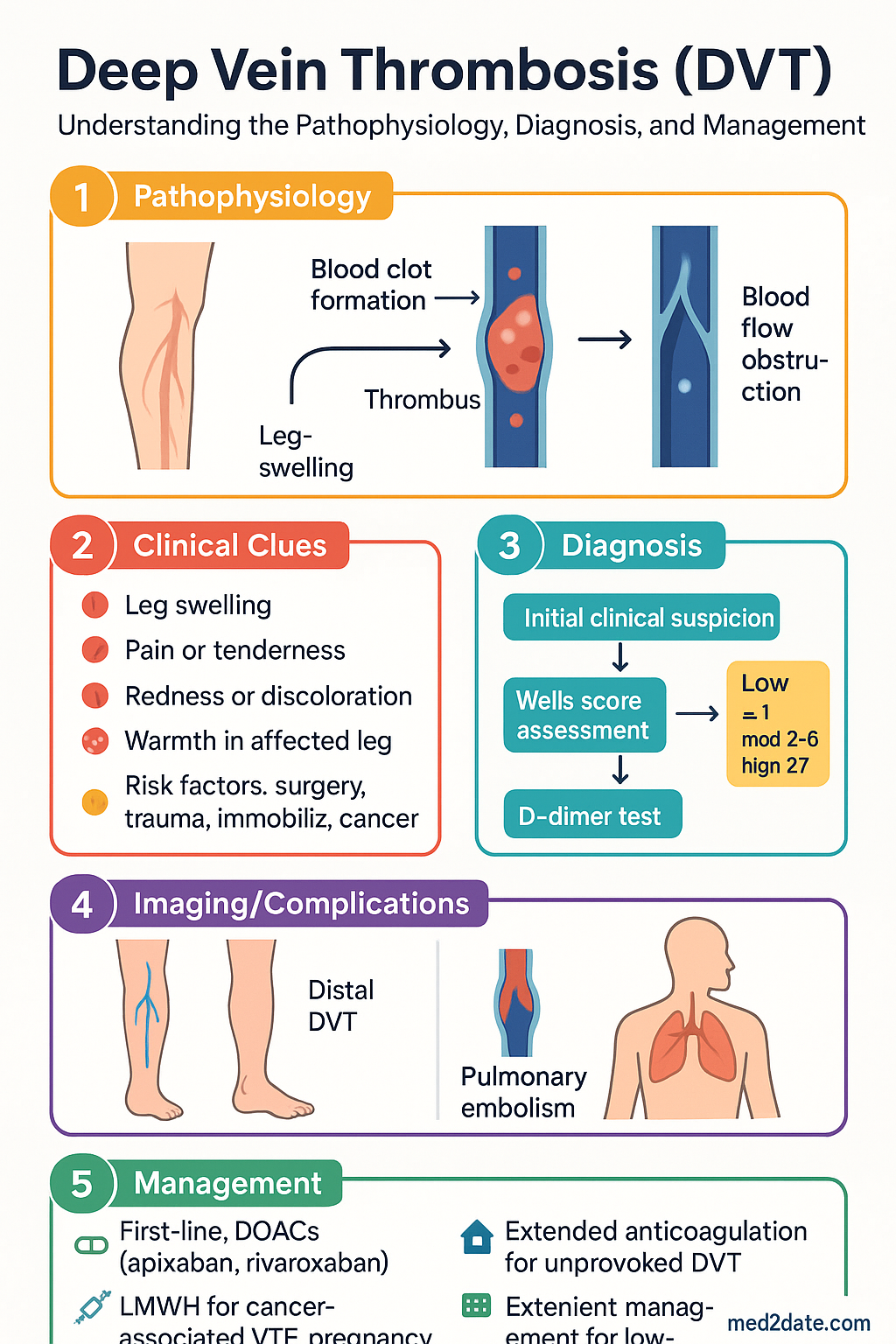
The diagnostic approach to suspected DVT integrates clinical probability assessment (Wells score), D-dimer testing, and compression ultrasound to efficiently confirm or exclude DVT while minimising unnecessary imaging.
Clinical Probability — Wells Score
The modified Wells score stratifies patients into low, moderate, or high clinical probability categories, guiding subsequent investigations.
| Clinical Feature | Points |
|---|---|
| Active cancer (treatment within 6 months or palliative) | +1 |
| Paralysis, paresis, or recent plaster immobilisation of lower limb | +1 |
| Recently bedridden ≥3 days or major surgery within 12 weeks | +1 |
| Localised tenderness along deep venous system | +1 |
| Entire leg swollen | +1 |
| Calf swelling ≥3 cm compared with asymptomatic leg (measured 10 cm below tibial tuberosity) | +1 |
| Pitting oedema confined to symptomatic leg | +1 |
| Collateral superficial veins (non-varicose) | +1 |
| Previously documented DVT | +1 |
| Alternative diagnosis at least as likely as DVT | −2 |
D-Dimer
High-sensitivity D-dimer (ELISA or immunoturbidimetric assay, threshold <500 µg/L FEU) is most useful for excluding DVT in low-to-moderate probability patients. A negative result (<500 µg/L) has a negative predictive value of >97%.
D-dimer is less specific in hospitalised patients, pregnant women, the elderly, post-operative patients, and those with active malignancy, infection, or inflammation. It should not be used as a standalone test to rule in DVT.
Compression Ultrasound (CUS)
Compression ultrasound is the gold-standard imaging modality for DVT diagnosis. It is non-invasive, widely available across Australian metropolitan and regional centres, and does not require contrast.
Proximal vs Distal DVT
| Feature | Proximal DVT | Distal (Calf) DVT |
|---|---|---|
| Location | Popliteal, femoral, iliac veins | Peroneal, posterior tibial, gastrocnemius, soleal veins |
| Proportion of all DVT | ~75% | ~25% |
| PE risk | High (25–50% if untreated) | Lower (~10%), but may extend proximally |
| Treatment | Anticoagulation recommended | Anticoagulation if symptomatic, high-risk features, or extension on serial CUS |
| Serial CUS | Not needed if positive | If not anticoagulating: repeat in 5–7 days to assess for proximal extension |
Diagnostic Algorithm
Acute Management
The goals of acute DVT management are to prevent thrombus extension, reduce PE risk, and minimise post-thrombotic syndrome. Treatment has been revolutionised by DOACs, which allow most patients to be managed entirely in the outpatient setting.
Anticoagulation Choice
Anticoagulation is the cornerstone of DVT treatment. The choice of agent depends on clinical context, renal function, drug interactions, patient preference, and cost.
Preferred Anticoagulation Strategy
Warfarin remains appropriate for patients with mechanical heart valves, antiphospholipid syndrome (triple-positive), severe renal impairment where DOACs are contraindicated, and patient preference. LMWH bridging is required when initiating warfarin.
Outpatient vs Inpatient Management
Evidence from the OTIS-DVT, OASIS, and other trials demonstrates that most patients with acute DVT can be safely managed as outpatients with equivalent outcomes to inpatient care.
| Criteria | Outpatient Suitable | Consider Inpatient |
|---|---|---|
| Haemodynamic status | Stable | Hypotension, shock (suspected PE) |
| Symptoms | Isolated leg DVT, manageable pain | Severe limb ischaemia (phlegmasia), massive iliofemoral DVT |
| Bleeding risk | Low bleeding risk | High bleeding risk, recent GI haemorrhage |
| Comorbidities | Few or well-managed | Decompensated heart failure, severe renal/hepatic disease |
| Social factors | Reliable, can attend follow-up | No fixed address, cognitive impairment, no access to medication |
| Anticoagulant plan | DOAC (no monitoring needed) | Warfarin initiation with LMWH bridging |
LMWH Bridging
LMWH bridging is required when warfarin is used rather than a DOAC. Enoxaparin (Clexane®) 1.5 mg/kg SC once daily or 1 mg/kg SC BD is commenced immediately and continued for a minimum of 5 days AND until the INR is ≥2.0 for at least 24 hours (typically 2 consecutive INR results in range).
Compression Therapy
Graduated compression stockings (GCS, 20–30 mmHg) have traditionally been recommended to reduce post-thrombotic syndrome. However, the SOX trial (2014) showed no significant reduction in PTS with compression stockings versus placebo. Current recommendations are nuanced:
- Symptom relief: GCS may reduce acute leg swelling, pain, and heaviness. They may be offered for symptom management on a case-by-case basis.
- PTS prevention: Routine use of GCS specifically to prevent PTS is no longer universally recommended (referenced in eTG and 2024 ISTH guidance).
- Application: 20–30 mmHg thigh-high stockings, fitted 1–2 weeks after acute DVT onset when swelling has improved. Wear during the day, remove at night.
- Contraindications: Peripheral arterial disease (ABPI <0.8), diabetic neuropathy with skin breakdown, severe peripheral oedema.
Adjunctive Measures
- Early mobilisation: Bed rest is NOT required. Patients should be encouraged to mobilise as tolerated from the time of diagnosis. A Cochrane review (2014) showed no increased PE risk with early ambulation.
- Elevation: Elevate the affected limb when resting to reduce swelling.
- Analgesia: Paracetamol, short-course NSAIDs (if no contraindication to concurrent anticoagulation), or mild opioids for severe pain.
- PESI/sPESI for concomitant PE: If PE is also present, use the Pulmonary Embolism Severity Index to guide disposition (outpatient management for class I–II).
Provoked vs Unprovoked DVT
The distinction between provoked and unprovoked DVT is fundamental to determining anticoagulation duration, the need for thrombophilia testing, and cancer screening.
Definitions
| Category | Examples | Anticoagulation Duration |
|---|---|---|
| Major provoked | Surgery (within 6 weeks), significant trauma (within 6 weeks), lower-limb plaster cast/immobilisation (within 6 weeks), hospitalisation with reduced mobility | 3 months (sufficient in most cases) |
| Minor provoked | Oestrogen therapy (HRT, OCP), long-haul travel (>8 hours), minor illness with reduced mobility, pregnancy/puerperium | 3 months (consider extension if ongoing risk factor) |
| Unprovoked | No identifiable provoking factor, or only minor transient risk factor (e.g., minor infection) | ≥3 months, then reassess for extended therapy |
| Cancer-associated | Active cancer (diagnosis within 6 months, metastatic, on treatment, or recurrent) | Minimum 6 months, ongoing as long as cancer active (LMWH or DOAC) |
Thrombophilia Testing
Thrombophilia testing is frequently over-requested. Routine testing after a first provoked DVT is not recommended, as it rarely changes management and can cause unnecessary anxiety.
- First unprovoked VTE in a young patient (<50 years) — if results would influence duration of anticoagulation
- Recurrent VTE, particularly if unprovoked
- VTE at unusual sites (cerebral, splanchnic, portal vein)
- Strong family history of VTE (first-degree relatives with VTE <50 years)
- Planning to stop anticoagulation — test while on treatment may be informative for genetic tests (Factor V Leiden, prothrombin G20210A); functional tests (protein C, S, antithrombin) should be deferred until ≥2 weeks after stopping anticoagulation
Cancer Screening After Unprovoked DVT
Approximately 7–12% of patients with unprovoked VTE will be diagnosed with cancer within 12 months. Age-appropriate cancer screening is recommended.
Anticoagulation Duration Decisions
The decision to continue or stop anticoagulation beyond the initial treatment period is one of the most important clinical decisions in DVT management.
When the provoking factor has resolved (e.g., post-surgical immobilisation ended, OCP ceased), the recurrence risk returns to baseline (~1–3% per year). Extended anticoagulation offers minimal additional benefit and exposes patients to bleeding risk. Discontinue anticoagulation at 3 months with a clear plan for patient education regarding recurrent symptoms.
Recurrence risk is ~10% per year if anticoagulation is stopped after 3 months. Factors favouring extended therapy: male sex, elevated D-dimer after stopping (D-dimer positive at 1 month off anticoagulation predicts high recurrence), persistent residual vein obstruction on CUS, post-thrombotic syndrome, and patient preference. Factors favouring stopping: female sex, high bleeding risk, falls risk, low D-dimer off anticoagulation, patient preference.
Recurrent VTE Prevention
After stopping anticoagulation for a first unprovoked DVT, recurrence rates are approximately 10% at 1 year, 25% at 5 years, and 36% at 10 years. Extended anticoagulation is highly effective at reducing recurrence, but the decision must balance this benefit against ongoing bleeding risk.
Extended Anticoagulation
For patients in whom the decision is made to continue anticoagulation indefinitely, reduced-dose regimens provide a favourable balance between efficacy and safety:
Risk–Benefit Assessment Tools
Several validated tools assist in the decision to extend or stop anticoagulation:
| Tool | Components | Application |
|---|---|---|
| D-dimer after stopping AC | Repeat D-dimer 1 month after stopping anticoagulation. If elevated, high recurrence risk (≥8%/yr) | If negative: consider stopping. If positive: consider restarting extended therapy |
| Vienna Prediction Model | D-dimer, sex, site of VTE, provoked status | Estimates recurrence risk to guide duration decision |
| HERDOO2 (women only) | Hyperpigmentation, oedema, D-dimer >250, obesity, age ≥65 | Women with ≤1 point may safely stop anticoagulation after 5–12 months |
| DOAC Score / VTE-BLEED | Bleeding risk factors including age, cancer, anaemia, renal disease, antiplatelet use | Assess bleeding risk to contextualise recurrence risk |
| Residual vein obstruction (RVO) | CUS at 3–6 months: presence of non-compressible residual thrombus | RVO modestly increases recurrence risk; used in some European protocols, less in Australian practice |
Thrombophilia Management
Thrombophilia-specific management considerations:
- Antiphospholipid syndrome (APS): Lifelong warfarin (target INR 2.0–3.0). DOACs are generally avoided in triple-positive APS based on the TRAPS trial (rivaroxaban was inferior with increased thrombotic events). May consider DOACs in single/double-positive APS if warfarin is impractical (shared decision-making).
- Factor V Leiden / Prothrombin mutation: Heterozygous — does not mandate lifelong therapy alone but supports extended therapy in unprovoked VTE. Homozygous or compound heterozygous — generally lifelong anticoagulation.
- Antithrombin deficiency: High thrombotic risk. Consider lifelong anticoagulation. LMWH or UFH during high-risk periods (surgery, pregnancy). Antithrombin concentrate available for critical situations (PPTA supply).
- Protein C / S deficiency: Moderate thrombotic risk. Consider extended therapy after unprovoked VTE. Warfarin-induced skin necrosis risk — always bridge with LMWH.
Special Situations
Upper Extremity DVT (UEDVT)
Upper extremity DVT accounts for ~10% of all DVT cases. It is classified as primary (effort-related/Paget-Schroetter syndrome or idiopathic) or secondary (catheter-related, malignancy-related, or due to thoracic outlet syndrome).
- Diagnosis: CUS of the axillary and subclavian veins has good sensitivity for axillary DVT but limited sensitivity for subclavian and innominate vein thrombosis. CT or MR venography is preferred for proximal UEDVT. D-dimer is less validated in this context.
- Treatment: Anticoagulation with a DOAC (apixaban or rivaroxaban) or LMWH/warfarin for a minimum of 3 months. If catheter-related and the catheter is still needed, anticoagulate with the catheter in situ; remove if no longer required.
- Thrombolysis: Consider catheter-directed thrombolysis or surgical decompression for severe effort-related UEDVT (Paget-Schroetter syndrome) with significant symptoms, particularly in young, active patients. Refer to vascular surgery.
- Paget-Schroetter syndrome: First rib resection should be considered after initial thrombolysis and anticoagulation to prevent recurrence, typically at 3–6 months. Refer to cardiothoracic/vascular surgery.
Superficial Thrombophlebitis (Superficial Vein Thrombosis — SVT)
Superficial vein thrombosis (SVT) is common and often self-limiting, but can be associated with concurrent DVT in 6–40% of cases and carries a risk of extension into the deep venous system.
| Risk Category | Features | Management |
|---|---|---|
| Low risk | SVT >3 cm from saphenofemoral junction (SFJ), no prior VTE, no active cancer, varicose vein-related | Compression stockings, NSAIDs, warm compresses. Consider fondaparinux 2.5 mg SC daily for 45 days (CALISTO trial) |
| High risk | SVT within 3 cm of SFJ, prior VTE, active cancer, SVT in non-varicose veins, extensive SVT (>5 cm), pregnancy | Anticoagulation as for DVT — therapeutic-dose DOAC or LMWH for 6 weeks–3 months. CUS to exclude DVT |
Catheter-Related Thrombosis (CRT)
Central venous catheters (CVCs), peripherally inserted central catheters (PICCs), port-a-caths, and haemodialysis catheters all carry VTE risk. CRT accounts for 30–60% of all UEDVT cases.
- Incidence: PICCs: 2–5%. CVCs: 2–30% depending on catheter type, patient factors, and surveillance method. Higher with femoral vein catheters.
- Diagnosis: CUS is the first-line investigation. If negative and clinical suspicion remains, CT venography is recommended.
- Treatment: Anticoagulation for as long as the catheter is in situ, plus at least 3 months after catheter removal. If the catheter is still required (e.g., chemotherapy, parenteral nutrition), anticoagulate without removing the catheter unless it is non-functional, infected, or no longer needed.
- Catheter removal: Not mandatory if the catheter is still needed and functional. Remove if possible, as this may reduce recurrence.
- Prophylaxis: Routine prophylactic anticoagulation for CVCs is not recommended (Cochrane review). Ensure proper catheter placement, flushing protocols, and aseptic technique.
Pregnancy-Associated VTE
VTE is a leading cause of maternal morbidity and mortality in Australia. The risk is 5–10× higher than in non-pregnant women of the same age, with the highest risk in the postpartum period (6-week postpartum window).
Pregnancy VTE Risk Stratification
| Risk Level | Features | Thromboprophylaxis |
|---|---|---|
| Low | No risk factors, or single minor transient risk factor | Early mobilisation, hydration, consider risk reassessment in each trimester |
| Moderate | Prior provoked VTE (not hormone-related), BMI >30, age >35, IVF, varicose veins, smoking | Consider LMWH 40 mg SC OD from first trimester |
| High | Prior unprovoked VTE, heritable thrombophilia with family history, antiphospholipid syndrome, multiple risk factors | LMWH 40 mg SC OD throughout pregnancy + 6 weeks postpartum |
| Very High | Prior VTE in pregnancy, known antithrombin deficiency, multiple thrombophilias | LMWH treatment dose throughout pregnancy + 6 weeks postpartum |
Phlegmasia Cerulea Dolens and Phlegmasia Alba Dolens
These are severe, limb-threatening presentations of extensive DVT requiring urgent intervention:
- Phlegmasia alba dolens: Extensive iliofemoral DVT with severe leg swelling, pain, and pallor but preserved arterial inflow. Treat with anticoagulation and consider catheter-directed thrombolysis.
- Phlegmasia cerulea dolens: Massive DVT with complete venous outflow obstruction leading to arterial compromise, cyanosis, and threatened limb. Medical emergency requiring immediate systemic anticoagulation, catheter-directed thrombolysis, or surgical thrombectomy. Fasciotomy may be needed for compartment syndrome. Refer urgently to vascular surgery.
Monitoring
Monitoring requirements vary by anticoagulant type. DOACs require less monitoring than warfarin but still warrant regular clinical review.
| Anticoagulant | Routine Monitoring | Frequency | Special Monitoring |
|---|---|---|---|
| Apixaban / Rivaroxaban | FBC, renal function (eGFR/CrCl), LFTs | Baseline, 1 month, then every 6–12 months | More frequently if CrCl 15–30 mL/min, age ≥80, weight <60 kg, drug interactions |
| Warfarin | INR (target 2.0–3.0) | Until stable: 2–3× weekly. Stable: every 2–4 weeks (up to 12 weeks if very stable per TTR >70%) | TTR (time in therapeutic range) calculation every 6 months; aim for >65% |
| LMWH | Anti-Xa levels, FBC, creatinine | Baseline, then as indicated | Anti-Xa if: CrCl <30 mL/min, weight <50 or >120 kg, pregnancy, extremes of age |
| UFH (IV) | APTT (target 1.5–2.5× control) | 6 hours after bolus, then every 6 hours during infusion, with each rate change | Platelet count at baseline and every 2–3 days (monitoring for HIT) |
Clinical Follow-Up Schedule
Special Populations
Pregnancy
Paediatrics
Elderly (≥75 years)
Renal Impairment
Hepatic Impairment
Immunocompromised
Aboriginal and Torres Strait Islander Health Considerations
Quick Reference — Anticoagulation Duration
📚 References
- 1. Lim W, Le Gal G, Bates SM, et al. American Society of Hematology 2018 guidelines for management of venous thromboembolism: diagnosis of venous thromboembolism. Blood Adv. 2018;2(22):3226-3256.
- 2. Kearon C, Akl EA, Ornelas J, et al. Antithrombotic therapy for VTE disease: CHEST guideline and expert panel report. Chest. 2016;149(2):315-352. Updated 2021.
- 3. Agnelli G, Buller HR, Cohen A, et al. Oral apixaban for the treatment of acute venous thromboembolism (AMPLIFY). N Engl J Med. 2013;369(9):799-808.
- 4. Bauersachs R, Berkowitz SD, Brenner B, et al. Oral rivaroxaban for symptomatic venous thromboembolism (EINSTEIN-DVT). N Engl J Med. 2010;363(26):2499-2510.
- 5. Weitz JI, Lensing AWA, Prins MH, et al. Rivaroxaban or aspirin for extended treatment of venous thromboembolism (EINSTEIN-CHOICE). N Engl J Med. 2017;376(13):1211-1222.
- 6. Agnelli G, Buller HR, Cohen A, et al. Apixaban for extended treatment of venous thromboembolism (AMPLIFY-EXT). N Engl J Med. 2013;368(8):699-708.
- 7. Kahn SR, Shapiro S, Wells PS, et al. Compression stockings to prevent post-thrombotic syndrome: a randomised placebo-controlled trial (SOX trial). Lancet. 2014;383(9920):880-888.
- 8. Righini M, Van Es J, den Exter PL, et al. Age-adjusted D-dimer cutoff levels to rule out pulmonary embolism: the ADJUST-PE study. JAMA. 2014;311(11):1117-1124.
- 9. Decousus H, Prandoni P, Mismetti P, et al. Fondaparinux for the treatment of superficial-vein thrombosis in the legs (CALISTO). N Engl J Med. 2010;363(13):1222-1232.
- 10. Lee AY, Levine MN, Baker RI, et al. Low-molecular-weight heparin versus a coumarin for the prevention of recurrent venous thromboembolism in patients with cancer (CLOT). N Engl J Med. 2003;349(2):146-153.
- 11. Raskob GE, van Es N, Verhamme P, et al. Edoxaban for the treatment of cancer-associated venous thromboembolism (Hokusai VTE Cancer). N Engl J Med. 2018;378(7):615-624.
- 12. Young AM, Marshall A, Thirlwall J, et al. Comparison of an oral factor Xa inhibitor with low molecular weight heparin in patients with cancer with venous thromboembolism: results of a randomized trial (SELECT-D). J Clin Oncol. 2018;36(20):2017-2023.
- 13. Carrier M, Lazo-Langner A, Shivakumar S, et al. Screening for occult cancer in unprovoked venous thromboembolism (SCREEN). N Engl J Med. 2015;373(8):697-704.
- 14. Anderson DR, Kahn SR, Rodger MA, et al. Computed tomographic pulmonary angiography vs ventilation-perfusion lung scanning in patients with suspected pulmonary embolism: a randomized controlled trial (PIOPED II). JAMA. 2007;298(23):2743-2753.
- 15. National Health and Medical Research Council (NHMRC). Clinical practice guideline for the prevention of venous thromboembolism in patients admitted to Australian hospitals. Melbourne: NHMRC; 2009. Updated by ACSQHC National VTE Prevention Clinical Care Standard 2018.
- 16. Australian Institute of Health and Welfare (AIHW). Venous thromboembolism in Australia. AIHW Cat. No. CVD 86. Canberra: AIHW; 2023.
- 17. Monagle P, Chan AKC, Goldenberg NA, et al. Antithrombotic therapy in neonates and children: Antithrombotic Therapy and Prevention of Thrombosis, 9th ed: ACCP Evidence-Based Clinical Practice Guidelines. Chest. 2012;141(2 Suppl):e737S-e801S.
- 18. Wells PS, Anderson DR, Rodger M, et al. Evaluation of D-dimer in the diagnosis of suspected deep-vein thrombosis. N Engl J Med. 2003;349(13):1227-1235.
- 19. Royal Australian College of General Practitioners (RACGP). Guideline for the management of venous thromboembolism in primary care. East Melbourne: RACGP; 2022.
- 20. Bates SM, Jaeschke R, Stevens SM, et al. Diagnosis of DVT: Antithrombotic Therapy and Prevention of Thrombosis, 9th ed: ACCP Guidelines. Chest. 2012;141(2 Suppl):e351S-e418S.