Home Renal & Nephrology Rapidly Progressive GN (RPGN) – Crescentic GN

Rapidly Progressive GN (RPGN) – Crescentic GN

📋 Key Information Summary

📋
  • Rapidly progressive glomerulonephritis (RPGN) / crescentic GN causes renal function loss over days to weeks; crescents on renal biopsy are the hallmark histological finding.
  • Three pathogenic types: Type I (anti-GBM disease, ~10%), Type II (immune complex, ~25–30%), Type III (pauci-immune / ANCA-associated, ~55–65%).
  • Anti-GBM disease requires urgent plasmapheresis + cyclophosphamide + high-dose corticosteroids; renal survival depends on starting treatment before dialysis-dependence.
  • Goodpasture syndrome = anti-GBM with pulmonary haemorrhage; smoking and hydrocarbon exposure increase lung involvement risk.
  • ANCA-associated vasculitis (AAV) includes granulomatosis with polyangiitis (GPA, c-ANCA / PR3), microscopic polyangiitis (MPA, p-ANCA / MPO), and eosinophilic granulomatosis with polyangiitis (EGPA).
  • Induction for severe AAV: IV pulse methylprednisolone 500–1000 mg × 3 days then oral prednisolone 1 mg/kg/day (max 60 mg) + cyclophosphamide or rituximab.
  • Rituximab (Rituxan® / Riximyo®) is preferred in relapsing GPA/MPA and in younger patients (fertility preservation); PBS Authority Required.
  • Plasmapheresis is indicated for pulmonary haemorrhage, dialysis-dependent RPGN, and dual-positive anti-GBM + ANCA disease.
  • Mycophenolate mofetil or azathioprine are used for maintenance after cyclophosphamide induction; rituximab is increasingly used as maintenance.
  • All suspected RPGN is a nephrology emergency — refer immediately for renal biopsy and specialist management.
  • Infection (especially Pneumocystis jirovecii) is the leading cause of death during immunosuppression; co-trimoxazole prophylaxis is mandatory.
  • Aboriginal and Torres Strait Islander peoples have higher rates of glomerulonephritis, later presentation, and barriers to specialist access — proactive screening and culturally safe pathways are essential.
  • Australian annual incidence of AAV is approximately 10–20 per million population, peaking in the 6th–7th decade; anti-GBM is rare (~1 per million).

Introduction & Australian Epidemiology

Rapidly progressive glomerulonephritis (RPGN), also termed crescentic glomerulonephritis, is a clinicopathological syndrome characterised by rapid deterioration of renal function over days to weeks, accompanied by the formation of cellular or fibrocellular crescents in >50% of glomeruli on renal biopsy. Without prompt treatment, progression to end-stage kidney disease (ESKD) within weeks is the norm.

RPGN is not a single disease but a final common pathway of several immune-mediated injuries to the glomerular basement membrane (GBM). The immunopathological mechanism determines classification into three types: anti-GBM antibody-mediated (Type I), immune complex-mediated (Type II), and pauci-immune / ANCA-associated (Type III).

Parameter Detail
Australian incidence (all RPGN)~12–20 per million per year
AAV (MPA, GPA, EGPA)~10–15 per million; most common cause of RPGN in Australia
Anti-GBM disease~0.5–1 per million; bimodal (20–30 yrs and >60 yrs)
Immune complex RPGNVariable; secondary to lupus nephritis, IgA vasculitis, infection-related
Peak age AAV60–70 years
Sex ratio AAVSlight male predominance (M:F ~1.3:1)
ATSI populationHigher rates of post-infectious GN and IgA nephropathy; later presentation of RPGN
⚠️
Nephrology emergency: Any patient presenting with acute kidney injury, active urine sediment (dysmorphic red cells, red cell casts), and proteinuria should be referred to a nephrologist urgently. A renal biopsy should be performed as soon as possible if RPGN is suspected — delaying biopsy worsens outcomes.
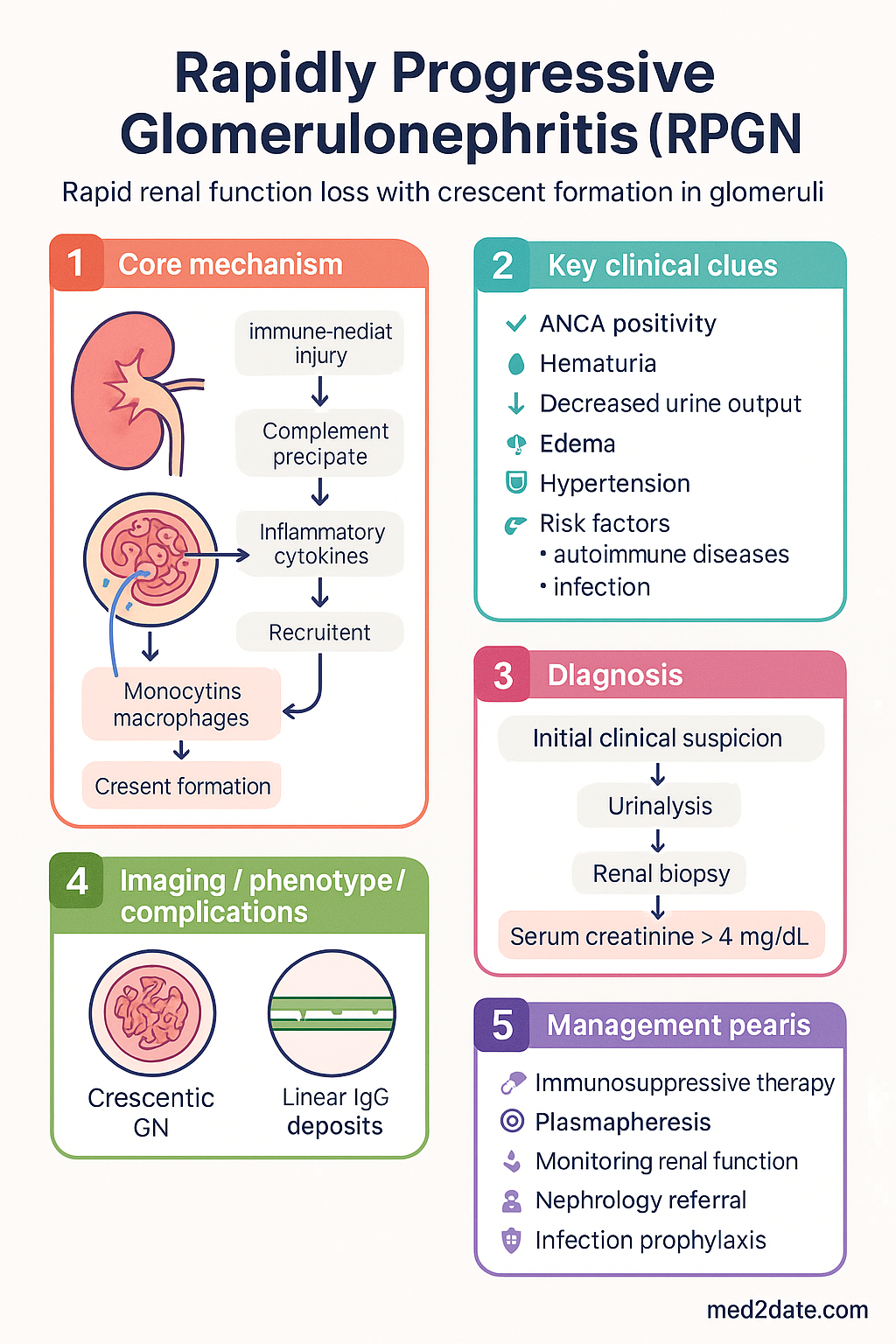
Rapidly Progressive GN (RPGN) – Crescentic GN clinical infographic — pathophysiology, clinical clues, diagnosis, imaging, and management
Tap or click image to enlarge — Rapidly Progressive GN (RPGN) – Crescentic GN: pathophysiology, clinical clues, diagnosis, imaging, and management.
Rapidly Progressive GN (RPGN) – Crescentic GN infographic, full size

Classification: Type I (Anti-GBM), Type II (Immune Complex), Type III (ANCA)

The immunofluorescence pattern on renal biopsy is the cornerstone of RPGN classification. Accurate typing guides therapy, as treatments differ substantially between categories.

Feature Type I (Anti-GBM) Type II (Immune Complex) Type III (Pauci-immune / ANCA)
MechanismAntibody to α3 chain of type IV collagenDeposition of immune complexes in glomeruliANCA-mediated neutrophil activation; minimal deposits
IF patternLinear IgG along GBMGranular IgG/IgA/IgM + C3Few or no immune deposits (pauci-immune)
Frequency~10%~25–30%~55–65%
SerologyAnti-GBM antibody positiveVariable; ANA, anti-dsDNA, low C3/C4c-ANCA (PR3) or p-ANCA (MPO)
Common associationsGoodpasture syndrome (lung haemorrhage)SLE (class IV LN), IgA vasculitis, post-streptococcal, cryoglobulinaemiaGPA, MPA, EGPA; drug-induced (hydralazine, allopurinol)
Prognosis if untreatedESKD in weeks; fatal pulmonary haemorrhageDepends on underlying cause and crescent %ESKD within months; systemic organ damage
🚨
Dual-positive disease (anti-GBM + ANCA): Approximately 30–40% of anti-GBM patients are also ANCA-positive (usually MPO-ANCA). These patients have worse renal outcomes and require combined therapy for both conditions. Always check both anti-GBM antibodies and ANCA in suspected RPGN.

Anti-GBM Disease & Goodpasture Syndrome

Anti-GBM disease is caused by autoantibodies directed against the non-collagenous domain (NC1) of the α3 chain of type IV collagen, which is abundant in glomerular and alveolar basement membranes. When both renal and pulmonary involvement occur, the syndrome is termed Goodpasture syndrome.

Clinical Features

  • Rapidly progressive glomerulonephritis: haematuria, oligoanuria, rising creatinine over days
  • Pulmonary haemorrhage (50–80% of patients): haemoptysis, dyspnoea, bilateral pulmonary infiltrates on CXR
  • Constitutional symptoms: malaise, arthralgia, fever
  • Smoking, hydrocarbon inhalation, and viral respiratory infections trigger alveolar capillary damage and clinical lung disease
  • Bimodal age distribution: young adults (20–30 years) and elderly (>60 years)
🚨
Pulmonary haemorrhage is life-threatening. Massive haemoptysis or respiratory failure requires ICU admission, urgent plasmapheresis, and airway management. Avoid renal biopsy until pulmonary haemorrhage is controlled if possible.

Diagnosis

  • Serum anti-GBM antibody (ELISA): positive in >90% of cases; titre correlates with disease activity
  • Renal biopsy (gold standard): linear IgG on immunofluorescence; crescentic GN on light microscopy
  • Always check concurrent ANCA — dual-positive patients require longer immunosuppression
  • Bronchoalveolar lavage (BAL): haemosiderin-laden macrophages confirm alveolar haemorrhage

Treatment

💉
Plasmapheresis
Therapeutic plasma exchange · Removes anti-GBM antibodies
Regimen Daily exchanges × 14 days or until anti-GBM undetectable; 60 mL/kg exchange with 5% albumin replacement (fresh frozen plasma if active bleeding)
Key indication All confirmed anti-GBM disease; pulmonary haemorrhage; dialysis-dependent RPGN
PBS status ✔ PBS General Benefit
💊
Cyclophosphamide
Endoxan® · Alkylating agent
IV pulse 15 mg/kg IV every 2–4 weeks (adjusted for age, renal function, WCC)
Oral 2–2.5 mg/kg/day (reduce by 25% if eGFR <30; reduce by 50% if age >60)
Duration 3–6 months (anti-GBM alone: shorter course; dual-positive: longer)
PBS status ✔ PBS General Benefit
💊
Methylprednisolone (IV pulse)
Solu-Medrol® · Corticosteroid
Induction 500–1000 mg IV daily × 3 days, then oral prednisolone 1 mg/kg/day (max 60 mg), taper over 6 months
PBS status ✔ PBS General Benefit
Anti-GBM is one of the few RPGN types where outcomes can be predicted by baseline renal function. Patients who present not requiring dialysis have >90% renal survival with treatment. Those who are dialysis-dependent at presentation and have >50% crescents (fibrocellular/fibrous) rarely recover renal function, but treatment is still given to prevent pulmonary haemorrhage.

ANCA-Associated Vasculitis (MPA, GPA, EGPA)

ANCA-associated vasculitis (AAV) is the most common cause of RPGN in Australia. The three syndromes — granulomatosis with polyangiitis (GPA), microscopic polyangiitis (MPA), and eosinophilic granulomatosis with polyangiitis (EGPA) — share a pauci-immune crescentic GN histology but differ in extrarenal manifestations.

Feature GPA (Wegener's) MPA EGPA (Churg-Strauss)
ANCA typec-ANCA / anti-PR3 (80–90%)p-ANCA / anti-MPO (70–80%)p-ANCA / anti-MPO (40–70%); often ANCA-negative
Classic triadUpper respiratory, lower respiratory, renalRenal ± pulmonary capillaritisAsthma, eosinophilia, vasculitis
ENTSaddle-nose deformity, sinusitis, otitis, subglottic stenosisUncommonNasal polyps, allergic rhinitis
LungNodules, cavitating lesions, alveolar haemorrhagePulmonary capillaritis, ILDTransient pulmonary infiltrates (Löffler-like)
RenalPauci-immune GN (50–80%)Pauci-immune GN (>90%)Pauci-immune GN (25–45%)
NerveMononeuritis multiplexMononeuritis multiplexMononeuritis multiplex (common)
SkinPalpable purpura, ulcersPalpable purpuraPalpable purpura, nodules
EosinophiliaNoNo>1000/µL (hallmark)

Five-Factor Score (FFS) — Revised 2009

The FFS predicts mortality in AAV and guides intensity of induction therapy. One point each for: age >65, cardiac involvement, GI involvement, renal insufficiency (creatinine >150 µmol/L), and absence of ENT manifestations. Score ≥2 = poor prognosis requiring more aggressive therapy.

Limited / Early
FFS 0, non-organ-threatening
Constitutional symptoms, ENT-only GPA, mild renal involvement (no crescents or crescentic <10%)
Setting: Outpatient nephrology/rheumatology
Generalised
FFS 0–1, organ-threatening
Crescentic GN (active), pulmonary capillaritis, mononeuritis multiplex, skin ulcers
Setting: Inpatient induction therapy
Severe / Refractory
FFS ≥2 or dialysis-dependent or alveolar haemorrhage
Rapidly progressive GN with dialysis, massive pulmonary haemorrhage, cardiac/GI involvement
Setting: ICU / HDU + intensive induction

Pathophysiology

The crescent formation in RPGN represents a severe inflammatory response within Bowman's space, driven by disruption of the glomerular basement membrane and entry of fibrin, macrophages, and T cells into the urinary space. Proliferation of parietal epithelial cells and infiltrating monocytes creates the cellular crescent, which over time becomes fibrocellular and then fibrous (irreversible).

Type I — Anti-GBM

  • Autoantibodies bind the NC1 domain of α3(IV) collagen in GBM and alveolar basement membrane
  • Fix complement → neutrophil recruitment → GBM disruption → fibrin leakage into Bowman's space → crescent formation
  • Genetic association: HLA-DR15 (susceptibility), HLA-DR7 (protection)

Type II — Immune Complex

  • Circulating or in-situ immune complexes deposit in the mesangium or subendothelial/subepithelial space
  • Complement activation, leucocyte infiltration, and GBM damage ensue
  • Causes: lupus nephritis class IV, IgA nephropathy/vasculitis, post-infectious GN, cryoglobulinaemic GN

Type III — Pauci-immune (ANCA-associated)

  • ANCA activate primed neutrophils → degranulation, ROS generation, NETosis → endothelial injury
  • Minimal immune deposits on IF/EM (hence "pauci-immune")
  • Complement alternative pathway amplification is increasingly recognised
  • Genetic: HLA-DP, HLA-DQ associations with PR3-ANCA; HLA-DQ with MPO-ANCA

Clinical Presentation & Diagnostic Criteria

Presenting Features

  • Acute nephritic syndrome: haematuria (often macroscopic), proteinuria (usually subnephrotic), oedema, hypertension, oliguria
  • Rapidly rising serum creatinine over days to weeks (doubling within 3 months is the classic definition)
  • Constitutional: malaise, fatigue, arthralgia, myalgia, fever, weight loss
  • Pulmonary: haemoptysis, dyspnoea (anti-GBM, AAV with alveolar haemorrhage)
  • Upper airway: crusting rhinitis, sinusitis, saddle-nose, hearing loss (GPA)
  • Skin: palpable purpura, livedo reticularis, digital infarcts, pyoderma gangrenosum
  • Neurological: mononeuritis multiplex, cranial nerve palsies
  • Ocular: scleritis, episcleritis, orbital pseudotumour (GPA)

Diagnostic Approach

1
Clinical Suspicion
AKI + active urine sediment (dysmorphic RBCs, RBC casts) + proteinuria = RPGN until proven otherwise.
2
Urgent Serology
Anti-GBM antibody, ANCA (PR3 and MPO ELISA), ANA, anti-dsDNA, complement (C3, C4), serum immunoglobulins, cryoglobulins, hepatitis B/C, blood cultures.
3
Renal Biopsy
Gold standard. Light microscopy (crescents, fibrinoid necrosis), immunofluorescence (linear/granular/pauci-immune), electron microscopy (substructure of deposits).
4
Classify & Treat
Type I → plasmapheresis + immunosuppression; Type II → treat underlying cause; Type III → AAV induction protocol.
⚠️
Do not delay treatment waiting for biopsy results if the clinical picture is strongly suggestive of RPGN — particularly if there is pulmonary haemorrhage or rapidly progressive AKI. Empirical pulse methylprednisolone can be commenced while awaiting biopsy and serology.

Investigations

Essential
Renal Biopsy
Gold standard. Light microscopy, IF, EM. Assess crescent %, fibrous vs cellular, tubulointerstitial fibrosis. Perform within 24–48 hours of clinical suspicion.
Essential
Anti-GBM Antibody (ELISA)
MBS Item 71128. Sensitivity >95%. Serum; results within 24–48 hours at major labs. Repeat to guide plasmapheresis duration.
Essential
ANCA (PR3 and MPO ELISA)
MBS Item 71119. Both PR3-ANCA and MPO-ANCA should be tested; IIF alone is insufficient. Sensitivity ~90% for generalised AAV.
Available
ANA, Anti-dsDNA, Complement (C3, C4)
MBS Items 71117, 71118. To evaluate SLE-related immune complex RPGN. Low C3 + low C4 suggests immune complex disease; normal complement in AAV/anti-GBM.
Available
Serum Immunoglobulins + SPEP
IgA elevation (IgA nephropathy/vasculitis), cryoglobulinaemia screening.
Available
Hepatitis B, C Serology; HIV
Infection-related GN, cryoglobulinaemia, and essential screening before immunosuppression.
Available
Blood / Urine Cultures
Post-infectious GN, endocarditis-related GN, shunt nephritis.
Available
FBC, CRP, ESR, LFT, LDH, Haptoglobin
Inflammatory markers, eosinophil count (EGPA), haemolysis screen.
Available
Urinalysis + Urine Protein:Creatinine Ratio
Dysmorphic red cells, red cell casts (active sediment), quantification of proteinuria. MBS Item 66816.
Referral
CT Chest / HRCT
Pulmonary haemorrhage assessment, GPA nodules/cavitation. Not a routine renal test — respiratory/radiology guided.
Specialist
Plasmapheresis Assessment
Vascular access assessment for therapeutic plasma exchange. Nephrology / haematology collaboration.

Management (Steroids, Cyclophosphamide, Rituximab, Plasmapheresis)

Management of RPGN has two phases: induction (achieving remission) and maintenance (preventing relapse). The intensity of induction depends on disease severity and type. All patients require infection prophylaxis and close monitoring.

Induction Therapy

💉
Methylprednisolone (IV Pulse)
Solu-Medrol® · Corticosteroid
Adult dose 500–1000 mg IV daily × 3 consecutive days
Then Oral prednisolone 1 mg/kg/day (max 60 mg), taper to 5 mg/day by 6 months
Paediatric dose 30 mg/kg IV pulse (max 1000 mg) × 3 days, then oral 2 mg/kg/day (max 60 mg)
Renal adjustment None required
PBS status ✔ PBS General Benefit
💊
Cyclophosphamide
Endoxan® · Alkylating agent
IV pulse (preferred) 15 mg/kg IV every 2–4 weeks × 6 pulses; dose adjusted for age >60 and eGFR <30 (reduce 25–50%)
Oral 2–2.5 mg/kg/day (max 150 mg/day) × 3–6 months; less preferred due to higher cumulative dose
Key toxicity Bone marrow suppression, haemorrhagic cystitis, gonadal toxicity, bladder cancer risk
Renal adjustment Reduce dose 25% if eGFR 10–30; reduce 50% if eGFR <10 or dialysis
PBS status ✔ PBS General Benefit
💊
Rituximab
Rituxan® / Riximyo® / Mabthera® · Anti-CD20 monoclonal antibody
Induction dose 375 mg/m² IV weekly × 4 weeks (RAVE regimen) or 1000 mg IV × 2 doses 2 weeks apart (RITUXVAS regimen)
Preferred in Relapsing GPA/MPA; women of childbearing age; patients intolerant of cyclophosphamide
Efficacy Non-inferior to cyclophosphamide for AAV induction (RAVE, RITUXVAS trials)
PBS status ⚠ PBS Authority Required — Specialist initiation for AAV
💉
Plasmapheresis (PLEX)
Therapeutic Plasma Exchange
Regimen 60 mL/kg per exchange, daily or on alternate days × 7–14 exchanges
Replacement 5% albumin; use FFP for last exchange if bleeding risk or anti-GBM
Strong indications Anti-GBM disease (all); dialysis-dependent AAV (PEXIVAS showed no benefit in non-dialysis AAV without pulmonary haemorrhage); severe pulmonary haemorrhage; dual-positive disease
PBS status ✔ PBS General Benefit
ℹ️
PEXIVAS Trial (2020, NEJM): Plasmapheresis did not reduce the composite of ESKD or death in AAV patients who were not dialysis-dependent and did not have alveolar haemorrhage. However, plasmapheresis remains standard of care for anti-GBM disease, dialysis-dependent AAV (with <50% fibrous crescents), and severe pulmonary haemorrhage.

Maintenance Therapy

💊
Azathioprine
Imuran® · Purine analogue
Dose 2 mg/kg/day (max 200 mg/day); check TPMT before commencing
Duration Minimum 18–24 months after cyclophosphamide induction; often longer for PR3-ANCA
PBS status ✔ PBS General Benefit
💊
Mycophenolate Mofetil
CellCept® · Inosine monophosphate dehydrogenase inhibitor
Dose 1–1.5 g BD (reduced if eGFR <25)
Role Alternative to azathioprine; IMPROVE trial showed higher relapse rate vs azathioprine in MPA; may be preferred in renal transplant patients
PBS status ✔ PBS General Benefit (Authority for non-transplant use may be required)
💊
Rituximab (Maintenance)
Rituxan® / Riximyo®
Dose 500 mg IV every 6 months × 2 years (MAINRITSAN regimen)
Efficacy MAINRITSAN trial: significantly lower relapse rate vs azathioprine (5% vs 29% at 28 months)
PBS status ⚠ PBS Authority Required

Infection Prophylaxis (Mandatory)

🛡️
Co-trimoxazole
Bactrim® · Pneumocystis jirovecii prophylaxis
Dose Single strength (400/80 mg) daily or double strength (800/160 mg) 3 times/week
Duration Throughout immunosuppression and for 3–6 months after stopping
PBS status ✔ PBS General Benefit
🛡️
Pantoprazole / Esomeprazole
Proton pump inhibitor · GI prophylaxis with corticosteroids
Dose 20–40 mg daily throughout corticosteroid use
PBS status ✔ PBS General Benefit

Monitoring

Disease Activity Monitoring

Test Frequency (Induction) Frequency (Maintenance) Purpose
eGFR, Creatinine, U&EWeeklyMonthly → 3-monthlyRenal function trajectory
Urinalysis + UPCWeeklyMonthly → 3-monthlyActive sediment (flares)
ANCA titre (PR3/MPO)Baseline, 3 monthsEvery 3–6 monthsRising titres may predict relapse (especially PR3)
Anti-GBM titreWeekly during PLEXMonthly × 12 monthsGuide PLEX cessation; detect relapse
FBCTwice weekly (CYC)Monthly (AZA/MMF)Bone marrow suppression
LFT, U&EFortnightlyMonthly → 3-monthlyDrug toxicity
Immunoglobulins (IgG)BaselineEvery 6 months (especially with rituximab)Hypogammaglobulinaemia risk
Hepatitis B (if HBsAg+)Monitor viral loadThroughoutReactivation prophylaxis

When to Repeat Biopsy

  • Failure to respond to induction therapy at 3–6 months
  • Suspected relapse with atypical features
  • To assess chronicity (fibrous crescents) before re-induction

Special Populations

🤰 Pregnancy
Cyclophosphamide: Contraindicated in pregnancy (teratogenic Category D). Discontinue ≥3 months before conception.
Rituximab: Category C; avoid if possible but may be used in life-threatening disease. B-cell depletion in neonate for months post-delivery.
Corticosteroids: Prednisolone is Category A (crosses placenta minimally). Can be used for induction/maintenance.
Azathioprine: Category D but considered acceptable in pregnancy for transplant and autoimmune disease (extensive safety data).
Mycophenolate: Contraindicated — teratogenic (Category D). Must stop ≥6 weeks before conception.
Plasmapheresis: Safe in pregnancy for anti-GBM and AAV.
Preferred regimen: Steroids + azathioprine (or rituximab if severe) during pregnancy; plan pregnancy during stable remission.
👶 Paediatrics
RPGN is rare in children; causes include IgA vasculitis, lupus nephritis, post-infectious GN, and anti-GBM (very rare).
Methylprednisolone pulse: 30 mg/kg (max 1000 mg) IV × 3 days.
Cyclophosphamide: 0.5–2 mg/kg/day oral or 500–1000 mg/m² IV pulse (dose-adjust for age).
Rituximab: 375 mg/m² IV weekly × 4 doses; used in refractory lupus nephritis and AAV.
Gonadal toxicity counselling is essential for adolescents receiving cyclophosphamide — consider fertility preservation (sperm/egg banking).
Refer to paediatric nephrology at a tertiary centre.
👴 Elderly (≥65 years)
AAV peaks in the 6th–7th decade; age >65 is a Five-Factor Score item (worse prognosis).
Cyclophosphamide: Reduce dose by 25–50% for age >60; IV pulse preferred over oral (lower cumulative dose).
Rituximab: Often preferred in elderly to avoid cyclophosphamide toxicity (infections, bladder cancer).
Higher infection risk: aggressive PJP prophylaxis, consider IVIg if hypogammaglobulinaemia develops.
Fall risk with corticosteroids; monitor bone density, consider bisphosphonate prophylaxis.
🩺 Renal Impairment / Dialysis
Dialysis-dependent patients: cyclophosphamide dose reduce 50% (eGFR <10).
Plasmapheresis can be performed in dialysis patients — coordinate with vascular access.
If fibrous crescents >50% and chronicity is advanced, renal recovery is unlikely — focus on extrarenal disease control.
Transplantation: defer until disease is in sustained remission (minimum 6–12 months, ideally 12 months off immunosuppression for anti-GBM; AAV remission >6 months).
🫁 Hepatic Impairment
Cyclophosphamide: hepatotoxic metabolites — monitor LFTs closely; avoid in severe hepatic dysfunction.
Azathioprine: hepatotoxicity risk; avoid if severe liver disease. Check TPMT.
Rituximab: safe in hepatic impairment (no hepatic metabolism); preferred agent.
🦠 Immunocompromised
Screen for hepatitis B/C and HIV before commencing immunosuppression.
HBsAg-positive: antiviral prophylaxis (entecavir or tenofovir) throughout immunosuppression and ≥12 months after.
Prior TB: screen with IGRA; treat latent TB before starting rituximab/cyclophosphamide.
Monitor IgG levels with rituximab — IVIg replacement if IgG <4 g/L and recurrent infections.

Aboriginal and Torres Strait Islander Health Considerations

Aboriginal and Torres Strait Islander Health
Epidemiology
Aboriginal and Torres Strait Islander peoples experience higher rates of glomerulonephritis, including post-infectious GN and IgA nephropathy, which may progress to RPGN. Chronic kidney disease prevalence is 2–3 times higher than non-Indigenous Australians (AIHW 2023).
Later Presentation
Patients in remote and very remote areas often present with more advanced renal disease due to delayed recognition and limited access to specialist nephrology services. A higher index of suspicion for RPGN is required in any Indigenous patient with unexplained AKI.
Remote Access Barriers
Renal biopsy requires transfer to a regional or metropolitan centre. Telehealth nephrology consultations (MBS Items 91822, 91823) should be used early. RFDS retrieval may be needed for urgent transfer. Plasmapheresis is available only in major centres.
Cultural Safety
Engage Aboriginal Health Workers and Liaison Officers. Provide information in plain language and in-language where possible. Respect Sorry Business and family decision-making structures. Use the AIDA Cultural Safety Framework.
Medication Access
Ensure PBS prescriptions are accessible through Section 100 (Remote Area Aboriginal Health Services) supply. Co-trimoxazole, prednisolone, and azathioprine are available as PBS General Benefits. Rituximab requires Authority — coordinate with metropolitan specialist if initiating remotely.
Comorbidities
High rates of diabetes, rheumatic heart disease, and chronic infections (H. pylori, strongyloides) complicate immunosuppression decisions. Screen for latent TB, hepatitis B/C, and strongyloides before induction therapy.
Follow-up
Long-term maintenance immunosuppression requires regular blood monitoring — utilise community health centres and Aboriginal Medical Services for blood draws, with results reviewed via telehealth. Address transport, housing, and social determinants that affect treatment adherence.

📚 References

  1. 1. Jennette JC, Xiao H, Falk RJ. Pathogenesis of vascular inflammation by anti-neutrophil cytoplasmic antibodies. J Am Soc Nephrol. 2006;17(5):1235–1242.
  2. 2. Jayne DRW, Gaskin G, Rasmussen N, et al. Randomized trial of plasma exchange or high-dosage methylprednisolone as adjunctive therapy for severe renal vasculitis. J Am Soc Nephrol. 2007;18(7):2180–2188.
  3. 3. Walsh M, Merkel PA, Peh CA, et al. Plasma exchange and glucocorticoid dosing in the treatment of anti-neutrophil cytoplasm antibody associated vasculitis (PEXIVAS): an open-label randomised trial. Lancet. 2020;395(10221):214–223.
  4. 4. Stone JH, Merkel PA, Spiera R, et al. Rituximab versus cyclophosphamide for ANCA-associated vasculitis (RAVE). N Engl J Med. 2010;363(3):221–232.
  5. 5. Jones RB, Tervaert JWC, Hauser T, et al. Rituximab versus cyclophosphamide in ANCA-associated renal vasculitis (RITUXVAS). N Engl J Med. 2010;363(3):211–220.
  6. 6. Guillevin L, Pagnoux C, Karras A, et al. Rituximab versus azathioprine for maintenance in ANCA-associated vasculitis (MAINRITSAN). N Engl J Med. 2014;371(19):1771–1780.
  7. 7. Kidney Disease: Improving Global Outcomes (KDIGO). KDIGO 2024 Clinical Practice Guideline for the Management of Glomerular Diseases. Kidney Int. 2024;105(4S):S1–S276.
  8. 8. Australian Institute of Health and Welfare (AIHW). Chronic kidney disease: Australian facts. Cat. no. PHE 228. Canberra: AIHW; 2023.
  9. 9. Australian Commission on Safety and Quality in Health Care (ACSQHC). National Safety and Quality Health Service Standards. 2nd ed. Sydney: ACSQHC; 2021.
  10. 10. Mukhtyar C, Guillevin L, Cid MC, et al. EULAR recommendations for the management of primary small and medium vessel vasculitis. Ann Rheum Dis. 2009;68(3):310–317.
  11. 11. Lyons PA, Rayner TF, Trivedi S, et al. Genetically distinct subsets within ANCA-associated vasculitis. N Engl J Med. 2012;367(3):214–223.
  12. 12. Hellmark T, Segelmark M. Diagnosis and classification of Goodpasture's disease (anti-GBM). J Autoimmun. 2014;48–49:108–112.
  13. 13. Torres M, Berger SP, Laga M, et al. Maintenance of remission with rituximab versus azathioprine for ANCA-associated vasculitis: a long-term follow-up of the MAINRITSAN trial. Ann Intern Med. 2020;173(6):436–443.