Home Renal & Nephrology Lupus Nephritis

Lupus Nephritis

📋 Key Information Summary

📋
  • Lupus nephritis (LN) develops in approximately 40–60% of systemic lupus erythematosus (SLE) patients, typically within the first 5 years of diagnosis.
  • ISN/RPS biopsy classification (Class I–VI) guides prognosis and treatment intensity; Class III (focal) and Class IV (diffuse) proliferative nephritis carry the highest risk of end-stage kidney disease (ESKD).
  • All patients with SLE and clinical renal involvement (proteinuria ≥0.5 g/day, active sediment, declining eGFR) require a renal biopsy for definitive classification.
  • Hydroxychloroquine (Plaquenil®) is recommended for ALL patients with LN regardless of class — reduces flares, thrombotic risk, and improves long-term renal survival.
  • Induction therapy for Class III/IV LN: mycophenolate mofetil (MMF) or intravenous cyclophosphamide (Euro-Lupus or NIH regimen) plus corticosteroids.
  • Maintenance therapy for Class III/IV: MMF (preferred) or azathioprine for at least 3–5 years after complete/partial response.
  • Class V (membranous) LN with nephrotic-range proteinuria: MMF + low-dose corticosteroids; add calcineurin inhibitor (tacrolimus) if suboptimal response.
  • Belimumab (Benlysta®) is PBS-authority approved as add-on therapy in active LN with inadequate response to standard immunosuppression.
  • Voclosporin (Lupkynis®) — a novel calcineurin inhibitor — has emerging evidence (AURORA trial) and TGA approval for Class III–V LN.
  • Anti-dsDNA titres and complement levels (C3/C4) correlate with disease activity but should be interpreted alongside clinical and histological findings.
  • All immunosuppressed patients require Pneumocystis jirovecii prophylaxis (trimethoprim-sulfamethoxazole) and cardiovascular risk factor optimisation.
  • Aboriginal and Torres Strait Islander peoples have higher SLE prevalence, later presentation, and worse renal outcomes — targeted screening and culturally safe care are essential.

Introduction & Australian Epidemiology

Lupus nephritis (LN) is one of the most serious and common organ-threatening manifestations of systemic lupus erythematosus (SLE), occurring in approximately 40–60% of SLE patients during the course of their disease. Renal involvement typically presents within the first five years of SLE diagnosis and is a major determinant of morbidity and mortality, with up to 10–30% of patients with proliferative LN progressing to end-stage kidney disease (ESKD) within 15 years.

In Australia, SLE affects an estimated 20–50 per 100,000 population, with significantly higher prevalence among women of childbearing age (female-to-male ratio approximately 9:1). Aboriginal and Torres Strait Islander Australians experience a disproportionate burden of autoimmune disease, including SLE and LN, with higher rates of renal disease, later presentation, and poorer long-term outcomes compared to non-Indigenous Australians. The AIHW reports that chronic kidney disease related to autoimmune glomerulonephritis contributes meaningfully to the ESKD burden in Indigenous communities.

The World Health Organization (WHO) classification of lupus nephritis, originally developed in 1974 and subsequently revised by the International Society of Nephrology/Renal Pathology Society (ISN/RPS) in 2003 and updated in 2018, provides a standardised histopathological framework that guides prognostication and therapeutic decision-making. The ISN/RPS classification (Class I–VI) remains the cornerstone of LN management in Australian nephrology and rheumatology practice.

Management of LN has evolved significantly over the past two decades. While intravenous cyclophosphamide combined with corticosteroids was historically the standard induction regimen, mycophenolate mofetil (MMF) has emerged as equally effective with a more favourable safety profile and is now the preferred first-line induction and maintenance agent in most Australian centres. Biologic therapies — particularly belimumab (Benlysta®), now PBS-listed for LN — and the calcineurin inhibitor voclosporin represent important additions to the therapeutic armamentarium. This guideline synthesises current Australian and international evidence for the diagnosis, classification, and management of lupus nephritis in the Australian healthcare context.
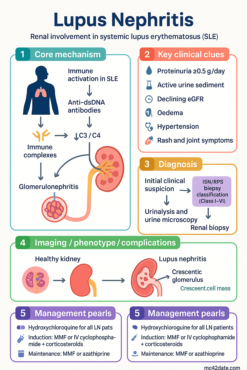
Lupus Nephritis clinical infographic — pathophysiology, clinical clues, diagnosis, imaging, and management
Tap or click image to enlarge — Lupus Nephritis: pathophysiology, clinical clues, diagnosis, imaging, and management.
Lupus Nephritis infographic, full size

ISN/RPS Classification (Class I–VI)

The ISN/RPS classification system requires adequate renal biopsy tissue (minimum 10 glomeruli on light microscopy, ideally ≥20) and mandates immunofluorescence and electron microscopy for complete evaluation. Classes are defined by the predominant histological pattern; however, mixed patterns and transformation between classes over time are well recognised.

Class Name Light Microscopy IF / EM Clinical Relevance
I Minimal mesangial LN Normal LM Mesangial immune deposits No clinical renal disease; no treatment required
II Mesangial proliferative LN Mesangial hypercellularity or matrix expansion Mesangial deposits Mild proteinuria (<1 g/day), no active sediment; treat SLE globally; monitor
III Focal LN Endocapillary or extracapillary proliferation in <50% of glomeruli Subendothelial ± mesangial deposits Active nephritis; requires immunosuppression; subclassified A (active), A/C, C (chronic)
IV Diffuse LN Proliferation in ≥50% of glomeruli (segmental [IV-S] or global [IV-G]) Subendothelial ± mesangial deposits Most aggressive proliferative class; highest ESKD risk; aggressive induction required
V Membranous LN Diffuse thickening of GBM with subepithelial deposits Diffuse subepithelial deposits Nephrotic syndrome common; may coexist with III or IV (mixed); different therapeutic approach
VI Advanced sclerosing LN ≥90% globally sclerosed glomeruli without residual activity Few residual deposits Irreversible; immunosuppression not indicated; manage as CKD; plan for RRT
⚠️
Beware mixed classes: Approximately 15–25% of biopsies show overlapping features (e.g., Class III/V or IV/V). Treatment follows the more aggressive proliferative component. The ISN/RPS 2018 update emphasises reporting the activity index (AI) and chronicity index (CI) to guide prognosis — high CI (>7) predicts poor renal recovery regardless of treatment.

Activity Index (AI) and Chronicity Index (CI): The NIH activity index scores glomerular proliferation, leucocyte infiltration, karyorrhexis, fibrinoid necrosis, cellular crescents, hyaline deposits, and interstitial inflammation (0–24). The chronicity index scores glomerular sclerosis, fibrous crescents, tubular atrophy, and interstitial fibrosis (0–12). An AI >12 suggests aggressive disease; a CI >7 portends poor renal recovery and progression to ESKD.

Clinical Features & Renal Presentation

The clinical presentation of lupus nephritis varies substantially depending on the histological class, ranging from asymptomatic urinary abnormalities detected on screening to rapidly progressive glomerulonephritis (RPGN) with acute kidney injury. In Australian practice, routine urinalysis and serum creatinine monitoring at every SLE review is standard of care.

Typical Clinical Presentations by Class

Class I–II
Mild / Subclinical
Asymptomatic proteinuria <1 g/day, microscopic haematuria, normal or near-normal eGFR. Often detected on screening urinalysis. Nephrotic syndrome rare.
Setting: Outpatient rheumatology/nephrology
Class III / V
Moderate Nephritis
Proteinuria 1–3 g/day, active urine sediment (RBC casts, dysmorphic RBCs), moderate haematuria, mild–moderate renal impairment. Class V may present with full nephrotic syndrome (oedema, hypoalbuminaemia, hyperlipidaemia).
Setting: Outpatient with close monitoring; consider admission if AKI
Class IV / Severe III
Severe Proliferative Nephritis
Nephrotic-range proteinuria (>3.5 g/day), active sediment with RBC casts, AKI (rising creatinine), hypertension, oliguria. May present as RPGN with crescentic disease. Extrarenal SLE flares common (serositis, cytopenias, rash).
Setting: Inpatient admission; urgent nephrology input; consider ICU if rapidly deteriorating

Key Clinical Signs to Assess

  • Oedema: Peripheral, periorbital, sacral — suggests nephrotic syndrome or fluid overload from AKI.
  • Hypertension: Present in 30–50% of LN at diagnosis; correlates with disease severity and chronicity.
  • Rash: Active malar rash or discoid lesions may parallel renal flare activity.
  • Joint symptoms: Synovitis may indicate concurrent systemic flare.
  • Serositis: Pleuritis or pericarditis suggests high systemic disease activity.
  • Cytopenias: Thrombocytopenia, leucopenia, or haemolytic anaemia may coexist with renal flare.
🚨
Red flags requiring urgent nephrology referral: Rapidly rising creatinine (>50% increase over days–weeks), nephrotic-range proteinuria with AKI, active urine sediment with RBC casts, macroscopic haematuria, or oligoanuria. These features suggest severe proliferative LN (Class III/IV) with crescents requiring urgent biopsy and aggressive immunosuppression.

Investigations (Biopsy, Complement, Anti-dsDNA)

Investigation of suspected lupus nephritis requires a combination of serological markers, urine studies, renal function assessment, and — critically — renal biopsy for histopathological classification. The following investigations should be performed in all patients with SLE and evidence of renal involvement.

Essential
Renal biopsy
Gold standard for ISN/RPS classification. Indicated when new-onset proteinuria ≥0.5 g/day, unexplained rise in creatinine, or active urinary sediment in a patient with SLE. Provides light microscopy, immunofluorescence, and electron microscopy. Adequate tissue: minimum 10 glomeruli (ideally ≥20). Reports AI and CI. Available at all tertiary centres; remote patients may require transfer. MBS item 13905 (renal biopsy).
Available
Anti-dsDNA antibodies
Sensitivity 50–70% for LN; titres correlate with disease activity and may rise preceding clinical flare (Farr radioimmunoassay or ELISA). Crithidia luciliae immunofluorescence test (CLIF) is more specific. Available in all Australian pathology services. MBS item 69476.
Available
Complement levels (C3, C4)
Low C3 and/or C4 indicates complement consumption via classical pathway activation and correlates with active LN. Serial monitoring aids flare detection. C4 more specific for classical pathway; C3 may be low in both active LN and hereditary deficiencies. Available in all Australian pathology services. MBS item 69446.
Available
Urinalysis and urine microscopy
Dipstick for proteinuria, haematuria, leucocyturia. Microscopy for RBC casts (pathognomonic of glomerulonephritis), dysmorphic RBCs, WBC casts. Spot urine protein-to-creatinine ratio (uPCR) or albumin-to-creatinine ratio (uACR) for quantification. Available at all sites including point-of-care. MBS item 69300.
Available
Serum creatinine and eGFR
Baseline and serial monitoring (CKD-EPI equation). Acute rise suggests active proliferative nephritis or crescentic disease. Progressive decline over months suggests chronicity. Available at all sites.
Available
24-hour urine protein or uPCR
Quantifies proteinuria; nephrotic range defined as ≥3.5 g/day. uPCR (mg/mmol) correlates well with 24-hour collection and is preferred for serial monitoring. MBS item 69300.
Available
ANA and ENA panel
ANA is almost universally positive in SLE. Anti-Smith and anti-dsDNA are highly specific. Anti-Ro/La may indicate secondary Sjögren overlap. MBS items 69476, 69479.
Available
Antiphospholipid antibodies
Lupus anticoagulant, anticardiolipin antibodies (IgG/IgM), anti-β2-glycoprotein I. Positive in 30–40% of SLE; associated with thrombotic microangiopathy, pregnancy complications, and accelerated atherosclerosis. Relevant to anticoagulation decisions. MBS item 69446.
Available
Renal ultrasound
Assesses kidney size, cortical thickness, hydronephrosis, and echogenicity. Small kidneys with increased echogenicity suggest chronicity. Do not delay biopsy if ultrasound is normal in the context of active serology. MBS item 55000.
Specialist
C1q antibodies
Strongly associated with proliferative LN (Class III/IV) and hypocomplementaemia. Not routinely available in all centres; typically ordered by nephrologist/rheumatologist. May be performed at reference laboratories (e.g., Royal College of Pathologists of Australasia).
ℹ️
Monitoring frequency in established LN: Anti-dsDNA, C3/C4, creatinine, uPCR, and FBC should be performed every 1–3 months during active treatment, then every 3–6 months during stable maintenance. A rising anti-dsDNA titre with falling complement may precede clinical flare by weeks — consider closer monitoring and rheumatology review.

Risk Stratification & Severity Scoring

Risk stratification in lupus nephritis integrates histological class, activity/chronicity indices, clinical severity markers, and patient factors to guide treatment intensity and prognostication.

Low Risk
Class I, II, or V (non-nephrotic)
Proteinuria <1 g/day, normal eGFR, no active sediment. Low CI (<3). Minimal fibrosis on biopsy.
HCQ + RAAS blockade; immunosuppression may not be required
Moderate Risk
Class III (≤50% glomeruli), Class V (nephrotic), mixed III/V
Proteinuria 1–3 g/day, mildly reduced eGFR, active sediment. AI 5–10, CI 3–7. Moderate fibrosis.
Standard induction (MMF) + corticosteroids; close monitoring q1–2 months
High Risk
Class IV (diffuse), severe Class III, mixed IV/V, crescentic disease
Nephrotic-range proteinuria (>3.5 g/day), AKI (creatinine rising), hypertension, RBC casts. AI >10, CI may be variable. Cellular crescents on biopsy.
Aggressive induction: MMF or IV cyclophosphamide + pulse methylprednisolone; consider add-on belimumab or voclosporin

Poor Prognostic Factors

  • Diffuse proliferative class (IV-S or IV-G) with crescents
  • Chronicity index >7 (tubular atrophy, interstitial fibrosis, global sclerosis)
  • Failure to achieve complete or partial response within 6–12 months
  • African, Hispanic, or Aboriginal Australian ethnicity (higher ESKD progression rates)
  • Delayed diagnosis (>6 months from onset of renal involvement to treatment)
  • Persistent nephrotic-range proteinuria despite induction therapy
  • Recurrent renal flares (≥2 flares associated with increased chronicity)
  • Non-adherence to hydroxychloroquine and immunosuppressive therapy

Management

Management of lupus nephritis involves a multiphase approach: induction of remission, maintenance of remission, and long-term supportive care. All patients require hydroxychloroquine, RAAS blockade (ACE inhibitor or ARB), cardiovascular risk optimisation, and bone protection. Immunosuppressive therapy is determined by ISN/RPS class, severity, and patient factors.

Universal Measures (All Classes)

  • Hydroxychloroquine: Recommended for ALL patients with SLE and LN regardless of class — reduces flares by 50%, decreases thrombotic risk, improves renal survival, and reduces mortality.
  • RAAS blockade: ACE inhibitor or ARB for proteinuria reduction and renoprotection. Target BP <130/80 mmHg (or <125/75 if proteinuria >1 g/day).
  • Statin therapy: If dyslipidaemia or cardiovascular risk factors present. Nephrotic syndrome itself increases CV risk.
  • Vitamin D and calcium: Supplementation in all patients receiving corticosteroids. Target 25-OH vitamin D >75 nmol/L.
  • PJP prophylaxis: Trimethoprim-sulfamethoxazole (TMP-SMX) 480 mg daily or 960 mg three times weekly if receiving significant immunosuppression (MMF, cyclophosphamide, or high-dose corticosteroids).
  • Vaccination: Influenza annually, pneumococcal (Prevenar 13 then Pneumovax 23), COVID-19 as per ATAGI. Avoid live vaccines on immunosuppression. Hepatitis B if not immune.

Hydroxychloroquine

💊
Hydroxychloroquine
Plaquenil® · Quinoric® · Antimalarial / immunomodulator
Adult dose 200–400 mg daily (≤5 mg/kg/day actual body weight)
Paediatric dose 3–5 mg/kg/day (max 400 mg/day)
Route Oral
Frequency Once daily (can split to BD if GI intolerance)
Duration Indefinite — lifelong unless significant toxicity
Renal adjustment No dose adjustment required; caution if eGFR <30 (limited data, monitor)
Key toxicity Retinal toxicity — baseline ophthalmology review, then annual screening from year 5 (or earlier if risk factors: high dose, renal impairment, concomitant tamoxifen)
PBS status ✔ PBS General Benefit

Class III/IV Proliferative LN — Induction Therapy

Two equally accepted induction regimens are used in Australian practice. The choice between MMF and IV cyclophosphamide depends on centre experience, patient factors (fertility, infection risk, prior intolerance), and access to infusion services.

First-line induction (preferred): Mycophenolate mofetil (MMF) + corticosteroids. MMF has equivalent efficacy to IV cyclophosphamide for induction (ALMS, MAINTAIN trials) with fewer adverse effects (less gonadotoxicity, less infection, no haemorrhagic cystitis).

Option A: Mycophenolate Mofetil (MMF) + Corticosteroids

💊
Mycophenolate mofetil (MMF)
CellCept® · Myfortic® · Inosine monophosphate dehydrogenase inhibitor
Adult induction dose 1 g BD (2 g/day), titrate up over 1–2 weeks from 500 mg BD
Maximum dose 3 g/day if suboptimal response and tolerated
Paediatric dose 600 mg/m²/day divided BD (max 2 g/day)
Route Oral
Induction duration 6 months (assess response at 6 months; continue if improving)
Renal adjustment If eGFR <25: max 1 g/day (limited data; monitor MPA levels if available)
Key toxicity GI disturbance (diarrhoea, nausea), leucopenia, teratogenicity — effective contraception essential
PBS status ⚠ PBS Authority Required

Corticosteroids (Induction Phase)

💊
Methylprednisolone (pulse)
Solu-Medrol® · Corticosteroid
Adult dose 500 mg–1 g IV daily for 3 days at initiation of induction
Route IV infusion over 30–60 minutes
Followed by Prednisolone 0.5–1 mg/kg/day (max 60 mg), taper over 3–6 months to ≤7.5 mg/day or off
PBS status ✔ PBS General Benefit

Option B: Intravenous Cyclophosphamide + Corticosteroids

💊
Cyclophosphamide (IV)
Endoxan® · Alkylating agent
Euro-Lupus regimen (preferred) 500 mg IV fortnightly × 6 doses (total 3 g) — lower toxicity, equivalent efficacy to high-dose NIH regimen
NIH regimen (if severe/crescentic) 500–1000 mg/m² IV monthly × 6 doses, then quarterly × 2 years
Route IV infusion with MESNA and aggressive hydration
Renal adjustment Reduce dose by 25–50% if eGFR <30; avoid if eGFR <10 without dialysis support
Key toxicity Gonadotoxicity (GnRH agonist co-administration for fertility preservation), haemorrhagic cystitis, leucopenia, infection, malignancy risk (dose-dependent)
PBS status ✔ PBS General Benefit
⚠️
Fertility counselling is mandatory before cyclophosphamide. Gonadotropin-releasing hormone (GnRH) agonist (goserelin/Zoladex® or leuprorelin/Lucrin®) should be administered monthly during cyclophosphamide treatment in women of reproductive age to reduce gonadotoxicity. Sperm banking should be discussed with all men prior to treatment.

Class V (Membranous) LN — Management

Pure Class V LN without proliferative component presents primarily with nephrotic syndrome. Treatment differs from proliferative classes.

  • Non-nephrotic Class V (proteinuria <3.5 g/day): Hydroxychloroquine + RAAS blockade + corticosteroids; immunosuppression may be deferred if eGFR stable.
  • Nephrotic Class V (proteinuria ≥3.5 g/day): MMF (2–3 g/day) + low-dose prednisolone (0.5 mg/kg, taper to ≤10 mg by 3 months). Add tacrolimus (0.05–0.1 mg/kg/day) if suboptimal response at 6 months.
  • Resistant Class V: Consider rituximab (off-label but widely used; increasing evidence from LUNAR trial subgroup analyses and real-world data).

Maintenance Therapy (Class III/IV/V after induction)

After achieving complete or partial response to induction, patients require prolonged maintenance immunosuppression to prevent relapse. The ALMS maintenance trial demonstrated superiority of MMF over azathioprine for preventing renal flares.

💊
Mycophenolate mofetil (Maintenance)
CellCept® · Preferred maintenance agent
Adult dose 1–2 g/day (500 mg–1 g BD)
Duration Minimum 3–5 years after complete response; many centres continue indefinitely in high-risk patients
PBS status ⚠ PBS Authority Required
💊
Azathioprine
Imuran® · Purine analogue
Adult dose 2 mg/kg/day (1.5–2.5 mg/kg/day based on weight and TPMT status)
Paediatric dose 2 mg/kg/day
Route Oral
Renal adjustment Reduce dose by 50% if eGFR <25; check TPMT/NUDT15 genotype before commencing
Key toxicity Myelosuppression (check TPMT/NUDT15), hepatotoxicity, GI intolerance, increased infection risk
PBS status ✔ PBS General Benefit

Biologic & Novel Therapies

Belimumab (Benlysta®)

💉
Belimumab
Benlysta® · Anti-BLyS (BAFF) monoclonal antibody
Indication Add-on therapy in active LN (Class III, IV, V) with inadequate response to standard immunosuppression
Adult dose 10 mg/kg IV every 4 weeks (after loading: weeks 0, 2, 4) OR 200 mg SC weekly (after loading 200 mg × 2)
Evidence BLISS-LN trial: superior complete renal response at 2 years vs placebo (43% vs 32%; p=0.03)
PBS status 🔴 PBS Authority Required (Specialist)

Voclosporin (Lupkynis®)

💊
Voclosporin
Lupkynis® · Calcineurin inhibitor (novel)
Adult dose 23.7 mg BD (taken 12-hourly, 1 hour before or 2 hours after food)
Evidence AURORA trial: complete renal response at 52 weeks 41% vs 23% (voclosporin + MMF + low-dose steroids vs MMF + steroids; p<0.001)
Key toxicity Hypertension, nephrotoxicity (monitor eGFR), tremor, gingival hyperplasia, QTc prolongation
Renal adjustment Avoid if eGFR <45 at initiation; reduce dose or discontinue if eGFR declines >20% from baseline
PBS status 🔴 Not PBS listed (as of 2024) — TGA approved

Treatment Response Definitions

Response Definition
Complete response Proteinuria <0.5 g/day (uPCR <50 mg/mmol), normal or stable eGFR (within 10% of baseline or eGFR ≥90), inactive urine sediment
Partial response ≥50% reduction in proteinuria from peak, stable or improved eGFR
Treatment failure Failure to achieve partial response by 6 months or complete response by 12 months; worsening proteinuria or eGFR despite therapy
Renal flare Reappearance of active sediment, rise in proteinuria ≥50%, or new/worsening renal impairment after prior response

Management of Treatment Failure / Refractory LN

  • If MMF induction fails at 6 months: switch to IV cyclophosphamide (or vice versa).
  • If both MMF and cyclophosphamide fail: consider rituximab (1 g IV × 2 doses, 2 weeks apart) — increasing evidence from observational studies and RCTs (LUNAR trial showed trend to benefit).
  • Add-on belimumab to standard therapy is supported by BLISS-LN data.
  • Voclosporin as add-on to MMF is an option (AURORA data) but access is limited by PBS status.
  • Re-biopsy should be considered if treatment failure persists — may reveal class transformation or increased chronicity.

Monitoring

Regular monitoring is essential to detect treatment response, disease flares, and medication toxicity. The following schedule applies to patients with active LN on immunosuppressive therapy:

Every 2–4 weeks (induction phase, first 3 months)
FBC, LFTs, creatinine/eGFR, uPCR, anti-dsDNA, C3/C4. MMF drug levels (if available). Corticosteroid side-effect assessment (glucose, BP, weight).
Every 1–3 months (induction months 3–6)
Same as above; assess treatment response at 6 months (complete vs partial vs failure). Cyclophosphamide: FBC at nadir (10–14 days post-dose).
Every 3 months (maintenance phase)
FBC, creatinine/eGFR, uPCR, anti-dsDNA, C3/C4. Renal function trajectory. Bone density (DEXA) if on corticosteroids >3 months.
Every 6–12 months (stable maintenance)
Comprehensive review including cardiovascular risk assessment, ophthalmology (HCQ screening from year 5), vaccination status, reproductive planning.

Drug-Specific Monitoring

Drug Monitoring Frequency
Hydroxychloroquine Ophthalmology review (OCT + visual fields) Baseline, then annually from year 5 (or earlier if risk factors)
MMF FBC, LFTs; consider MPA-AUC levels FBC every 2 weeks × 3 months, then monthly; levels if suboptimal response
Cyclophosphamide FBC at nadir, urinalysis (haematuria), LFTs FBC days 10–14 post each infusion; urinalysis at each visit
Azathioprine FBC, LFTs; TPMT/NUDT15 before initiation FBC every 2 weeks × 2 months, then monthly
Belimumab Infection screening, FBC Prior to each infusion; infection assessment at each visit
Voclosporin eGFR, BP, lipid panel, potassium eGFR weekly × 4 weeks, then every 2 weeks × 2 months, then monthly

Special Populations

🤰
Pregnancy
Hydroxychloroquine
Safe in pregnancy — CONTINUE throughout pregnancy and breastfeeding. Discontinuation increases flare risk.
Azathioprine
Safe in pregnancy — the preferred maintenance immunosuppressant in pregnancy. Continue at pre-pregnancy dose.
Prednisolone
Safe at ≤20 mg/day. Pulse methylprednisolone may be used for severe flares in pregnancy.
Mycophenolate mofetil
TERATOGENIC — STOP at least 6 weeks before conception. Switch to azathioprine before planned pregnancy.
Cyclophosphamide
CONTRAINDICATED in pregnancy. Teratogenic and gonadotoxic. Avoid unless life-threatening maternal disease.
Belimumab
Limited human data — avoid in pregnancy if possible. If inadvertent exposure occurs, report to pregnancy registry.
ACE inhibitors / ARBs
CONTRAINDICATED in 2nd and 3rd trimester. Stop at positive pregnancy test. Switch to labetalol, methyldopa, or nifedipine for BP control.
Tacrolimus
May be continued in pregnancy if essential — monitor levels closely. Associated with prematurity and low birth weight.
👶
Paediatrics
Childhood-onset SLE (cSLE) accounts for 15–20% of all SLE cases. Renal involvement is more common and more severe in paediatric SLE than adult-onset disease.
MMF: 600 mg/m²/day divided BD (max 2 g/day). Cyclophosphamide: Euro-Lupus dosing applicable; growth and puberty monitoring essential.
Hydroxychloroquine: 3–5 mg/kg/day. Ophthalmology screening from age 6 or after 5 years of therapy, whichever is sooner.
Transition to adult nephrology/rheumatology should be planned from age 14–16 with a structured programme to reduce loss to follow-up.
👴
Elderly
Late-onset SLE (>50 years) tends to have milder renal disease but higher infection risk with immunosuppression. Seronegative presentations are more common.
Lower threshold for PJP prophylaxis. Corticosteroid side effects (osteoporosis, diabetes, falls) are amplified — use lowest effective dose and taper aggressively.
Renal dosing adjustments more frequently required; consider frailty and polypharmacy in treatment decisions.
🫘
Renal Impairment
MMF: reduce to max 1 g/day if eGFR <25. Cyclophosphamide: reduce by 25–50% if eGFR <30. Azathioprine: reduce by 50% if eGFR <25.
Voclosporin: avoid if eGFR <45 at initiation. Tacrolimus: dose adjust based on trough levels and renal function.
If ESKD develops: immunosuppression generally discontinued. Renal transplant is the preferred RRT modality with acceptable outcomes (5-year graft survival ~80%).
🫁
Hepatic Impairment
MMF: no hepatic adjustment required but monitor LFTs closely as hepatotoxicity reported. Azathioprine: hepatotoxic — avoid in severe liver disease; check LFTs regularly.
Cyclophosphamide: metabolised hepatically; reduce dose in severe hepatic impairment. Belimumab: no hepatic adjustment specified.
Autoimmune hepatitis may coexist with SLE (overlap syndrome); manage with specialist hepatology input.
🛡️
Immunocompromised
LN patients are already immunocompromised from SLE itself and its treatment. Additional immunosuppression (e.g., for organ transplant) requires careful specialist coordination.
Screen for latent TB (IGRA), hepatitis B/C, HIV, and strongyloides before commencing immunosuppression. PJP prophylaxis is standard during induction.
Live vaccines (MMR, varicella, yellow fever, BCG) are contraindicated on immunosuppression. Ensure household contacts are vaccinated.

Aboriginal and Torres Strait Islander Health Considerations

Aboriginal and Torres Strait Islander Health
Disease burden
Aboriginal and Torres Strait Islander Australians experience a significantly higher burden of SLE and lupus nephritis compared to non-Indigenous Australians. AIHW data demonstrate that autoimmune glomerulonephritis, including LN, contributes disproportionately to ESKD rates in Indigenous communities. Late presentation with advanced renal disease is more common.
Remote & rural access
Many Aboriginal and Torres Strait Islander people live in remote or very remote communities with limited access to nephrology, rheumatology, and renal biopsy services. Telehealth consultations (MBS items 91822, 91823) and visiting specialist outreach programmes are essential. Patients may require aeromedical retrieval for renal biopsy and acute stabilisation.
Cultural safety
Treatment decisions should be made collaboratively with patients, families, and Aboriginal Health Workers/Practitioners (AHW/AHPs). Use of Aboriginal interpreter services is mandatory when English is not the patient's first language. Respect for kinship obligations, sorry business, and connection to country should inform care planning and appointment scheduling.
Medication adherence
Complex multi-drug regimens present challenges in remote settings. PBS co-payment reductions (Closing the Gap PBS Co-payment) reduce financial barriers. Blister-packing of medications, medication management reviews (MBS item 900), and community pharmacist engagement improve adherence. Consider depot or long-acting formulations where available.
Comorbidities
Higher rates of diabetes, hypertension, obesity, and smoking in some communities compound renal damage from LN. Integrated chronic disease management (Aboriginal Medical Services, MBS 715 health checks) should address cardiovascular risk factors alongside immunosuppressive therapy. Rheumatic fever prophylaxis may be required concurrently.
Pregnancy & maternal health
LN in pregnancy requires close monitoring at tertiary centres where possible. Remote-living pregnant women with LN should be relocated to a birthing centre near a hospital with obstetric and neonatal facilities at least 2–4 weeks before expected delivery. Shared-care models between remote GPs, AHWs, and tertiary specialists improve maternal and foetal outcomes.
Data & surveillance
Accurate epidemiological data on LN in Aboriginal and Torres Strait Islander peoples remain limited. ANZDATA and AIHW CKD registers should be used to track outcomes. Research partnerships guided by NHMRC ethical guidelines for Indigenous health research (Values and Ethics framework) are encouraged to address evidence gaps.

📚 References

  1. 1. Bajema IM, Wilhelmus S, Alpers CE, et al. Revision of the International Society of Nephrology/Renal Pathology Society classification for lupus nephritis: clarification of definitions, and modified National Institutes of Health activity and chronicity indices. Kidney Int. 2018;93(4):789–796.
  2. 2. Appel GB, Contreras G, Dooley MA, et al. Mycophenolate mofetil versus cyclophosphamide for induction treatment of lupus nephritis. J Am Soc Nephrol. 2009;20(5):1103–1112. (ALMS trial)
  3. 3. Houssiau FA, Vasconcelos C, D'Cruz D, et al. The 10-year follow-up data of the Euro-Lupus Nephritis Trial comparing low-dose and high-dose intravenous cyclophosphamide. Ann Rheum Dis. 2010;69(1):61–64.
  4. 4. Dooley MA, Jayne D, Ginzler EM, et al. Mycophenolate versus azathioprine as maintenance therapy for lupus nephritis. N Engl J Med. 2011;365(20):1886–1895. (ALMS Maintenance trial)
  5. 5. Furie R, Rovin BH, Houssiau F, et al. Two-year, randomized, controlled trial of belimumab in lupus nephritis. N Engl J Med. 2020;383(12):1117–1128. (BLISS-LN trial)
  6. 6. Rovin BH, Teng YKO, Ginzler EM, et al. Efficacy and safety of voclosporin versus placebo for lupus nephritis (AURORA 1): a double-blind, randomised, multicentre, placebo-controlled, phase 3 trial. Lancet. 2021;397(10289):2070–2080.
  7. 7. Hahn BH, McMahon MA, Wilkinson A, et al. American College of Rheumatology guidelines for screening, treatment, and management of lupus nephritis. Arthritis Care Res. 2012;64(6):79