Home Renal & Nephrology Minimal Change Disease

Minimal Change Disease

📋 Key Information Summary

📋
  • Minimal change disease (MCD) is the most common cause of nephrotic syndrome in children (80–90%) and accounts for 10–15% of adult nephrotic syndrome cases in Australia.
  • MCD is characterised by diffuse podocyte foot process effacement on electron microscopy (EM) with normal light microscopy (LM) and negative immunofluorescence.
  • Nephrotic syndrome presentation: heavy proteinuria (≥3.5 g/day or urine protein:creatinine ratio ≥350 mg/mmol), hypoalbuminaemia, oedema, and hyperlipidaemia.
  • Over 90% of children and 70–80% of adults respond to oral corticosteroids — typically prednisolone — making MCD the most steroid-responsive glomerulopathy.
  • Children usually respond within 4 weeks; adults may require up to 16 weeks of initial steroid therapy before classifying as steroid-resistant.
  • Relapse rate is high (60–90% in children, 50–70% in adults); frequently relapsing (FRNS) and steroid-dependent (SDNS) patterns are common.
  • Oral cyclophosphamide is the second-line steroid-sparing agent for FRNS/SDNS in both children and adults; calcineurin inhibitors (ciclosporin, tacrolimus) are alternatives.
  • Supportive care with loop diuretics (furosemide ± spironolactone), sodium restriction, and compression stockings for oedema management.
  • Thromboprophylaxis with low-molecular-weight heparin (LMWH) is recommended for adults with serum albumin <25 g/L due to increased thromboembolic risk.
  • ACE inhibitors or ARBs should be initiated to reduce proteinuria and provide renoprotection once the acute nephrotic episode is controlled.
  • Secondary MCD must be excluded — drug-induced (NSAIDs, lithium, interferon), malignancy (Hodgkin lymphoma), and infection-associated aetiologies.
  • Kidney biopsy is essential in all adults and in children who are steroid-resistant or relapsing <1 year after first presentation.
  • Infection is the leading cause of death in paediatric MCD — pneumococcal and varicella infections carry significant risk; immunisation status must be maintained.

Introduction & Australian Epidemiology

Minimal change disease (MCD), also termed minimal change nephropathy or nil disease, is the most common cause of nephrotic syndrome in childhood and a clinically significant cause in adulthood. It is characterised by the nephrotic syndrome — heavy proteinuria, hypoalbuminaemia, oedema, and hyperlipidaemia — with the distinctive histopathological finding of diffuse podocyte foot process effacement on electron microscopy, while light microscopy appears essentially normal and immunofluorescence is negative.

The hallmark of MCD is its excellent response to corticosteroid therapy, with complete remission rates exceeding 90% in children and 70–80% in adults. However, relapse is frequent, and many patients require multiple courses of immunosuppression over their lifetime. MCD is generally associated with a favourable long-term renal prognosis, progressing to end-stage kidney disease (ESKD) in fewer than 5% of cases, though the morbidity from relapsing nephrotic syndrome, treatment side effects, and complications is substantial.

Epidemiological Feature Detail
Childhood nephrotic syndrome cause 80–90% of cases
Adult nephrotic syndrome cause 10–15% of cases
Peak paediatric incidence 2–6 years; male predominance (2:1)
Adult presentation age All ages; mean 30–40 years
Steroid response rate (children) >90%
Steroid response rate (adults) 70–80%
Relapse rate 60–90% children; 50–70% adults
Progression to ESKD <5%
⚠️
Clinical alert: Infection remains the leading cause of mortality in children with MCD. All patients must have up-to-date pneumococcal and varicella immunisation, and families should be counselled about early presentation of febrile illness during nephrotic relapse.
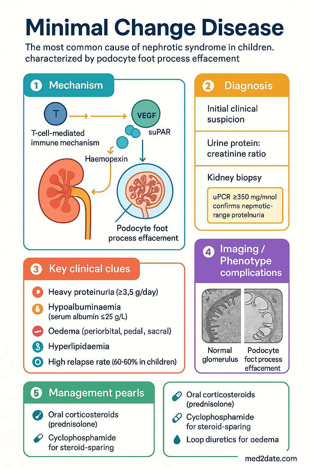
Minimal Change Disease clinical infographic — pathophysiology, clinical clues, diagnosis, imaging, and management
Tap or click image to enlarge — Minimal Change Disease: pathophysiology, clinical clues, diagnosis, imaging, and management.
Minimal Change Disease infographic, full size

Pathophysiology & Associations

Pathogenesis

The fundamental lesion in MCD is diffuse effacement (flattening) of the podocyte foot processes — the interdigitating extensions of glomerular visceral epithelial cells that form the slit diaphragm, a critical component of the glomerular filtration barrier. Loss of foot process architecture leads to increased permeability of the glomerular basement membrane (GBM) to albumin and other plasma proteins, producing selective proteinuria dominated by albumin.

The aetiology of podocyte injury in primary (idiopathic) MCD remains incompletely understood. Current evidence supports a T-cell-mediated immune mechanism producing one or more circulating permeability factors ("circulating factors") that directly injure podocytes. The rapid response to corticosteroids — often within days — and association with Hodgkin lymphoma and T-cell lymphomas are consistent with this immune-mediated pathogenesis. Proposed factors include haemopexin, vascular endothelial growth factor (VEGF), cardiotrophin-like cytokine factor 1 (CLCF-1), and soluble urokinase plasminogen activator receptor (suPAR), though none has been definitively established.

Additional pathogenic mechanisms under investigation include:

  • Podocyte cytoskeletal dysregulation: Altered expression of synaptopodin, nephrin, and podocin leading to impaired slit diaphragm integrity.
  • B-cell involvement: Recent evidence of podocyte autoantibodies (anti-nephrin) suggests a humoral component alongside T-cell dysregulation.
  • Toll-like receptor activation: Innate immune triggers (viral infections) may initiate relapse via TLR signalling on podocytes and immune cells.
  • Cytokine milieu: Interleukin-13 (IL-13) overproduction has been linked to podocyte injury and altered glomerular charge selectivity.

Secondary Causes & Associations

While most cases are idiopathic (primary MCD), secondary forms must be actively excluded, particularly in adults and atypical presentations.

Category Specific Associations
Drugs NSAIDs (most common), lithium, interferon-α, bisphosphonates (pamidronate), penicillamine, mercury (skin-lightening creams), rifampicin
Malignancy Hodgkin lymphoma (strongest association), non-Hodgkin lymphoma, thymoma, renal cell carcinoma
Infections HIV, syphilis, hepatitis B (rare), Mycoplasma
Allergic / Atopic Eczema, allergic rhinitis, food allergy (historical association, mechanism unclear)
Other Graft-versus-host disease post-allogeneic stem cell transplant, vaccinations (rare temporal association)
🚨
Malignancy screen: In all adults presenting with new-onset MCD, particularly those >40 years, a malignancy screen is mandatory — at minimum age-appropriate cancer screening, CT chest/abdomen/pelvis, LDH, and peripheral blood film. Hodgkin lymphoma can precede, coincide with, or follow MCD diagnosis by months to years.

Clinical Features (Nephrotic Syndrome)

Defining Nephrotic Syndrome

MCD typically presents with the full or partial nephrotic syndrome, defined by the following cardinal features:

Core Feature
Heavy Proteinuria
≥3.5 g/day (adults) or urine protein:creatinine ratio (uPCR) ≥350 mg/mmol on a random spot sample. In children, ≥2+ protein on dipstick for 3 consecutive days or uPCR ≥200 mg/mmol.
Diagnostic threshold
Core Feature
Hypoalbuminaemia
Serum albumin <25 g/L (severe <15 g/L). Results from massive urinary albumin losses exceeding hepatic synthetic capacity. Drives oedema formation.
Drives complications
Core Feature
Oedema
Peripheral oedema (periorbital, pedal, sacral), often progressing to ascites and pleural effusions. May be massive in children. Causes significant morbidity.
Presenting symptom in most

Clinical Presentation

Children typically present between ages 2–6 years with insidious onset of periorbital oedema, often worse in the morning and precipitated by intercurrent viral infections. Boys are affected twice as frequently as girls before puberty. The oedema may progress rapidly to generalised anasarca, with ascites, scrotal or vulval swelling, and pleural effusions.

Adults present at any age with gradual onset of dependent oedema, weight gain, and frothy urine. Presentation is less dramatic than in children, and proteinuria may be detected incidentally on routine urinalysis. Adults more commonly present with microscopic haematuria (20–30% vs. <20% in children) and are more likely to have hypertension (30–40%) and renal impairment (20–30%) at diagnosis.

Characteristic Features of MCD Nephrotic Syndrome

  • Selective proteinuria: Predominantly albumin (high selectivity index) — more common in MCD than in other glomerulopathies.
  • Absence or mild haematuria: Microscopic haematuria occurs in <20% of children and 20–30% of adults; gross haematuria is rare and should prompt consideration of IgA nephropathy or FSGS.
  • Normal or near-normal GFR: eGFR is typically preserved; transient acute kidney injury may occur with severe hypovolaemia or diuretic overuse.
  • Hypertension: Present in 10–20% of children, 30–40% of adults at presentation.
  • Hyperlipidaemia: Elevated total cholesterol and LDL; triglycerides variably elevated. Secondary to hepatic lipoprotein synthesis in response to hypoalbuminaemia.
  • Hypercoagulability: Particularly when serum albumin <20–25 g/L; renal vein thrombosis, deep vein thrombosis, pulmonary embolism, and cerebral venous sinus thrombosis (especially in children) are recognised complications.
⚠️
Thromboembolic risk: Nephrotic syndrome is a prothrombotic state due to urinary loss of antithrombin III, protein C and S, increased hepatic fibrinogen synthesis, and platelet hyperactivation. Adults with serum albumin <25 g/L should receive thromboprophylaxis with LMWH (enoxaparin 40 mg SC daily) unless contraindicated. Consider prophylaxis in children with severe hypoalbuminaemia <15 g/L.

Complications of Nephrotic Syndrome

Complication Mechanism / Notes
Infection Urinary loss of immunoglobulins (especially IgG), complement factors; peritonitis (Spontaneous Bacterial Peritonitis — pneumococcus most common); cellulitis; sepsis. Leading cause of death in paediatric MCD.
Thromboembolism Renal vein thrombosis (5–10% in adults), DVT, PE, cerebral venous thrombosis (children). Risk highest with albumin <20 g/L.
Hyperlipidaemia Accelerated atherosclerosis risk with prolonged nephrotic syndrome; statin therapy (atorvastatin) considered if persistently elevated after 3 months of nephrotic state.
Acute kidney injury Hypovolaemia, diuretic overuse, NSAIDs, ACE inhibitor initiation during severe oedema; usually reversible.
Protein malnutrition Negative nitrogen balance despite adequate intake; muscle wasting in prolonged nephrotic state.
Vitamin D deficiency Urinary loss of vitamin D binding protein; monitor 25-OH vitamin D levels and supplement as needed.
Growth retardation (children) Chronic disease, steroid use, nutritional losses; monitor growth velocity.

Investigations

Investigation of suspected MCD should confirm nephrotic syndrome, exclude secondary causes, assess complications, and confirm the diagnosis histologically where indicated.

Laboratory Investigations

Essential
Urine protein:creatinine ratio (uPCR)
MBS Item 66540. Random spot urine uPCR ≥350 mg/mmol confirms nephrotic-range proteinuria. Preferred over 24-hour urine collection (MBS Item 66545) for convenience.
Essential
Serum albumin
MBS Item 66511. <25 g/L confirms hypoalbuminaemia; <20 g/L indicates severe nephrotic state with increased thromboembolic risk.
Essential
Serum creatinine and eGFR
MBS Item 66511. Usually normal in MCD; elevated creatinine warrants consideration of AKI, tubular injury, or misdiagnosis (FSGS on biopsy).
Essential
Lipid profile
MBS Item 66511. Total cholesterol, LDL, HDL, triglycerides — hyperlipidaemia is a cardinal feature; total cholesterol typically >8 mmol/L in severe nephrotic state.
Essential
Urinalysis (dipstick + microscopy)
MBS Item 66536. Confirm proteinuria (≥2+ on dipstick). Assess for haematuria — significant glomerular haematuria is atypical for MCD and should prompt biopsy or alternative diagnosis.
Available
Serum lipids — Selectivity index
Calculates selectivity of proteinuria (IgG clearance ÷ transferrin clearance). Highly selective proteinuria (selectivity index <0.1) supports MCD over FSGS. Available at tertiary centres.
Essential
Serum immunoglobulins (IgG, IgA, IgM, IgE)
MBS Item 66511. IgG often reduced (urinary loss); IgE may be elevated in atopic patients. IgA elevation may suggest Henoch-Schönlein nephritis mimicking MCD.
Essential
Complement levels (C3, C4)
MBS Item 66511. Normal in MCD; low C3/C4 suggests membranous nephropathy, lupus nephritis, or MPGN.
Essential
ANA, dsDNA, ANCA (PR3/MPO)
MBS Item 66511. Exclude lupus nephritis and vasculitis. Typically negative in MCD.
Essential
Hepatitis B, Hepatitis C, HIV serology
MBS Item 69497 (Hep B), MBS Item 69490 (HCV), MBS Item 69486 (HIV). Mandatory to exclude secondary causes.
Available
Serum electrophoresis / free light chains
MBS Item 66511. Exclude myeloma-associated amyloid or monoclonal gammopathy if adult >40 years.
Essential
Full blood examination
MBS Item 66511. Eosinophilia may suggest allergic/drug-induced MCD; thrombocytosis occurs in nephrotic state.
Available
Antithrombin III level
May be low due to urinary loss; correlate with thrombotic risk. Available at major pathology providers.

Kidney Biopsy

Renal biopsy is the definitive diagnostic investigation and is required in all adults with new-onset nephrotic syndrome. In children, empirical steroid therapy is often commenced without biopsy for first episode, with biopsy reserved for steroid resistance, frequent relapse, or atypical features.

ℹ️
Biopsy indications in children: Age <1 year or >12 years at first presentation; steroid resistance at 4 weeks; atypical features (haematuria, hypertension, renal impairment); relapse within 6–12 months of completing first steroid course; or before initiating second-line immunosuppression (cyclophosphamide, calcineurin inhibitors).

Histopathological Findings in MCD

Modality Findings
Light Microscopy (LM) Normal or near-normal glomerular architecture. No proliferation, no thickening of GBM, no sclerosis. Occasional mild mesangial hypercellularity or increased mesangial matrix may be seen but is not prominent. Tubular epithelial cells may show reabsorption droplets (protein resorption droplets) in the cytoplasm.
Immunofluorescence (IF) Negative for all immunoglobulins (IgG, IgA, IgM) and complement (C3, C1q). Weak IgM positivity in mesangium may be seen in a minority of cases and may predict steroid resistance.
Electron Microscopy (EM) Diagnostic — diffuse podocyte foot process effacement. Complete flattening of foot processes (villous transformation) covering >80% of the GBM surface area. No electron-dense deposits. GBM thickness is normal. Mesangial areas may show mild increase in matrix. This is the key differentiating feature from early FSGS (where foot process effacement is also present but focal segmental sclerosis is visible on LM).
⚠️
Biopsy sampling error: MCD and FSGS may coexist, and FSGS lesions can be focal and missed by sampling. If steroid resistance is encountered in presumed MCD, repeat biopsy should be considered to detect evolving FSGS. The Columbia classification distinguishes MCD from FSGS by the absence of segmental sclerosis on LM — the foot process effacement seen on EM is shared by both entities.

Management

Management of MCD involves initial remission induction with corticosteroids, management of nephrotic syndrome complications, and long-term strategies to minimise relapse and steroid toxicity. The treatment approach varies between children and adults, and between first episode and relapsing disease.

Treatment Definitions

Term Definition
Complete remission uPCR <50 mg/mmol (or urine protein <0.3 g/day) for 3 consecutive days
Partial remission uPCR 50–200 mg/mmol with serum albumin improvement
Steroid resistance Failure to achieve complete remission after 16 weeks (adults) or 4 weeks (children) of daily prednisolone
Relapse uPCR >200 mg/mmol (or ≥3+ on dipstick) after having achieved remission
Frequently relapsing (FRNS) ≥2 relapses within 6 months of initial response, or ≥4 relapses in any 12-month period
Steroid dependent (SDNS) Relapse during steroid taper or within 2 weeks of stopping steroids

First-Line Therapy: Corticosteroids

💊
Prednisolone — Adults
Solone® · Panafcortelone® · Generic · Corticosteroid
Adult dose — Induction 1 mg/kg/day (max 60–80 mg) PO daily until complete remission (typically 4–16 weeks), then maintained at full dose for a further 2 weeks after remission
Adult dose — Taper Reduce by 5–10 mg every 2–4 weeks to reach alternate-day dosing (0.25–0.5 mg/kg on alternate days), then taper off over 3–6 months
Paediatric dose 60 mg/m²/day (max 60 mg) PO daily for 4–6 weeks, then 40 mg/m² on alternate days for 4–6 weeks; total initial course 8–12 weeks
Route Oral
Renal adjustment No dose adjustment; monitor for fluid retention. Use with caution if eGFR <30 mL/min.
Hepatic adjustment No adjustment; prednisolone is the active form (does not require hepatic conversion unlike prednisone)
PBS status ✔ PBS General Benefit
⚠️
Steroid side effects: Monitor closely for weight gain, glycaemic control (HbA1c every 3 months), blood pressure, bone density (DEXA if >3 months cumulative exposure), cataracts, avascular necrosis, growth suppression in children, and mood disturbance. All patients on prolonged steroids should receive calcium + vitamin D supplementation (Caltrate® 600 mg + cholecalciferol 1000 IU daily) and consider proton pump inhibitor prophylaxis.

Management of Complications

💊
Furosemide
Urex® · Frusemide Sandoz® · Generic · Loop diuretic
Adult dose 40–80 mg PO daily, titrate to effect. IV furosemide 40–80 mg for severe oedema (poor oral absorption in intestinal oedema).
Paediatric dose 1–2 mg/kg/dose PO BD (max 6 mg/kg/day). Monitor weight and electrolytes.
PBS status ✔ PBS General Benefit
💊
Spironolactone
Aldactone® · Generic · Potassium-sparing diuretic
Adult dose 25–50 mg PO daily as adjunct to furosemide to reduce hypokalaemia risk.
PBS status ✔ PBS General Benefit
💊
Enoxaparin (Thromboprophylaxis)
Clexane® · Generic · Low-molecular-weight heparin
Adult dose 40 mg SC daily when serum albumin <25 g/L. Continue until albumin >25 g/L or nephrotic state resolves. Renal dose adjustment if CrCl <30 mL/min: 20 mg SC daily.
PBS status ✔ PBS General Benefit
💊
Perindopril (ACE inhibitor)
Coversyl® · Generic · ACE inhibitor
Adult dose 2.5–5 mg PO daily; titrate to 10 mg daily. Reduces proteinuria by 30–50%. Avoid initiation during severe oedema or AKI — start after remission induction stabilises.
PBS status ✔ PBS General Benefit
💊
Atorvastatin
Lipitor® · Generic · HMG-CoA reductase inhibitor
Adult dose 10–20 mg PO daily. Consider if LDL >4.0 mmol/L persists for >3 months despite steroid-induced remission. Discontinue once nephrotic state resolves and lipids normalise.
PBS status ✔ PBS General Benefit

Second-Line Therapy: Steroid-Sparing Agents for FRNS/SDNS

💊
Cyclophosphamide
Endoxan® · Generic · Alkylating agent
Adult dose 2–2.5 mg/kg/day PO for 8–12 weeks (cumulative dose ≤200 mg/kg). Should be given with concomitant oral prednisolone at remission-inducing dose, then taper.
Paediatric dose 2 mg/kg/day PO for 8–12 weeks (cumulative dose ≤200 mg/kg). Higher efficacy when given during or immediately after steroid-induced remission.
Route Oral
Key side effects Gonadal toxicity (dose-dependent — >250 mg/kg cumulative risk of infertility); myelosuppression (FBC weekly); haemorrhagic cystitis (maintain hydration ≥2 L/day); increased infection risk.
Monitoring FBC weekly (WBC nadir at 10–14 days); renal function; urinalysis for haematuria. Aim for WBC >3.5 × 10⁹/L — reduce dose or hold if WBC <3.0.
PBS status ⚠ PBS Authority Required
💊
Ciclosporin
Neoral® · Sandimmun® · Generic · Calcineurin inhibitor
Adult dose 3–5 mg/kg/day PO in 2 divided doses (BD). Target trough level 100–200 ng/mL. Duration: 1–2 years with slow taper.
Paediatric dose 3–5 mg/kg/day PO in 2 divided doses. Monitor trough levels.
Key side effects Nephrotoxicity (chronic interstitial fibrosis — protocol biopsies recommended if use >2 years); hypertension; gingival hyperplasia; hypertrichosis; hyperlipidaemia.
PBS status ⚠ PBS Authority Required
💊
Tacrolimus
Prograf® · Generic · Calcineurin inhibitor
Adult dose 0.05–0.1 mg/kg/day PO in 2 divided doses. Target trough 5–10 ng/mL. Alternative to ciclosporin with less cosmetic side effects.
Key side effects Nephrotoxicity, neurotoxicity (tremor, headache), new-onset diabetes mellitus, alopecia.
PBS status ⚠ PBS Authority Required
💊
Mycophenolate mofetil (MMF)
CellCept® · Generic · Antiproliferative immunosuppressant
Adult dose 1–1.5 g PO BD. Used off-label for steroid-dependent or frequently relapsing MCD as alternative to alkylating agents.
Key side effects GI upset (nausea, diarrhoea), myelosuppression, teratogenicity (pregnancy category D — mandatory contraception).
PBS status ✖ Not PBS for MCD indication
💊
Levamisole
Ergamisol® · Immunomodulator
Paediatric dose 2.5 mg/kg on alternate days or 2–3 times per week. Used as steroid-sparing agent for FRNS in children; evidence from small RCTs and cohort studies. Australian access may require compounding.
Key side effects Reversible neutropenia (monitor FBC monthly); rash; GI upset. Rare: agranulocytosis, vasculitis.
PBS status ✖ Not PBS listed

Treatment Algorithm

1
First Episode — Induction
Oral prednisolone 1 mg/kg/day (adults) or 60 mg/m²/day (children) until complete remission + 2 weeks, then taper over 3–6 months. Supportive care (diuretics, salt restriction, thromboprophylaxis if albumin <25 g/L).
2
Steroid Resistance (no remission at 16 weeks adult / 4 weeks child)
Re-biopsy to exclude FSGS or other glomerulopathy. Consider calcineurin inhibitor (ciclosporin or tacrolimus) ± low-dose prednisolone. Refer to nephrologist.
3
Frequently Relapsing / Steroid Dependent
Oral cyclophosphamide 2–2.5 mg/kg/day for 8–12 weeks (first choice if fertility concerns addressed). Alternatives: ciclosporin, tacrolimus, or MMF if alkylating agents contraindicated.
4
Second or Subsequent Relapses
Repeat course of cyclophosphamide (cumulative lifetime dose <250 mg/kg), or switch to calcineurin inhibitor for prolonged maintenance. Rituximab (off-label, PBS Authority Required) considered in refractory SDNS.

Monitoring

During Active Nephrotic Syndrome

Daily
Urine dipstick (protein) — home monitoring for children; weight and fluid balance recording.
Weekly
Serum albumin, electrolytes, renal function (eGFR/creatinine), FBC if on cyclophosphamide.
2–4 weekly
uPCR, lipids, blood pressure, weight, growth parameters (children).

During Steroid Therapy

Parameter Frequency
Blood glucose (fasting or HbA1c) Every 3 months; more frequently if risk factors
Blood pressure Every visit
Weight / BMI Every visit
Ophthalmology review Baseline, then annually if cumulative >6 months exposure
Bone density (DEXA) If cumulative prednisolone >3 months; repeat every 1–2 years
25-OH Vitamin D Baseline and every 6 months; supplement if <50 nmol/L
Growth (children) Height velocity every 3–6 months; plot on growth chart

During Cyclophosphamide Therapy

Essential
Full blood examination (FBC)
Weekly — monitor WBC (target >3.5 × 10⁹/L), platelets, haemoglobin. Hold drug if WBC <3.0 × 10⁹/L or ANC <1.5 × 10⁹/L.
Essential
Urinalysis for haematuria
Weekly — early detection of haemorrhagic cystitis. Maintain adequate hydration (≥2 L/day oral fluids).
Available
Fertility counselling
Pre-treatment counselling is mandatory, particularly in adolescents and young adults. Sperm banking / oocyte cryopreservation should be discussed when cumulative dose may approach or exceed 150 mg/kg.

Long-Term Follow-Up

  • Nephrology review every 3–6 months during stable remission; more frequently during relapse or active taper.
  • Annual urine protein assessment (uPCR), serum creatinine/eGFR, lipids, and blood pressure.
  • Maintain lifelong awareness of relapse risk — patients should have urine dipstick testing available at home and know to seek medical review for frothy urine, oedema, or weight gain >1 kg/day.
  • Annual influenza vaccination (funded under NIP); pneumococcal vaccination (PCV13 + PPSV23) and varicella vaccination (if non-immune) are essential.
  • Avoid nephrotoxic agents: NSAIDs, aminoglycosides, iodinated contrast (unless essential with hydration protocol).

Special Populations

🤰 Pregnancy
Pre-conception: Achieve stable remission (ideally >6 months) before conception. Stop cyclophosphamide at least 3 months before planned pregnancy (teratogenic — Category D).
Management: Prednisolone is considered safe in pregnancy (Category A). Azathioprine is the preferred steroid-sparing agent in pregnancy if required. Ciclosporin and tacrolimus may be used with monitoring.
Complications: Increased risk of relapse during pregnancy. Pre-eclampsia may be difficult to distinguish from nephrotic syndrome relapse. Thromboprophylaxis with LMWH (enoxaparin) is indicated for albumin <25 g/L — safe in pregnancy.
Monitor uPCR, renal function, blood pressure every 2–4 weeks during pregnancy. Multidisciplinary obstetric–nephrology care recommended.
👶 Paediatrics
First episode: Empirical prednisolone 60 mg/m²/day (max 60 mg) × 4–6 weeks, then 40 mg/m² alternate days × 4–6 weeks. Biopsy not required unless steroid resistance or atypical features.
Relapse protocol: Prednisolone 60 mg/m²/day until remission + 3 days, then 40 mg/m² alternate days × 4 weeks.
Infection prevention: Varicella zoster immunoglobulin (Varizig®) or aciclovir prophylaxis for varicella exposure. Pneumococcal vaccination: PCV13 (NIP funded) + PPSV23 (recommended). No live vaccines during immunosuppression.
Growth: Alternate-day steroid regimens reduce growth suppression. Monitor height velocity; refer to paediatric endocrinologist if growth faltering persists.
School/exercise: Children in remission should attend school normally. During oedema phase, restrict sodium intake. Exercise is not restricted once oedema resolves.
👴 Elderly
Diagnosis: Kidney biopsy always indicated in adults, including elderly. Higher threshold for malignancy-associated MCD in patients >60 years.
Steroid use: Lower starting doses (0.5–0.8 mg/kg/day) may be considered. Increased risk of steroid complications: diabetes, osteoporosis, infection, psychosis.
Thromboprophylaxis: More aggressive — LMWH recommended for all elderly nephrotic patients with albumin <25 g/L; assess bleeding risk with HAS-BLED score.
Fall prevention measures essential. Review concurrent medications — discontinue NSAIDs. Polypharmacy review by pharmacist.
🫘 Renal Impairment
Acute kidney injury: May complicate MCD due to hypovolaemia or diuretic overuse. Avoid nephrotoxins. IV colloid (human albumin 20% + furosemide) may be used for refractory oedema — only in specialist setting.
Chronic kidney disease: Atypical — if CKD present, strongly consider FSGS on biopsy rather than pure MCD.
Drug adjustments: Enoxaparin: 20 mg SC daily if CrCl <30 mL/min. Ciclosporin: increased nephrotoxicity risk — monitor closely.
🫁 Hepatic Impairment
Steroids: Prednisolone (active form) preferred over prednisone; no adjustment needed. However, hypoalbuminaemia from hepatic dysfunction may be confused with nephrotic hypoalbuminaemia — use uPCR for definitive assessment.
Cyclophosphamide: Requires hepatic activation to aldophosphamide; use with caution in severe hepatic impairment.
Ciclosporin: Metabolised hepatically via CYP3A4 — trough level monitoring essential; avoid concomitant CYP3A4 inhibitors (erythromycin, azoles, grapefruit).
🛡️ Immunocompromised
HIV-associated MCD: Recognised association; responds to antiretroviral therapy (ART). Steroids may be added for refractory cases. Biopsy essential to exclude HIV-associated nephropathy (collapsing FSGS).
Post-transplant: MCD recurrence post-renal transplant is rare (unlike FSGS). If recurrence, treated with plasma exchange and high-dose steroids.
Concomitant immunosuppression: In patients already on immunosuppression (e.g., post-organ transplant), steroid dose adjustments should be made in consultation with transplant team.

Aboriginal and Torres Strait Islander Health Considerations

Aboriginal and Torres Strait Islander Health
Epidemiological context
Aboriginal and Torres Strait Islander Australians experience higher rates of CKD and ESKD than non-Indigenous Australians (AIHW 2023). While MCD itself is not specifically overrepresented, nephrotic syndrome presentations may be complicated by concurrent infections (post-streptococcal GN, hepatitis B) that are more prevalent in Indigenous communities, potentially confusing the diagnostic picture.
Access barriers
Renal biopsy services are limited in remote and rural Australia. Patients in remote communities may need aeromedical retrieval (RFDS) to access tertiary nephrology centres in Darwin, Alice Springs, Townsville, or capital cities. Telehealth nephrology consultations are available through the Australian Government telehealth programme (MBS items 99, 110, 112).
Medication access
PBS medications require access to pharmacies, which may be limited in remote communities. Closing the Gap PBS co-payment measure ensures Aboriginal and Torres Strait Islander patients pay reduced or no PBS co-payments for eligible medications. Remote Area Aboriginal Health Services can hold medication stocks under Section 100 arrangements.
Infection risk
Higher rates of pneumococcal disease, hepatitis B, and varicella in some communities. Ensure pneumococcal vaccination (PCV13 + PPSV23) is up to date; varicella vaccination status should be confirmed. Spontaneous bacterial peritonitis prophylaxis with penicillin V may be considered during active nephrotic episodes in high-risk settings.
Cultural safety
Engage Aboriginal Health Workers and Aboriginal Liaison Officers in care planning. Respect cultural obligations and family structures. Provide culturally appropriate patient education materials (Kidney Health Australia resources available in Aboriginal languages). Yarning-based education approaches improve understanding and adherence.
Comorbidity burden
Higher prevalence of diabetes, hypertension, and obesity in Indigenous Australians may confound nephrotic syndrome presentation and management. Glycaemic control is particularly important during steroid therapy — monitor BGLs closely and consider early endocrinology input for Aboriginal patients with pre-existing diabetes.
Follow-up
Care coordination through Aboriginal Community Controlled Health Organisations (ACCHOs) improves follow-up rates. The Nephrology and Aboriginal Health initiative and RHDAustralia guidelines provide frameworks for chronic disease management in remote settings. Consider shared care models between ACCHOs and tertiary nephrology services.

📚 References

  1. 1. Kidney Disease: Improving Global Outcomes (KDIGO). KDIGO 2021 Clinical Practice Guideline for the Management of Glomerular Diseases. Kidney Int. 2021;100(4S):S1–S276.
  2. 2. Cattran DC, Brenchley PE. Minimal change disease. In: Feehally J, Floege J, Tonelli M, Johnson RJ, eds. Comprehensive Clinical Nephrology. 6th ed. Philadelphia: Elsevier; 2019:285–298.
  3. 3. Vivarelli M, Massella L, Ruggiero B, Emma F. Minimal change disease. Clin J Am Soc Nephrol. 2017;12(2):332–345.
  4. 4. Australian Institute of Health and Welfare (AIHW). Chronic kidney disease: Australian facts