📋 Key Information Summary
- Facial pain encompasses a broad differential — always distinguish dental/orofacial causes from neurologic, vascular, and sinister aetiologies before initiating treatment.
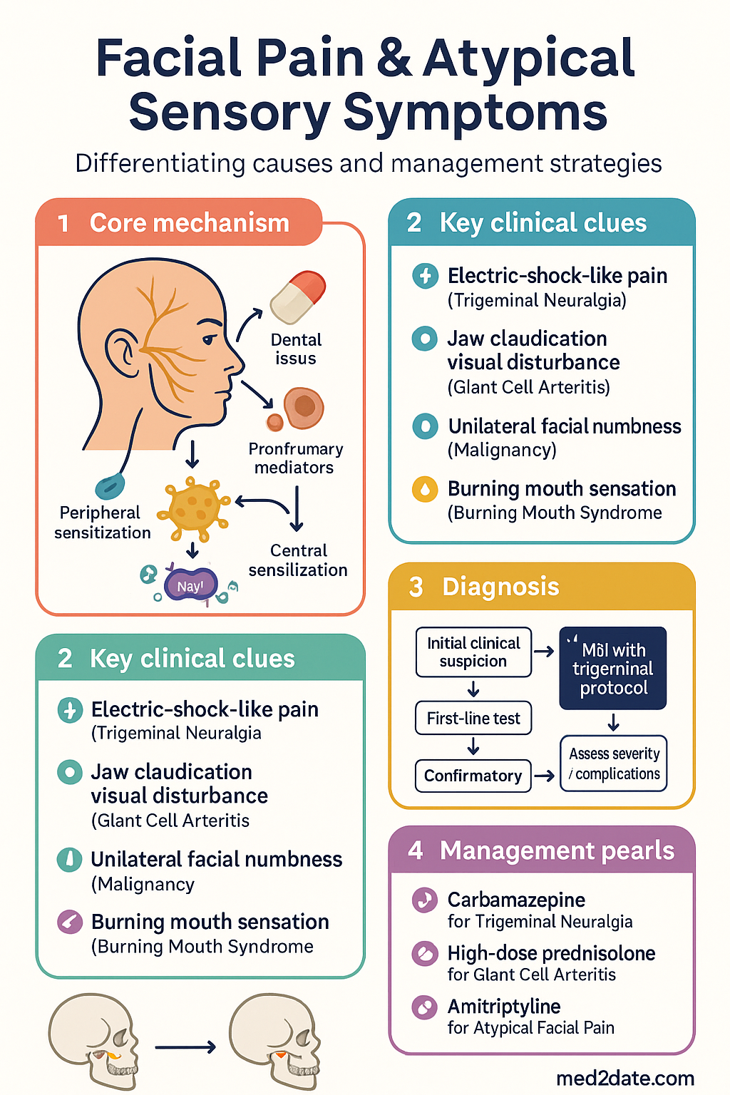
- Red flags requiring urgent referral include new-onset temporal headache in patients >50 years (giant cell arteritis), unilateral facial numbness with proptosis (malignancy/cavernous sinus lesion), progressive cranial nerve palsies, and jaw claudication.
- Trigeminal neuralgia (TN) is the most common neurologic cause of facial pain — characterised by brief, electric-shock-like paroxysms in V2/V3 distribution, often triggered by light touch, eating, or wind.
- Carbamazepine (200–1200 mg/day PO) remains first-line for classical TN; oxcarbazepine (300–1200 mg/day) is an effective alternative with a better side-effect profile. Both require HLA-B*1502 screening where feasible.
- Temporomandibular disorders (TMD) are the most common cause of non-dental orofacial pain — managed with conservative measures (patient education, jaw exercises, occlusal splints), short-course NSAIDs, and physiotherapy.
- Giant cell arteritis (GCA) is a medical emergency — initiate high-dose prednisolone (50–100 mg/day PO) immediately on clinical suspicion; do not wait for temporal artery biopsy to confirm.
- Atypical facial pain (persistent idiopathic facial pain / burning mouth syndrome) is a diagnosis of exclusion — requires systematic elimination of dental, neurologic, vascular, and sinister causes before labelling.
- MRI brain with trigeminal protocol is the investigation of choice for suspected TN, to exclude secondary causes (MS plaque, posterior fossa tumour, vascular loop compression).
- Amitriptyline (10–75 mg nocte PO) is first-line prophylactic therapy for atypical facial pain and persistent idiopathic facial pain; duloxetine (30–60 mg/day PO) is a second-line option.
- Aboriginal and Torres Strait Islander peoples experience disproportionately high rates of untreated dental disease contributing to orofacial pain — culturally safe oral health programmes and timely specialist access are essential.
- Neurosurgical options (microvascular decompression, percutaneous rhizotomy, gamma knife radiosurgery) are reserved for medically refractory TN and require specialist referral.
- Multidisciplinary pain services are integral for refractory atypical facial pain — combining pharmacotherapy, cognitive-behavioural therapy, and interventional procedures.
Introduction & Australian Epidemiology
Facial pain is a heterogeneous symptom complex that draws from dental, neurologic, musculoskeletal, vascular, and psychogenic domains. The face is among the most densely innervated regions of the body, supplied by the three divisions of the trigeminal nerve (V1 ophthalmic, V2 maxillary, V3 mandibular), the facial nerve (CN VII), and contributions from cervical nerve roots. This neuroanatomic complexity creates a wide differential diagnosis and significant potential for referred pain.
In Australia, orofacial pain affects an estimated 5–7% of the adult population at any given time, with temporomandibular disorders (TMD) accounting for the majority of non-dental cases. Trigeminal neuralgia has an incidence of approximately 12.6 per 100,000 person-years, with higher prevalence in women and those aged over 50 years. Aboriginal and Torres Strait Islander Australians experience orofacial pain at approximately twice the rate of the non-Indigenous population, driven largely by higher rates of untreated dental caries and periodontal disease (AIHW 2023).
Atypical facial pain — now classified under the International Classification of Headache Disorders, 3rd edition (ICHD-3) as persistent idiopathic facial pain (PIFP) — accounts for up to 10% of all facial pain presentations to neurology clinics. The condition is characterised by daily or near-daily facial pain that does not fulfil criteria for another cranial neuralgia and for which no structural cause has been identified. Burning mouth syndrome, another atypical sensory condition, predominantly affects post-menopausal women and has an estimated prevalence of 1–5% in the general population.
The diagnostic challenge lies in distinguishing benign, self-limiting causes from life-threatening conditions such as giant cell arteritis, nasopharyngeal carcinoma, or intracranial neoplasms. A structured, systematic approach to evaluation — incorporating history, examination, and targeted investigation — is essential to avoid both under-diagnosis of sinister pathology and over-investigation of benign conditions.
Orofacial Pain Evaluation
The evaluation of orofacial pain requires a structured history and examination to categorise the pain as dental, neurologic, musculoskeletal, vascular, or secondary to systemic disease. The approach must be systematic to avoid premature closure of diagnosis.
History — Key Domains
- Onset and duration: Acute (<3 months) vs chronic (>3 months); sudden vs gradual.
- Character: Sharp, electric-shock-like (suggests neuralgia), dull aching (dental/TMD), throbbing (vascular), burning (neuropathic/small fibre).
- Location and radiation: Unilateral vs bilateral; specific nerve distribution (V1, V2, V3); radiation to ear, temple, or neck.
- Triggers and relieving factors: Light touch, wind, chewing (TN); jaw movement (TMD); hot/cold foods (dental); position (sinusitis).
- Associated symptoms: Jaw claudication, visual disturbance (GCA); hearing change, facial weakness (acoustic neuroma); nasal obstruction, epistaxis (nasopharyngeal carcinoma); skin vesicles (herpes zoster).
- Dental history: Recent procedures, multiple dental consultations without resolution, missing teeth, ill-fitting dentures.
- Psychosocial factors: Depression, anxiety, sleep disturbance, medicolegal claims, illness behaviour — these are common in chronic facial pain and do not exclude organic disease but should be documented.
Examination — Structured Approach
- Inspection: Facial symmetry, skin lesions (vesicles in zoster, erythema in cellulitis), temporal artery (tenderness, thickening, reduced pulse), nasal cavity (if accessible), oral mucosa.
- Palpation: Temporal arteries bilaterally, parotid glands, TMJ (click, crepitus, tenderness), masticatory muscles (masseter, temporalis, medial/lateral pterygoids), trigger points in cervical and cranial musculature.
- Neurologic examination: Testing of all three trigeminal divisions — light touch (cotton), pinprick, corneal reflex (V1), jaw jerk reflex, motor function of CN V (jaw opening/closing against resistance) and CN VII (forehead sparing vs complete facial palsy).
- TMD assessment: Maximum inter-incisal opening (normal >40 mm), lateral excursion, joint sounds (clicking, crepitus), deviation on opening.
- Dental examination: Percussion of individual teeth, vitality testing (if available), assessment for caries, abscess, cracked tooth syndrome.
- Cranial nerve examination: Full screen of CN II–XII, with particular attention to V, VII, and VIII.
Distinguishing Dental vs Neurologic Causes
| Feature | Dental / Odontogenic | Neurologic | Musculoskeletal (TMD) |
|---|---|---|---|
| Pain character | Dull, aching, throbbing | Electric, shooting, lancinating; or burning | Dull, aching, worse with jaw use |
| Location | Localised to a specific tooth or quadrant | Follows dermatomal distribution (V1–V3) | Pre-auricular, masseter, temporal region |
| Triggers | Hot/cold, sweet, biting pressure | Light touch, wind, talking, eating (TN) | Chewing, yawning, jaw clenching |
| Duration | Minutes to hours, may be constant | Seconds (TN); constant (PIFP, neuropathic) | Hours, worsens throughout the day |
| Associated signs | Swelling, caries, abscess, failed dental treatment | Sensory loss, trigger zones, corneal hyporeflexia | TMJ click/crepitus, limited opening, bruxism |
| Response to local anaesthetic | Pain resolves with dental block | Pain may persist or follow deeper nerve | Partial relief with trigger point injection |
Temporomandibular Disorders (TMD)
TMD encompasses a group of musculoskeletal conditions affecting the masticatory muscles, temporomandibular joint, and associated structures. It is the most common non-dental cause of orofacial pain in Australian adults, with a peak incidence between ages 20 and 45, and a female-to-male ratio of approximately 3:1.
Classification (DC/TMD criteria):
- Group I — Myofascial pain: Pain in the masticatory muscles with trigger points; most common subtype (approximately 45% of TMD cases).
- Group II — Disc displacement: With or without reduction; presents with clicking, locking, or restricted opening.
- Group III — Arthralgia, osteoarthritis, osteoarthrosis: Inflammatory or degenerative joint disease; crepitus, pain on jaw movement.
Management of TMD:
- Patient education and self-management: Soft diet, avoidance of excessive jaw opening (yawning widely, chewing gum), warm compresses, jaw relaxation exercises, sleep hygiene, and stress management. This is the foundation of all TMD management.
- Occlusal splint therapy: Hard acrylic stabilisation splint worn at night; reduces bruxism-related muscle fatigue. Fitted by dentist or prosthodontist.
- Physiotherapy: Jaw exercises, manual therapy, postural correction, dry needling of trigger points. Referral to a physiotherapist experienced in orofacial pain.
- Pharmacotherapy: Short-course NSAIDs (ibuprofen 400 mg PO TDS with food, 7–14 days), or naproxen 250–500 mg PO BD. Muscle relaxants (cyclobenzaprine 5–10 mg PO nocte) may be considered for myofascial pain but evidence is limited. Avoid chronic benzodiazepine use.
- Invasive treatments: Arthrocentesis, botulinum toxin injection (off-label), and arthroscopy are reserved for refractory cases and require specialist (oral and maxillofacial surgery) referral.
Red Flags — When to Investigate Urgently
- Giant cell arteritis: Age >50, new temporal headache, jaw claudication, visual symptoms, ESR >50 mm/hr, weight loss, scalp tenderness. Treat empirically with high-dose prednisolone — do NOT await biopsy.
- Malignancy: Unilateral facial numbness (especially V2 or V3), proptosis, unexplained cranial nerve palsy, nasal obstruction with epistaxis, unexplained weight loss, lymphadenopathy, non-healing oral ulcer >3 weeks.
- Acute herpes zoster (shingles): Prodromal pain followed by vesicular eruption in a dermatomal distribution (V1 most common with risk to eye); requires urgent ophthalmology referral if V1 involved.
- Cavernous sinus pathology: Multiple cranial nerve palsies (III, IV, V1, V2, VI), proptosis, chemosis — urgent MRI.
- Cerebral aneurysm / SAH: Thunderclap facial or head pain, neck stiffness, loss of consciousness — emergency CT head ± LP.
- Posterior fossa lesion: Facial pain with ipsilateral hearing loss, ataxia, or other cranial nerve signs — urgent MRI.
Atypical Facial Pain
Atypical facial pain is a term that encompasses several conditions in which facial pain does not conform to the diagnostic criteria of established cranial neuralgias, dental pathology, or identifiable structural disease. Under the ICHD-3 classification, the primary entities are persistent idiopathic facial pain (PIFP, previously "atypical facial pain") and burning mouth syndrome.
Persistent Idiopathic Facial Pain (PIFP)
PIFP is characterised by facial pain that is deep, poorly localised, constant or near-constant, and does not follow a peripheral nerve distribution. It is by definition a diagnosis of exclusion — all other facial pain aetiologies must be systematically ruled out.
ICHD-3 Diagnostic Criteria for PIFP:
- A: Facial pain fulfilling criteria B and C
- B: Recurring daily for >2 hours/day for >3 months
- C: Pain is poorly localised, not following the distribution of a peripheral nerve, and is typically deep, dull, aching, or burning
- D: Not better accounted for by another ICHD-3 diagnosis
Burning Mouth Syndrome (BMS)
BMS presents as a burning, scalding, or tingling sensation of the oral mucosa — typically affecting the tongue, palate, and lips — in the absence of identifiable oral or systemic pathology. It predominantly affects post-menopausal women (female:male ratio approximately 7:1) and is thought to involve small-fibre neuropathy of intraoral sensory afferents.
Key features:
- Burning pain that typically worsens throughout the day, often absent on waking and maximal by evening
- Xerostomia (subjective dry mouth) in up to 70% of patients, though salivary flow is often objectively normal
- Dysgeusia (altered taste — commonly metallic or bitter) in 50–65%
- Oral mucosa appears clinically normal on examination
- Significant psychological comorbidity — depression and anxiety are present in up to 50–70%
Secondary causes to exclude before diagnosing BMS:
- Oral candidiasis (especially in denture wearers, inhaled corticosteroid users, immunocompromised patients)
- Nutritional deficiencies: iron, folate, vitamin B12, zinc — request serum levels
- Medication-related: ACE inhibitors, angiotensin receptor blockers, antiretrovirals, chemotherapeutic agents
- Sjögren syndrome — consider anti-Ro (SSA) and anti-La (SSB) antibodies, Schirmer test
- Contact allergy (dental materials, toothpaste ingredients — sodium lauryl sulphate)
- Endocrine: diabetes mellitus, thyroid dysfunction — request HbA1c, TSH
Trial Treatments for Atypical Facial Pain
Management of PIFP and BMS requires a structured, stepped approach combining pharmacotherapy with non-pharmacological strategies.
Step 1 — Non-Pharmacological Interventions
- Reassurance and education: Validation of the patient's pain experience, explanation that PIFP/BMS is a recognised neurobiological condition (not "imaginary"), and that effective treatments exist.
- Cognitive-behavioural therapy (CBT): Addresses pain catastrophising, maladaptive coping, and mood disturbance. Best delivered by a psychologist with experience in chronic pain. Available through some publicly funded chronic pain programmes.
- Physiotherapy: Orofacial relaxation techniques, jaw and neck exercises, posture correction — particularly when there is comorbid TMD or bruxism.
- Topical capsaicin (0.025–0.075% cream): Applied to the affected area of the face 3–4 times daily. Evidence is modest but may provide relief via desensitisation of TRPV1 receptors. Requires several weeks of consistent use before benefit is apparent.
Step 2 — First-Line Pharmacotherapy
Step 3 — Second-Line Pharmacotherapy
Step 4 — Referral to Neurology or Pain Medicine
Referral is indicated when:
- Diagnosis remains uncertain after comprehensive primary care evaluation
- Two or more adequate pharmacotherapy trials (minimum 8 weeks at target dose) have failed
- Significant diagnostic red flags require further investigation (MRI, nerve conduction studies)
- There is diagnostic concern for secondary TN (MS, posterior fossa tumour)
- Severe functional impairment with inability to work or maintain social participation
- Significant psychological comorbidity requiring integrated management
Interventional Pain Procedures (Specialist Setting)
| Procedure | Indication | Setting | Notes |
|---|---|---|---|
| Sphenopalatine ganglion block | PIFP, cluster headache, facial neuropathic pain | Pain medicine specialist / ENT | Trans-nasal approach; can be performed in clinic; repeated blocks may be needed |
| Trigger point injections | Myofascial TMD, referred facial pain | GP, dentist, pain specialist | Local anaesthetic ± corticosteroid; MBS Item 18356 |
| Botulinum toxin type A | Refractory TMD, chronic migraine with facial pain | Neurologist, pain specialist | Off-label for TMD; TGA-approved for chronic migraine (≥15 days/month); PBS authority for chronic migraine |
| Peripheral nerve stimulation | Refractory neuropathic facial pain | Pain medicine specialist | Implanted; requires trial stimulation period; limited availability in Australia |
Pathophysiology
Trigeminal Neuralgia — Classical
Classical TN is most commonly caused by neurovascular compression of the trigeminal nerve root entry zone (REZ) by the superior cerebellar artery (SCA) or, less commonly, the anterior inferior cerebellar artery (AICA). Pulsatile arterial contact demyelinates the nerve at the REZ, generating ephaptic transmission — ectopic impulse spread between adjacent demyelinated axons. This produces the characteristic paroxysmal, electric-shock-like pain triggered by innocuous stimuli (light touch, chewing, wind).
The pain-free refractory period following an attack is thought to result from post-excitatory hyperpolarisation of damaged neurons. Over time, repeated paroxysms can lead to central sensitisation in the trigeminal nucleus caudalis, resulting in background aching between paroxysms — a feature of long-standing, untreated TN.
Secondary TN
Secondary TN may result from demyelination (multiple sclerosis — TN occurs in 2–5% of MS patients, often bilateral), posterior fossa tumours (schwannoma, meningioma), arteriovenous malformations, or pontine infarction. These structural causes disrupt normal nerve conduction through direct compression or demyelination.
Persistent Idiopathic Facial Pain
The pathophysiology of PIFP is incompletely understood but is thought to involve a combination of peripheral and central mechanisms:
- Peripheral sensitisation: Local tissue injury or inflammation (dental procedures, sinusitis, trauma) may initiate nociceptive input that persists after the inciting event has resolved.
- Central sensitisation: Prolonged nociceptive input to the trigeminal nucleus caudalis produces neuroplastic changes (wind-up, expanded receptive fields, lowered activation thresholds) that maintain pain independently of peripheral input.
- Descending modulation dysfunction: Impaired function of the periaqueductal grey–rostral ventromedial medulla–dorsal horn pathway reduces endogenous pain inhibition.
- Psychological factors: Catastrophising, hypervigilance, and mood disturbance amplify pain perception through limbic system engagement — these are not causative but are powerful modulators.
Burning Mouth Syndrome
BMS is thought to involve small-fibre neuropathy of intraoral trigeminal afferents (C-fibres and Aδ-fibres) with subsequent central disinhibition. Neuroimaging studies demonstrate altered activity in the trigeminal sensory nuclei and insular cortex. The circadian pattern (worsening through the day) may relate to progressive depletion of inhibitory neurotransmitters (GABA, endogenous opioids) with sustained sensory input.
Giant Cell Arteritis
GCA is a granulomatous vasculitis affecting medium and large arteries, particularly the extracranial branches of the aortic arch. The temporal, vertebral, and ophthalmic arteries are commonly involved. Intimal hyperplasia leads to luminal stenosis and tissue ischaemia. Facial pain in GCA results from ischaemia of the masticatory muscles (jaw claudication) or ischaemic neuropathy of cranial nerves. Untreated, GCA can cause sudden irreversible blindness via posterior ciliary artery occlusion.
Clinical Presentation & Diagnostic Criteria
Trigeminal Neuralgia — ICHD-3 Criteria
- A: Recurrent paroxysms of unilateral facial pain in the distribution of one or more divisions of the trigeminal nerve, with no radiation beyond the trigeminal distribution
- B: Pain has at least 3 of the following: (1) paroxysms lasting from a fraction of a second to 2 minutes; (2) severe intensity; (3) electric-shock-like, shooting, stabbing, or sharp character; (4) precipitated by innocuous stimuli to the affected side of the face
- C: Not better accounted for by another ICHD-3 diagnosis
Classical TN: Caused by vascular compression (MRI evidence of neurovascular contact); may have sensory deficit on examination.
Secondary TN: Caused by an identifiable underlying disease (MS, tumour, structural lesion).
Idiopathic TN: No demonstrable cause on imaging or investigation.
TN vs Symptomatic TN — Clinical Differentiation
| Feature | Classical TN | Secondary / Symptomatic TN |
|---|---|---|
| Age of onset | Typically >50 years | Any age (younger in MS) |
| Side | Almost always unilateral; bilateral in <5% | Bilateral more common (especially MS) |
| Triggers | Well-defined trigger zones on face | May be spontaneous; triggers less consistent |
| Neurologic signs | Examination normal (though up to 30% may have subtle sensory loss) | Sensory loss, corneal hyporeflexia, motor weakness, other cranial nerve signs |
| Response to carbamazepine | Excellent (diagnostic aid) | Partial or poor response |
| MRI findings | Neurovascular contact at REZ | Structural lesion, MS plaque, tumour |
Other Cranial Neuralgias
| Neuralgia | Nerve | Pain Distribution | Distinguishing Features |
|---|---|---|---|
| Glossopharyngeal | CN IX | Posterior tongue, tonsillar fossa, ear | Triggered by swallowing, talking; may cause syncope (cardioinhibitory reflex) |
| Geniculate (Nervus intermedius) | CN VII sensory component | Deep ear, periauricular | Paroxysmal ear pain; may have herpetic vesicles in ear canal (Ramsay Hunt) |
| Occipital | Greater/lesser occipital nerves (C2–C3) | Posterior scalp, behind ear, periorbital referral | Tenderness over occipital nerve; may mimic frontal facial pain |
| Post-herpetic neuralgia | Any trigeminal division (V1 most common) | Dermatomal, within prior zoster distribution | Pain persisting >90 days after zoster rash; allodynia and burning |
Giant Cell Arteritis — Diagnostic Criteria
ACR 1990 criteria (≥3 of 5 required for diagnosis):
- Age at onset ≥50 years
- New-onset or new-type headache
- Temporal artery tenderness or reduced pulse
- ESR ≥50 mm/hr (Westergren)
- Abnormal temporal artery biopsy (showing necrotising arteritis with mononuclear cell infiltrate or granulomatous inflammation, usually with giant cells)
Note: Jaw claudication (pain in the jaw muscles with chewing, resolving with rest) is the most specific symptom for GCA but is not included in ACR criteria. Its presence markedly increases the likelihood of GCA.
Investigations
Investigation should be guided by clinical suspicion. Not all patients with facial pain require neuroimaging — those with a classic TN presentation in the correct age group and no red flags may be trialled on carbamazepine with imaging arranged electively. However, any atypical features mandate neuroimaging before treatment.
Risk Stratification & Severity

Empirical Therapy
Trigeminal Neuralgia — First-Line
Acute Dental / Odontogenic Pain
Acute / Temporising Analgesia for All Facial Pain
- Paracetamol: 1 g PO QDS (max 4 g/day) — foundation analgesic; safe in most populations. PBS General Benefit.
- NSAIDs: Ibuprofen 400 mg PO TDS or naproxen 250–500 mg PO BD with food. Short course only. Avoid in renal impairment, GI bleeding risk, and anticoagulant use.
- Topical agents: Lignocaine 5% medicated plasters (Versatis®) — cut to size and applied to the affected area of the face for up to 12 hours/day. Useful for localised neuropathic pain. PBS Authority Required.
Directed / Pathogen-Specific Therapy
TN — Second-Line (Carbamazepine Inadequate Response or Intolerance)
Neurosurgical Options for Refractory TN
| Procedure | Mechanism | Success Rate | Recurrence | Setting |
|---|---|---|---|---|
| Microvascular decompression (MVD) | Surgical separation of compressing vessel from nerve REZ via posterior fossa craniotomy | 70–90% pain-free at 1 year | 15–25% at 10 years | Neurosurgery (major centres); MBS Item 39214 |
| Percutaneous balloon compression | Meckel's cave catheter balloon compression of Gasserian ganglion | 80–90% initial relief | 20–30% at 3 years | Neurosurgery / interventional radiology |
| Stereotactic radiosurgery (Gamma Knife) | Focused radiation to trigeminal REZ | 60–80% significant pain relief | 30–50% at 3–5 years | Specialist radiosurgery centres (limited availability in Australia) |
| Percutaneous radiofrequency thermocoagulation | Controlled thermal lesion of Gasserian ganglion | 85–95% initial relief | 25–50% at 5 years | Neurosurgery |
Referral for surgery: Indicated when two or more adequate medical therapies have failed or are intolerable, and quality of life is significantly impacted. Microvascular decompression offers the best long-term outcomes for classical TN with confirmed neurovascular contact on MRI. Percutaneous procedures (balloon compression, RF thermocoagulation) are preferred in elderly or medically unfit patients. Referral to a neurosurgeon with TN expertise is essential.
Giant Cell Arteritis — Directed Therapy
Post-Herpetic Neuralgia (Facial)
Monitoring
Trigeminal Neuralgia — Carbamazepine / Oxcarbazepine Monitoring
| Timepoint | Investigations | Clinical Assessment |
|---|---|---|
| Baseline (pre-treatment) | FBE, LFTs, UEC, serum sodium (if oxcarbazepine), HLA-B*1502 (if applicable) | Pain characterisation, dental examination, neurologic examination, MRI brain (trigeminal protocol) |
| 2 weeks | FBE, LFTs | Side-effect screen, dose titration progress, pain response |
| 1 month | FBE, LFTs, UEC, sodium | Pain diary review, functional assessment |
| 3-monthly (first 12 months) | FBE, LFTs, UEC | Ongoing response, side effects, need for dose adjustment or second-line agent |
| Annually (ongoing) | FBE, LFTs, UEC | Review need for ongoing therapy; consider trial reduction if pain-free >6–12 months |
Atypical Facial Pain — Monitoring Framework
- 4–6 weekly reviews during medication titration phase — assess pain intensity (NRS 0–10), functional status (work, sleep, social), side effects, and mood.
- Validate treatment response at 8–12 weeks: If no clinically meaningful improvement (≥30% reduction in pain intensity or meaningful functional improvement) at adequate dose, consider dose escalation or switch to second-line agent.
- Reassess diagnosis at 6 months: If pain persists and is unresponsive to treatment, reconsider the diagnosis — request neuroimaging (if not already performed), consider specialist referral. New neurologic signs may have emerged.
- Long-term follow-up: 3-monthly during the first year; 6-monthly thereafter. Monitor for depression (PHQ-9), anxiety (GAD-7), sleep disturbance, and substance use.
- Pain diary: Encourage patients to keep a simple daily pain diary recording intensity, triggers, medication use, and functional impact — invaluable for identifying patterns and assessing treatment response.
Giant Cell Arteritis — Monitoring
- ESR and CRP at weeks 2, 4, 8, and then monthly during the steroid taper.
- Rising ESR/CRP during taper suggests relapse — increase steroid dose and seek rheumatology guidance.
- Blood glucose monitoring (steroid-induced hyperglycaemia); BP monitoring; DEXA at 12 months.
- Relapse rate is approximately 50% during the first 2 years — prolonged treatment and close monitoring are essential.
Special Populations
Pregnancy
Paediatrics
Elderly
Renal Impairment
Hepatic Impairment
Immunocompromised
Aboriginal and Torres Strait Islander Health
Aboriginal and Torres Strait Islander Australians experience significantly higher rates of orofacial pain compared to the non-Indigenous population. According to the AIHW (2023), Indigenous Australians are approximately 2.5 times more likely to report toothache and 1.6 times more likely to have untreated dental decay. Access to timely dental care remains a major barrier, particularly in regional and remote communities.
📚 References
- 1. Headache Classification Committee of the International Headache Society (IHS). The International Classification of Headache Disorders, 3rd edition. Cephalalgia. 2018;38(1):1–211. doi:10.1177/0333102417738202.
- 2. Cruccu G, Di Stefano G, Truini A. Trigeminal neuralgia. N Engl J Med. 2020;383(8):754–762. doi:10.1056/NEJMra1914484.
- 3. Bendtsen L, Zakrzewska JM, Abbott J, et al. European Academy of Neurology guideline on trigeminal neuralgia. Eur J Neurol. 2019;26(6):831–849. doi:10.1111/ene.13950.
- 4. Hronis T, Roberts H, Engel J. Orofacial pain: an update for general dental practice. Aust Dent J. 2020;65(S1):S56–S65. doi:10.1111/adj.12757.
- 5. Benoliel R, Gaul C. Persistent idiopathic facial pain. Cephalalgia. 2017;37(7):680–691. doi:10.1177/0333102417706311.
- 6. Hutton A, Smith L, Wray A. Burning mouth syndrome: an update. Aust Dent J. 2021;66(3):248–256. doi:10.1111/adj.12846.
- 7. Hunder GG, Bloch DA, Michel BA, et al. The American College of Rheumatology 1990 criteria for the classification of giant cell arteritis. Arthritis Rheum. 1990;33(8):1122–1128. doi:10.1002/art.1780330810.
- 8. Stone JH, Tuckwell K, Dimonaco S, et al. Trial of tocilizumab in giant-cell arteritis. N Engl J Med. 2017;377(4):317–328. doi:10.1056/NEJMoa1613849.
- 9. Australian Institute of Health and Welfare (AIHW). Oral health and dental care in Australia. Canberra: AIHW; 2023. Available at: www.aihw.gov.au.
- 10. Royal Australian College of General Practitioners (RACGP). Guidelines for preventive activities in general practice (Red Book). 9th edition. Melbourne: RACGP; 2016 (updated 2023). Chapter 10: Oral health.
- 11. Schiffman E, Ohrbach R, Truelove E, et al. Diagnostic Criteria for Temporomandibular Disorders (DC/TMD) for Clinical and Research Applications. J Oral Facial Pain Headache. 2014;28(1):6–27. doi:10.11607/ofp.1500.
- 12. Therapeutic Goods Administration (TGA). Medicines Safety Update: Carbamazepine and HLA-B*1502 in patients of Asian ancestry. Canberra: Department of Health and Aged Care; 2020. Available at: www.tga.gov.au.
- 13. Australian Pain Society. Pain in the community: a national strategy for pain management in Australia. Sydney: Australian Pain Society; 2019.
- 14. De Rossi SS, Greenberg MS. Burning mouth syndrome. Dent Clin North Am. 2019;63(3):387–398. doi:10.1016/j.cden.2019.02.003.
- 15. Royal Australian and New Zealand College of Psychiatrists (RANZCP). Management of patients with pain and comorbid mental health conditions. Melbourne: RANZCP; 2020.