Home Immunology B Cell Maturation

B Cell Maturation

📋 Key Information Summary

📋
  • B cell maturation begins in the bone marrow from haematopoietic stem cells and progresses through pro-B → pre-B → immature B cell stages, each defined by rearrangement status of immunoglobulin heavy and light chain genes.
  • V(D)J recombination, catalysed by RAG1/RAG2 recombinases, generates the diverse B cell receptor (BCR) repertoire; deficiencies in this process cause severe combined immunodeficiency (SCID).
  • Central tolerance in the bone marrow eliminates or edits self-reactive BCRs through receptor editing (secondary light chain rearrangement) and clonal deletion (apoptosis of high-affinity self-reactive clones).
  • Peripheral tolerance checkpoints include anergy induction, follicular exclusion, and Fas-mediated deletion — failure of these mechanisms underpins systemic lupus erythematosus and other autoimmune diseases.
  • Germinal centre maturation in secondary lymphoid organs drives somatic hypermutation (SHM) and class-switch recombination (CSR), both requiring activation-induced cytidine deaminase (AID).
  • T follicular helper (Tfh) cells provide critical CD40L and IL-21 signals to germinal centre B cells, licensing affinity maturation and selection by follicular dendritic cells.
  • Plasma cell differentiation is governed by the transcriptional network BLIMP-1 (PRDM1) → XBP-1, which extinguishes PAX5 and BCL6 to permit antibody secretion and survival in bone marrow niches.
  • In Australia, primary immunodeficiencies affecting B cell development (XLA, CVID, hyper-IgM syndromes) have a combined prevalence estimated at 1 in 10 000–25 000; ATSI populations may have delayed diagnosis due to limited specialist access in remote areas.
  • Flow cytometry immunophenotyping (CD19, CD20, CD10, CD38, CD138, surface immunoglobulin) is the standard investigation and is available through major Australian hospital laboratories (MBS item 65090).
  • Therapeutic targeting of B cell lineages — rituximab (anti-CD20), belimumab (anti-BAFF), CAR-T cells, and bispecific antibodies — exploits knowledge of maturation-stage surface antigen expression.
  • Immunoglobulin replacement therapy (subcutaneous or IV) is PBS Authority Required for patients with confirmed primary antibody deficiency; monitoring trough IgG every 3–6 months is recommended.
  • Distinguishing benign reactive B cell expansions from lymphoproliferative disorders requires integration of morphology, immunophenotyping, and molecular studies (e.g. clonality testing for IGH rearrangement).

Introduction & Australian Epidemiology

B cell maturation encompasses the entire developmental trajectory from multipotent haematopoietic stem cells in the bone marrow through peripheral selection, activation, somatic diversification, and terminal differentiation into antibody-secreting plasma cells or long-lived memory B cells. This process is fundamental to adaptive humoral immunity and forms the biological basis for vaccination, immunodeficiency, autoimmunity, and B cell malignancies.

In Australia, disorders of B cell maturation contribute significantly to clinical practice across multiple specialties. Primary immunodeficiency diseases (PIDs) affecting B cell development — including X-linked agammaglobulinaemia (Bruton disease), common variable immunodeficiency (CVID), and hyper-IgM syndromes — have an estimated combined prevalence of 1 in 10 000 to 1 in 25 000 live births. The Australian Institute of Health and Welfare (AIHW) reports that non-Hodgkin lymphoma, many of which arise from aberrant germinal centre B cell maturation, is the sixth most common cancer nationally, with approximately 6 300 new diagnoses per year.

Understanding normal B cell maturation is essential for interpreting flow cytometry results, diagnosing immunodeficiency, selecting targeted therapies (rituximab, CAR-T, bispecific antibodies), and recognising the pathogenesis of autoimmune and lymphoproliferative conditions. This guideline provides a comprehensive framework of normal B cell maturation with clinical correlates relevant to Australian practice.

Key Terminology

Term Definition Clinical Relevance
V(D)J recombination Somatic rearrangement of variable (V), diversity (D), and joining (J) gene segments to generate unique immunoglobulin genes Defects → SCID (RAG1/2 mutations); errors → B cell lymphomas (translocations)
RAG1 / RAG2 Recombination-activating genes encoding endonucleases essential for V(D)J recombination Homozygous loss-of-function → T−B−NK+ SCID
Somatic hypermutation (SHM) Introduction of point mutations in immunoglobulin variable regions during germinal centre reaction AID deficiency → Hyper-IgM syndrome type 2; aberrant SHM → lymphoma
Class-switch recombination (CSR) DNA recombination event that changes the immunoglobulin heavy-chain constant region (e.g. IgM → IgG, IgA, or IgE) Defects → elevated IgM with low IgG/IgA (hyper-IgM syndromes)
AID (AICDA) Activation-induced cytidine deaminase; enzyme essential for both SHM and CSR Loss-of-function → Hyper-IgM syndrome type 2
Follicular dendritic cells (FDCs) Stromal cells in germinal centres that present native antigen on their surface to B cells Mediate affinity-based selection; long-term antigen retention supports memory
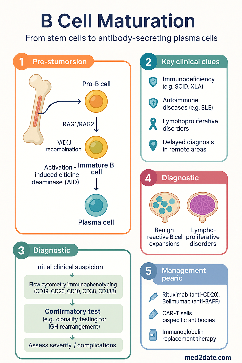
B Cell Maturation clinical infographic — pathophysiology, clinical clues, diagnosis, imaging, and management
Tap or click image to enlarge — B Cell Maturation: pathophysiology, clinical clues, diagnosis, imaging, and management.
B Cell Maturation infographic, full size

Pro-B to Immature B Cell Development

B lymphopoiesis in humans occurs predominantly in the bone marrow throughout life, though the site of production shifts from foetal liver in utero to bone marrow after birth. The developmental programme from haematopoietic stem cell (HSC) to immature B cell takes approximately 2–3 weeks and is governed by ordered transcription factor expression, cytokine signalling, and the sequential rearrangement of immunoglobulin gene segments.

Developmental Stages & Immunophenotype

Stage Surface Markers (CD) Gene Rearrangement Key Transcription Factors Functional Checkpoint
Common lymphoid progenitor (CLP) CD34+, CD38+, CD10+, CD45RA+ Germline configuration EBF1, PAX5 (upregulated), E2A Commitment to B lineage; PAX5 represses alternative lineage genes
Early pro-B cell CD34+, CD10+, CD19+ D–JH rearrangement on chromosome 14 PAX5, EBF1, Ikaros Initiation of heavy chain recombination
Late pro-B cell CD34+/−, CD10+, CD19+ V–DJH rearrangement PAX5, FOXO1 Completion of heavy chain VDJ; surrogate light chain pairing tested
Large pre-B cell CD10+, CD19+, CD34−, cytoplasmic μ heavy chain+ Heavy chain rearrangement complete; light chain in germline PAX5, STAT5 (IL-7R signalling) Pre-BCR checkpoint: μ heavy chain pairs with VpreB/λ5 surrogate light chain → signals proliferation (6–8 cell divisions)
Small pre-B cell CD10+, CD19+, CD34−, surface Ig− V–Jκ or V–Jλ light chain rearrangement IRF4, downregulation of FOXO1 Pre-BCR signals cease; cells exit cycle and rearrange light chain
Immature B cell CD10+, CD19+, CD20lo, surface IgM+, IgD− Functional BCR (IgM) expressed PAX5 (maintained), BCL6 (nascent) Central tolerance checkpoint: cells tested for self-reactivity in bone marrow

The Pre-BCR Checkpoint

The pre-BCR — comprising a μ heavy chain non-covalently associated with the surrogate light chain (VpreB and λ5/IGLL1) — represents the first major quality-control checkpoint in B cell development. Successful pre-BCR assembly signals the cell to proliferate (clonal expansion of ~6–8 divisions), downregulate RAG1/RAG2, and then exit the cell cycle to commence light chain rearrangement. Failure to produce a functional heavy chain (or surrogate light chain) results in developmental arrest and apoptosis.

⚠️
Clinical correlate: Mutations in the μ heavy chain gene (IGHM), λ5 (IGLL1), or the signal transduction molecule Igα (CD79a) cause autosomal recessive agammaglobulinaemia. X-linked agammaglobulinaemia (XLA) due to BTK (Bruton tyrosine kinase) mutations blocks pre-BCR signalling downstream, resulting in maturation arrest at the pre-B cell stage with absent peripheral B cells (CD19+ count <1%) and profoundly low immunoglobulins. Prevalence in Australia: approximately 1 in 100 000 male births.

V(D)J Recombination — Molecular Detail

V(D)J recombination is initiated by the RAG1/RAG2 recombinase complex, which recognises recombination signal sequences (RSS) flanking each V, D, and J gene segment. RAG introduces a double-strand break at the RSS, and the non-homologous end joining (NHEJ) pathway — involving Ku70/Ku80, DNA-PKcs, Artemis, XRCC4, and DNA ligase IV — resolves the break to create a coding joint (expressed) and a signal joint (excised as a signal circle). This process is:

  • Ordered: Heavy chain D–J before V–DJ; heavy chain before light chain; κ before λ
  • Stochastic: each allele rearranges independently (allelic exclusion ensures only one specificity per cell)
  • Diverse: combinatorial diversity (number of V, D, J segments) plus junctional diversity (P-nucleotides, N-nucleotide additions by TdT, exonuclease trimming) yields >1011 possible specificities
🔴
Safety-critical: RAG1/RAG2 deficiency causes T−B−NK+ SCID (OMIM #601457, #608958) — a paediatric emergency requiring urgent referral to a paediatric immunologist and consideration of haematopoietic stem cell transplant (HSCT). In Australia, SCID newborn screening using T-cell receptor excision circles (TRECs) has been incorporated into the national NBS programme since 2018 (available in all states/territories).

Critical Cytokines & Transcription Factors

  • IL-7: The dominant cytokine for early B lymphopoiesis in mice; in humans, IL-7 is important but less exclusively required — human B cell development can partially proceed in IL-7 deficiency, though T cell development is severely impaired.
  • SCF (Kit ligand): Supports pro-B cell survival and proliferation via c-Kit (CD117).
  • FLT3 ligand: Acts on CLPs and early progenitors.
  • PAX5: Master regulator of B lineage commitment; activates B cell–specific genes (CD19, BLNK) and represses alternative lineage programmes (Notch1, M-CSFR). Loss of PAX5 → lineage infidelity.
  • EBF1: Cooperates with E2A (TCF3) to activate PAX5 and downstream B cell genes.
  • Ikaros (IKZF1): Chromatin remodelling factor required for lymphoid specification; IKZF1 deletions are associated with high-risk B-ALL in paediatric patients.

Central & Peripheral Tolerance

Because V(D)J recombination is a stochastic process, it inevitably generates BCRs that recognise self-antigens. Multiple tolerance checkpoints eliminate or silence these self-reactive clones, both in the bone marrow (central tolerance) and in the periphery (peripheral tolerance). Failure of tolerance is a central mechanism in systemic autoimmune diseases.

Central Tolerance (Bone Marrow)

Immature B cells that express surface IgM are tested against self-antigens in the bone marrow microenvironment. The outcome depends on the affinity and valency of the self-antigen encounter:

Low affinity
Ignored
Weak self-reactivity below the threshold for tolerance induction; clone proceeds to periphery as a potentially useful receptor with residual self-reactivity.
Outcome: Passive survival
Moderate affinity
Receptor Editing
RAG1/RAG2 re-expressed; secondary V–J light chain rearrangement (usually κ locus, then λ) generates a new BCR. If editing fails after multiple attempts, apoptosis ensues.
Outcome: New specificity or death
High affinity / multivalent
Clonal Deletion
Strong BCR cross-linking by abundant self-antigen delivers an apoptotic signal. Editing may be attempted first, but high-affinity anti-DNA or anti-MHC reactivities are typically deleted. BIM (BCL2L11) is the key pro-apoptotic mediator.
Outcome: Apoptosis
ℹ️
Receptor editing statistics: Approximately 25–50% of newly formed human immature B cells undergo receptor editing in the bone marrow, making it the dominant mechanism of central tolerance rather than clonal deletion.

Transitional B Cells

Immature B cells that survive central tolerance exit the bone marrow as transitional B cells (T1 and T2 stages in the spleen). These are the final gate before entry into the mature naive B cell pool.

Stage Phenotype Location Key Features
T1 transitional IgMhi, IgDlo, CD21lo, CD23−, CD38+ Splenic red pulp / marginal zone Highly susceptible to BCR-mediated apoptosis; short-lived (~3 days)
T2 transitional IgMhi, IgDhi, CD21+, CD23+, CD38+ Splenic follicles BAFF-receptor (BAFFR/BR3) expression enables BAFF-mediated survival; precursor to mature follicular or marginal zone B cells

Peripheral Tolerance Mechanisms

Self-reactive B cells that escape central tolerance may encounter self-antigen in the periphery. Multiple mechanisms restrain them:

  • Anergy: Chronic low-level BCR engagement without T cell help renders B cells functionally unresponsive. Anergic B cells express reduced surface IgM, fail to signal normally through the BCR, and have shortened half-lives (reduced BAFFR expression).
  • Follicular exclusion: Self-reactive B cells are excluded from B cell follicles (and hence from T cell help and germinal centre entry) by competition with non-self-reactive cells for limited BAFF and CXCL13 niches.
  • Fas-mediated deletion: If a self-reactive B cell receives BCR and CD40 signals but with inappropriate or absent cytokine support, Fas (CD95) engagement by FasL on T cells induces apoptosis.
  • Regulatory B cells (Bregs): A subset of B cells (typically CD19+CD24hiCD38hi or CD19+CD5+CD1dhi) that produce IL-10 and TGF-β, suppressing T cell and dendritic cell activation. Breg deficiency is implicated in SLE, rheumatoid arthritis, and multiple sclerosis.
⚠️
Autoimmune clinical correlate: Defective receptor editing is documented in patients with systemic lupus erythematosus (SLE). Polymorphisms in PTPN22, BLK, and BANK1 — genes involved in BCR signalling thresholds — are enriched in Australian SLE cohorts (ACR/EULAR 2019 criteria applicable). In children, SLE with B cell tolerance defects often presents with high-titre anti-dsDNA antibodies and lupus nephritis; rheumatology referral within 4 weeks of suspected diagnosis is recommended (ARA guidelines).

Clinical Implications of Tolerance Failure

Tolerance Defect Mechanism Associated Condition Autoantibody Example
Impaired receptor editing Reduced RAG re-expression or altered chromatin accessibility SLE Anti-dsDNA, anti-Smith
Excessive BAFF / BAFFR signalling Rescue of anergic self-reactive B cells SLE, Sjögren syndrome Anti-SSA/Ro, anti-SSB/La
Defective Fas–FasL pathway Failure to delete germinal centre autoreactive clones Autoimmune lymphoproliferative syndrome (ALPS) Direct Coombs (anti-RBC), anti-phospholipid
Breg deficiency Reduced IL-10–mediated suppression MS, RA, type 1 diabetes Variable; disease-specific

Germinal Centre Maturation

The germinal centre (GC) reaction in secondary lymphoid organs (lymph nodes, spleen, mucosa-associated lymphoid tissue) is the engine of antibody affinity maturation and class-switch recombination. This process underpins effective vaccination and is also the site of origin for the majority of aggressive B cell non-Hodgkin lymphomas in Australia.

Initiation of the Germinal Centre Reaction

The GC reaction is initiated when a mature follicular B cell encounters its cognate antigen presented by subcapsular sinus macrophages or follicular dendritic cells, internalises it via the BCR, processes it, and presents peptide–MHC II complexes to primed CD4+ T cells at the T–B border. This cognate interaction provides three essential signals:

  1. Signal 1 — BCR engagement: Antigen binding triggers BCR cross-linking and internalisation.
  2. Signal 2 — CD40/CD40L: CD40L (CD154) on activated T follicular helper (Tfh) cells engages CD40 on B cells — essential for GC formation; CD40 deficiency causes Hyper-IgM syndrome type 3.
  3. Signal 3 — Cytokines: IL-21 (the dominant GC cytokine), IL-4, and IL-6 drive B cell proliferation, AID expression, and differentiation.

Germinal Centre Architecture — Dark Zone & Light Zone

🌑 Dark Zone

Dense packing of rapidly dividing B cells (centroblasts) with CXCL12-producing reticular cells. Centroblasts express AID and undergo somatic hypermutation at a rate of ~10−3 mutations per base pair per cell division. Each division introduces 1–2 mutations in the variable region.

🌕 Light Zone

Less dense; contains follicular dendritic cells (FDCs) displaying immune complexes, Tfh cells, and non-dividing centrocytes. Centrocytes test their mutated BCR against antigen on FDCs. High-affinity clones capture more antigen, present more peptide–MHC II to Tfh, and receive survival signals (CD40L, IL-21). Low-affinity or self-reactive centrocytes die by apoptosis.

Somatic Hypermutation (SHM)

SHM is initiated by activation-induced cytidine deaminase (AID, encoded by AICDA), which deaminates cytosine to uracil in single-stranded DNA at the immunoglobulin variable region. Error-prone repair by mismatch repair (MMR) and base excision repair (BER, involving UNG and REV1) introduces mutations at and around the original lesion. Key features:

  • Targeted to the rearranged V(D)J region (not the constant region)
  • Requires transcription through the target locus (RNA pol II–associated cofactors recruit AID)
  • Hotspot motifs: WRCY (W = A/T, R = A/G, C, Y = C/T) on the coding strand; complementary motif on the non-coding strand
  • Approximately 50% of mutations are synonymous (no change to amino acid); the rest are non-synonymous, creating a diverse pool for selection

Class-Switch Recombination (CSR)

CSR changes the immunoglobulin heavy chain constant region from Cμ (IgM) to Cγ (IgG1–4), Cα (IgA1–2), or Cε (IgE), altering antibody effector function without changing antigen specificity. CSR requires:

  • AID: Deaminates cytosines in switch (S) regions upstream of each constant region gene
  • UNG and MMR: Process U:A mismatches into double-strand breaks in donor and acceptor S regions
  • NHEJ: Joins the broken ends, looping out the intervening DNA as a switch circle
  • Cytokine direction: IL-4 → IgE and IgG4; IFN-γ → IgG1 and IgG3; TGF-β → IgA; IL-21 promotes IgG1 and IgG3 in humans
⚠️
Hyper-IgM syndromes: AID deficiency (Hyper-IgM type 2) and UNG deficiency (Hyper-IgM type 5) both present with elevated IgM, absent IgG/IgA/IgE, normal or elevated lymphocyte counts, and susceptibility to encapsulated bacteria and opportunistic infections (Cryptosporidium in CD40L deficiency). In Australia, these patients require immunoglobulin replacement therapy (PBS Authority Required, criteria per PBAC) and infectious disease co-management.

T Follicular Helper (Tfh) Cells — The Orchestration of GC Selection

Tfh cells are specialised CD4+ T cells characterised by expression of CXCR5 (follicular homing), PD-1, ICOS, BCL6 (master transcription factor), and production of IL-21. Their role in the GC includes:

  • Affinity selection: Centrocytes that capture more antigen from FDCs present more peptide–MHC II to Tfh, receiving stronger CD40L and IL-21 survival signals.
  • Class-switch licensing: Tfh-derived cytokines (IL-4, IL-21) direct CSR.
  • Prevention of autoimmunity: Tfh cells that fail to receive adequate co-stimulation become T follicular regulatory (Tfr) cells, suppressing excessive GC reactions.

Clinical correlate — Tfh dysregulation: Excessive Tfh activity is observed in SLE (elevated circulating Tfh, CXCR5+PD-1+CD4+ T cells), Castleman disease (IL-6–driven Tfh expansion), and angioimmunoblastic T cell lymphoma (AITL, a Tfh-derived malignancy). Deficient Tfh function underlies impaired vaccine responses in patients on anti-CD20 therapy (rituximab); vaccination ≥6 months after rituximab cessation is recommended (ASCIA guidelines).

Germinal Centre Output — Memory B Cells vs Plasma Cells

The GC produces two functionally distinct populations:

Feature Memory B Cells Plasma Cells (Short-lived GC-derived)
Surface markers IgG/IgA/IgE+, CD27+, CD20+, CD38lo CD38hi, CD138+, CD20−, surface Iglo/−
Function Rapid recall response upon re-encounter with antigen; can re-enter GC for further maturation Immediate antibody secretion; migrate to medullary cords / mucosal sites
Location Circulation, marginal zone, mucosal tissues Lymph node medullary cords, splenic red pulp, bone marrow (if long-lived)
Half-life Decades (self-renewing) Days to weeks (short-lived); long-lived plasma cells can persist years in bone marrow niches
Clinical targeting Anti-CD20 (rituximab) depletes CD20+ memory B cells but spares plasma cells Anti-CD38 (daratumumab) and anti-CD138 target plasma cells; proteasome inhibitors (bortezomib) induce apoptosis

Plasma Cell Differentiation

Plasma cell differentiation is the terminal stage of B cell maturation, transforming a B cell from a signalling lymphocyte into a dedicated antibody-secreting factory capable of producing thousands of immunoglobulin molecules per second. This process is governed by a coordinated transcriptional switch that extinguishes the B cell identity programme and activates the unfolded protein response (UPR) to manage the enormous secretory load.

Transcriptional Regulation of Plasma Cell Differentiation

1
IRF4 (high levels)
Sustained high expression of IRF4 — driven by CD40, BCR, and IL-21 signalling — is the molecular switch that initiates the plasma cell programme. Low IRF4 maintains B cell identity; high IRF4 activates BLIMP-1.
2
BLIMP-1 (PRDM1)
B lymphocyte–induced maturation protein 1 is the master regulator of plasma cell differentiation. BLIMP-1 represses PAX5, BCL6, CIITA, and SPIB — effectively silencing the B cell gene expression programme, including MHC II expression (plasma cells are poor antigen presenters).
3
XBP-1
X-box binding protein 1 (spliced form, XBP-1s) is the central transcription factor of the unfolded protein response. Activated by IRE1α splicing of XBP1 mRNA, XBP-1s upregulates ER chaperones, ERAD components, and lipid biosynthesis enzymes to expand the endoplasmic reticulum for massive immunoglobulin secretion.
4
ATF6 & PERK branches of UPR
In addition to the IRE1α–XBP-1 pathway, ATF6 (chaperone induction) and PERK (transient translational attenuation via eIF2α phosphorylation) contribute to ER homeostasis. Chronic UPR activation (as in myeloma) can trigger apoptosis — exploited therapeutically by proteasome inhibitors (bortezomib, carfilzomib).

Short-Lived vs Long-Lived Plasma Cells

Property Short-Lived Plasma Cells Long-Lived Plasma Cells (LLPCs)
Origin Extrafollicular response; early GC output GC-derived; selected high-affinity centrocytes
Location Secondary lymphoid organs, inflamed tissues Bone marrow survival niches (CXCL12-producing stromal cells, APRIL, IL-6)
Lifespan Days to ~2 weeks Months to decades
Function Early wave of antigen clearance Sustained protective serum antibody titres (basis of long-term vaccine protection)
Susceptibility to rituximab Resistant (CD20−) Resistant (CD20−); explains persistence of protective antibodies post-rituximab in most patients
Therapeutic targeting Corticosteroids, cyclophosphamide Anti-CD38 (daratumumab, isatuximab), anti-SLAMF7 (elotuzumab), anti-BCMA (belantamab mafodotin), proteasome inhibitors

Bone Marrow Survival Niches

Long-lived plasma cells home to the bone marrow where they reside in specialised survival niches. These niches provide:

  • CXCL12 (SDF-1): Chemokine produced by CXCR4+ stromal cells; mediates homing and retention (CXCR4 antagonism with plerixafor mobilises plasma cells)
  • APRIL (TNFSF13): Binds BCMA (TNFRSF17) and TACI on plasma cells → activates NF-κB survival signalling
  • IL-6: From mesenchymal stromal cells and osteoclasts → STAT3 activation → MCL-1 and BCL-2 upregulation
  • CD44–hyaluronic acid: Adhesion interaction anchoring plasma cells in niches
  • Contact with eosinophils: Eosinophils in the bone marrow produce APRIL and IL-6, supporting LLPC survival
ℹ️
Clinical relevance — multiple myeloma: Multiple myeloma is a malignancy of bone marrow plasma cells, representing 1.8% of all cancers in Australia (~1 800 new cases/year, AIHW 2023). The tumour cells hijack the bone marrow survival niche. Therapeutic strategies directly informed by plasma cell biology include: daratumumab (anti-CD38), isatuximab (anti-CD38), belantamab mafodotin (anti-BCMA antibody–drug conjugate), idecabtagene vicleucel (anti-BCMA CAR-T), and elranatamab (anti-BCMA × CD3 bispecific).

Antibody Isotype Functions (Post-CSR Output)

Isotype Subclasses Key Functions Half-Life (days)
IgM First responder; pentameric form excellent at complement activation (classical pathway) 5
IgG IgG1, IgG2, IgG3, IgG4 IgG1/3: opsonisation, ADCC, complement; IgG2: anti-polysaccharide (encapsulated bacteria); IgG4: anti-inflammatory (blocks rather than activates effector functions) 21 (IgG1/2/4), 7 (IgG3)
IgA IgA1, IgA2 Mucosal immunity; secretory IgA (dimeric, J chain + secretory component) neutralises pathogens at mucosal surfaces 6
IgE Allergic and anti-parasitic responses; binds FcεRI on mast cells/basophils 2
IgD Co-expressed with IgM on naive B cells; role in B cell activation and upper respiratory mucosal immunity (incompletely understood) 3

Pathophysiology — Disorders of B Cell Maturation

Disruptions at each stage of B cell maturation produce distinct clinical syndromes. Understanding the stage of maturation arrest is critical for diagnosis and targeted management.

Maturation Arrests & Associated Conditions

Stage of Arrest Genetic Defect Condition Key Laboratory Findings
HSC / CLP Reticular dysgenesis (AK2) Severe combined immunodeficiency Absent T, B, NK cells; neutropenia
Pro-B / Pre-B BTK (XLA), IGHM, IGLL1, CD79a/b, BLNK Agammaglobulinaemia CD19+ B cells <1%; all Ig classes profoundly low
Immature B cell PIK3CD (gain-of-function) Activated PI3Kδ syndrome (APDS) Elevated transitional B cells; reduced class-switched memory; lymphoproliferation
GC reaction — SHM/CSR AICDA (AID), UNG Hyper-IgM syndrome types 2 & 5 Elevated IgM; absent IgG, IgA, IgE; enlarged lymph nodes with GC hyperplasia
GC reaction — T cell help CD40LG, CD40 Hyper-IgM syndrome types 1 & 3 Elevated IgM; absent IgG/IgA; neutropenia (CD40LG); opportunistic infections
Post-GC / plasma cell Multifactorial (CVID) Common variable immunodeficiency Low IgG + IgA and/or IgM; reduced class-switched memory B cells; diagnosed after age 2 years

Investigations

Evaluation of B cell maturation disorders requires a tiered approach, beginning with widely available screening tests and progressing to specialised immunological and molecular studies.

Essential
Serum immunoglobulin levels (IgG, IgA, IgM, IgE)
MBS item 65070 (immunoglobulin quantitation). Widely available in all Australian pathology laboratories. Establishes the presence and isotype of hypogammaglobulinaemia.
Essential
Full blood examination with differential lymphocyte count
MBS item 65060. Absolute lymphocyte count <1.5 × 10⁹/L (adults) or age-appropriate paediatric lower limit raises concern for lymphopenia including B cell deficiency.
Available
B cell immunophenotyping by flow cytometry
MBS item 65090 (lymphocyte subset analysis). Available through major hospital and private reference laboratories (Sullivan Nicolaides, Douglass Hanly Moir, Melbourne Pathology). Panel typically includes CD19, CD20, CD27, CD38, CD10, IgD, IgM to enumerate: total B cells, transitional (T1/T2), naive mature, class-switched memory, marginal zone-like, and plasmablasts.
Available
Specific antibody responses (vaccine challenge)
Anti-pneumococcal serotype IgG (pre- and post-Pneumovax 23® or Prevenar 13®); anti-tetanus IgG; anti-HBs (hepatitis B). Adequate response = ≥4-fold rise or protective level. Assesses functional antibody production despite normal total Ig levels.
Specialist
Molecular genetic testing
Targeted gene panels (BTK, AICDA, CD40LG, RAG1/RAG2, PIK3CD, PIK3R1) or whole-exome sequencing. Available through Victorian Clinical Genetics Services (VCGS), SA Pathology Genetics, Sonic Genetics. Medicare-funded under MBS item 73295 (genomic testing) for suspected inborn errors of immunity via state-based services.
Specialist
Bone marrow biopsy with plasma cell quantitation
Required for suspected myeloma or plasma cell neoplasm; includes morphology, flow cytometry (CD38, CD138, CD56, CD19, CD45), cytogenetics/FISH (t(11;14), t(4;14), del17p), and serum free light chain assay (MBS item 66882).
Referral
TREC / KREC newborn screening
Dried blood spot assay for T-cell receptor excision circles (TRECs) and Kappa-deleting recombination excision circles (KRECs). Part of Australian national NBS programme (since 2018 for TRECs). KRECs assess B cell output. Positive screen → urgent immunology referral.

Risk Stratification & Severity Scoring

Risk stratification in B cell disorders is guided by the degree of immunodeficiency, presence of end-organ damage, and association with lymphoproliferative or autoimmune complications.

Mild
Selective IgA Deficiency / Partial IgG Subclass Deficiency
IgA <0.07 g/L with normal IgG and IgM; or isolated IgG2/IgG3 subclass deficiency. Most patients are asymptomatic. Monitor for progression to CVID (~10% of selective IgA deficiency).
Setting: GP monitoring, annual immunoglobulin levels, vaccine response testing
Moderate
CVID Without Complications / XLA on Replacement
Hypogammaglobulinaemia requiring immunoglobulin replacement therapy (IVIg 400–600 mg/kg every 3–4 weeks, or SCIg 100–200 mg/kg weekly). Recurrent sinopulmonary infections controlled on prophylaxis. No autoimmune or granulomatous complications.
Setting: Immunologist oversight every 6–12 months; PBS Authority for immunoglobulin
Severe
SCID / CVID with Complications / Lymphoproliferative Transformation
SCID: absent T and/or B cells, life-threatening infections — emergency HSCT. CVID with granulomatous-lymphocytic interstitial lung disease (GLILD), splenomegaly, autoimmune cytopenias, or lymphoma (risk 2–8× general population). Requires multidisciplinary management.
Setting: Tertiary/quaternary centre; immunology, haematology, respiratory MDT

Therapeutic Considerations

Management of B cell disorders is tailored to the underlying maturation defect and its clinical consequences. The following drug cards address the major therapeutic agents targeting B cell lineages in current Australian practice.

💊
Intravenous Immunoglobulin (IVIg)
Intragam® P / Privigen® / Octagam®
Adult dose 400–600 mg/kg IV every 3–4 weeks; titrate to trough IgG ≥7 g/L
Paediatric dose 400–600 mg/kg IV every 3–4 weeks (same as adult); neonates: 500 mg/kg
Route Intravenous infusion (hospital or home)
Renal adjustment Use sucrose-free preparations if eGFR <30; rate-limit to ≤2 mg/kg/min; monitor creatinine
Hepatic adjustment No adjustment required
PBS status Authority Required — confirmed primary antibody deficiency or secondary hypogammaglobulinaemia with recurrent infections
💊
Subcutaneous Immunoglobulin (SCIg)
Cuvitru® / Hizentra® / HyQvia®
Adult dose 100–200 mg/kg SC weekly (Cuvitru/Hizentra); or 400 mg/kg SC every 4 weeks (HyQvia with rHuPH20)
Paediatric dose As per adult dosing on mg/kg basis; suitable from age 2 years (Cuvitru/Hizentra)
Route Subcutaneous infusion via pump or rapid push
Renal adjustment No specific adjustment; preferred over IVIg in patients with renal impairment
PBS status Authority Required — same criteria as IVIg; patient preference or IV access issues
💊
Rituximab
MabThera® / Riximyo® · Anti-CD20 monoclonal antibody
Adult dose 375 mg/m² IV weekly × 4 (lymphoma); or 1000 mg IV × 2 doses, 2 weeks apart (RA/SLE)
Mechanism Depletes CD20+ B cells (pre-B through mature/memory B cells); spares pro-B cells (CD20−) and plasma cells (CD20−)
Renal adjustment No adjustment required
PBS status PBS Restricted Benefit — NHL, CLL, RA (DMARD failure), ANCA vasculitis, pemphigus vulgaris
💊
Belimumab
Benlysta® · Anti-BAFF (BLyS) monoclonal antibody
Adult dose 10 mg/kg IV every 4 weeks (after loading at weeks 0, 2, 4); or 200 mg SC once weekly
Mechanism Neutralises BAFF, reducing survival of transitional and naive B cells; modulates B cell tolerance by removing BAFF-mediated rescue of anergic self-reactive clones
Renal adjustment No dose adjustment; caution in severe renal impairment
PBS status PBS Restricted Benefit — SLE with inadequate response to standard therapy; lupus nephritis (PBS-listed 2023)

Monitoring

Every 3–6 months
Trough serum IgG levels (target ≥7 g/L or individualised based on infection frequency) in patients on immunoglobulin replacement therapy.
Every 6–12 months
Lymphocyte subset panel (CD3, CD4, CD8, CD19, CD16/56) — MBS item 65090 — to assess B cell reconstitution after rituximab or to monitor for progressive decline in CVID.
Annually
Spirometry and HRCT (if indicated) for GLILD screening in CVID. Chest X-ray and lung function testing per Australasian Society of Clinical Immunology and Allergy (ASCIA) consensus.
Post-rituximab: 6-monthly
CD19+ B cell count to document repletion (typically 6–9 months post-treatment). Revaccination programme once CD19+ count >0.1 × 10⁹/L and ≥6 months post last rituximab dose.
As indicated
Quantitative vaccine responses (pneumococcal serotypes, tetanus, hepatitis B) to assess functional antibody production — essential before and after rituximab therapy.

Special Populations

🤰 Pregnancy
Immunoglobulin replacement
SCIg preferred during pregnancy (lower peak-trough fluctuation, fewer systemic reactions). Continue at usual dose; monitor trough IgG monthly in 2nd/3rd trimester. Safe in all trimesters (Category A — no teratogenicity).
Rituximab
Avoid if possible; crosses placenta (actively transported via FcRn from 2nd trimester). If essential, administer before week 16. Monitor neonatal B cells and immunoglobulins for 6 months post-delivery (risk of transient neonatal B cell depletion).
Belimumab
Limited human data; use only if benefit clearly outweighs risk. Animal data show no malformations at subclinical doses. Register with the Australian Pregnancy Registry for autoimmune conditions.
👶 Paediatrics
XLA (Bruton disease)
Typically presents at 6–12 months (after maternal IgG wanes) with recurrent bacterial otitis media, pneumonia, and GI infections. TREC screening may not detect (T cells are normal). Confirm with BTK gene sequencing. Lifelong immunoglobulin replacement and antibiotic prophylaxis required. Refer to paediatric immunologist at tertiary children's hospital.
SCID
Newborn screening (TRECs) enables pre-symptomatic diagnosis. Absolute lymphocyte count <3.0 × 10⁹/L at birth is a red flag. Confirm with lymphocyte subsets (absent T cells ± absent B/NK cells). HSCT is curative; transplant within first 3.5 months of life yields best outcomes (>90% survival in matched related donor). Refer urgently to SCID transplant centres (Royal Children's Hospital Melbourne, Children's Hospital Westmead).
Transient hypogammaglobulinaemia of infancy (THI)
IgG below age-appropriate normal with normal B cell numbers. Self-limiting — typically resolves by age 4 years. Monitor 6-monthly; avoid unnecessary immunoglobulin replacement unless significant recurrent infections.
👴 Elderly
Immunosenescence
Age-related decline in naive B cell output (reduced bone marrow haematopoiesis) and accumulation of age-associated B cells (ABCs, CD21−CD11c+). Results in impaired vaccine responses and increased susceptibility to infection. CVID diagnosis should be considered in elderly patients with new-onset hypogammaglobulinaemia.
CLL/SLL
Median age at diagnosis: 70 years. The most common leukaemia in Australia (~1 500 new cases/year). Accumulation of CD5+CD19+CD23+ B cells with impaired antibody function — hypogammaglobulinaemia develops in 30–50% over disease course. Consider IVIg if IgG <4 g/L and recurrent infections.
🫘 Renal Impairment
IVIg preparation
Use sucrose-free IVIg preparations (Intragam P, Privigen) in patients with eGFR <30 mL/min/1.73 m² or on dialysis. Sucrose-containing products carry osmotic nephrosis risk. Infuse at ≤2 mg/kg/min. Monitor serum creatinine 24–48 hours post-infusion.
🫁 Hepatic Impairment
Immunoglobulin and B cell therapies
No dose adjustments required for IVIg, SCIg, or rituximab in hepatic impairment. However, patients with chronic liver disease may have secondary hypogammaglobulinaemia; differentiate from primary immunodeficiency by B cell subset analysis.
🦠 Immunocompromised
Rituximab-induced hypogammaglobulinaemia
Repeated rituximab cycles (e.g. for lymphoma or autoimmune disease) may cause cumulative B cell depletion and secondary hypogammaglobulinaemia (IgG <4 g/L in ~15% after ≥5 cycles). Monitor IgG before each cycle. If IgG <4 g/L with recurrent infections, initiate immunoglobulin replacement per ASCIA guidelines.
Post-COVID B cell dysregulation
Emerging evidence of altered B cell subset distribution (expanded atypical memory B cells, impaired GC responses) in some patients post-SARS-CoV-2 infection. Clinical significance under investigation; monitor for new-onset hypogammaglobulinaemia or autoimmune features.

Aboriginal and Torres Strait Islander Health Considerations

Aboriginal and Torres Strait Islander Health
Prevalence & burden
ATSI Australians experience a higher burden of infectious diseases, including recurrent respiratory and ear infections in children, which may mask underlying primary immunodeficiency. The AIHW reports that ATSI children have 2–3 times the rate of hospitalisation for lower respiratory tract infections compared with non-Indigenous children, potentially delaying recognition of B cell maturation defects such as XLA or CVID.
Diagnostic delay
Access to specialist immunology services is limited in remote and very remote communities. Paediatric immunologists are concentrated in major capital cities. Telehealth (Medicare item 91822) can facilitate initial assessment, but flow cytometry and molecular testing require sample transport to metropolitan reference laboratories, adding 5–10 business days to turnaround.
Immunoglobulin access
SCIg (subcutaneous immunoglobulin) is the preferred modality in remote settings due to lower infrastructure requirements (no IV cannulation, home administration possible after training). PBS Authority approval is required; Aboriginal Health Workers can be trained to support SCIg administration with appropriate supervision. Plasma supply is dependent on Australian donor population — blood supply diversification programmes should be culturally inclusive.
Newborn screening
National SCID screening via TRECs is available across all states and territories. However, completeness of dried blood spot collection in remote communities requires ongoing quality assurance by jurisdictional newborn screening laboratories. Awareness among community health staff of the significance of abnormal TREC results is critical for timely referral.
Cultural safety
Genetic testing (including for XLA, SCID, and other B cell maturation defects) raises specific cultural considerations. Genetic counselling must be provided in a culturally safe manner, with engagement of Aboriginal and Torres Strait Islander liaison officers where available. Kinship systems and family structure should inform communication of X-linked inheritance patterns. Consent processes must be culturally appropriate and documented in accordance with NHMRC ethical guidelines for research involving ATSI peoples (where applicable to research participation).
Immunisation
ATSI children with B cell immunodeficiency require enhanced immunisation schedules per the Australian Immunisation Handbook. Live vaccines (MMR, varicella, BCG) are contraindicated in severe B cell deficiency (SCID, XLA, CVID). Coordination with Aboriginal Medical Services and the National Immunisation Programme is essential to ensure appropriate vaccine selection and timing.

📚 References

  1. 1. Pieper K, Grimbacher B, Eibel H. B-cell biology and development. J Allergy Clin Immunol. 2013;131(4):959–971. doi:10.1016/j.jaci.2013.01.046
  2. 2. Nemazee D. Mechanisms of central tolerance for B cells. Nat Rev Immunol. 2017;17(5):281–294. doi:10.1038/nri.2017.19
  3. 3. Victora GD, Nussenzweig MC. Germinal centers. Annu Rev Immunol. 2022;40:413–442. doi:10.1146/annurev-immunol-120419-022408
  4. 4. Nutt SL, Hodgkin PD, Tarlinton DM, Corcoran LM. The generation of antibody-secreting plasma cells. Nat Rev Immunol. 2015;15(3):160–171. doi:10.1038/nri3795
  5. 5. Tangye SG, Al-Herz W, Bousfiha A, et al. Human inborn errors of immunity: 2022 update on the classification from the International Union of Immunological Societies Expert Committee. J Clin Immunol. 2022;42(7):1473–1507. doi:10.1007/s10875-022-01289-3
  6. 6. Australasian Society of Clinical Immunology and Allergy (ASCIA). ASCIA Guidelines for Immunoglobulin Replacement Therapy in Primary Immunodeficiency. Sydney: ASCIA; 2023. Available at: https://www.allergy.org.au
  7. 7. Slade CA, Bosco JJ, Unglik GA, Bleasel KF, Naglik M, Gaff JM. Deficiency in antibody responses to immunisation in older adults. Clin Exp Immunol. 2023;211(1):1–11. doi:10.1093/cei/uxac115
  8. 8. Rawlings DJ, Metzler G, Wray-Dutra M, Jackson SW. Altered B cell signalling in autoimmunity. Nat Rev Immunol. 2017;17(7):421–436. doi:10.1038/nri.2017.24
  9. 9. Australian Institute of Health and Welfare (AIHW). Cancer Data in Australia. Canberra: AIHW; 2024. Cat. no. CAN 140. Available at: https://www.aihw.gov.au/reports/cancer/cancer-data-in-australia
  10. 10. Bousfiha A, Moundir A, Tangye SG, et al. The 2022 update of IUIS phenotypical classification for human inborn errors of immunity. J Clin Immunol. 2022;42(7):1508–1520. doi:10.1007/s10875-022-01352-z
  11. 11. Rook AH, Bousfiha A, Conley ME, et al. The 2019 American College of Rheumatology/European League Against Rheumatism classification criteria for systemic lupus erythematosus. Arthritis Rheumatol. 2019;71(9):1400–1412. doi:10.1002/art.40930
  12. 12. National Health and Medical Research Council (NHMRC). Ethical Conduct in Research with Aboriginal and Torres Strait Islander Peoples and Communities: Guidelines for Researchers and Stakeholders. Canberra: NHMRC; 2018.
for PBS-listed medicines at participating pharmacies.
Cultural safety
Engagement with Aboriginal Community Controlled Health Organisations (ACCHOs) is essential. Cultural safety training for non-Indigenous clinicians, use of Aboriginal Health Workers and Liaison Officers, and incorporation of traditional healing practices alongside Western medicine improve treatment adherence and outcomes. Avoidance of eye contact, respect for gender-sensitive examination practices, and understanding of sorry business protocols are critical elements of culturally safe care.
Medication adherence
Complex DMARD regimens with frequent monitoring requirements present adherence challenges. Long-acting depot injections (e.g., methotrexate SC) may improve adherence compared to oral regimens. Community pharmacy partnerships through the Indigenous Pharmacy Programmes improve medication management.
Specific conditions
Rheumatic heart disease (RHD) requires secondary prophylaxis with benzathine penicillin G (BPG) 1.2 MU IM every 3–4 weeks for a minimum of 10 years or until age 21 (whichever is longer). RHD registers (e.g., NT RHD Register) facilitate recall and follow-up. The Australian RHD Endgame Strategy targets elimination by 2031.
Referral pathways
Referral through ACCHOs and Aboriginal Hospital Liaison Officers (AHLOs) improves engagement. The Specialist Outreach Assistance Programme provides funded specialist visits to remote communities. NT, WA, and QLD have specific rheumatology outreach programmes targeting Indigenous communities.

📚 References

  1. 1. Australian Institute of Health and Welfare (AIHW). Autoimmune disease in Australia. Cat. no. PHE 312. Canberra: AIHW; 2023.
  2. 2. Fraenkel L, Bathon JM, England BR, et al. 2021 American College of Rheumatology guideline for the treatment of rheumatoid arthritis. Arthritis Care Res. 2021;73(7):924–939.
  3. 3. Fanouriakis A, Kostopoulou M, Alber K, et al. 2019 update of the EULAR recommendations for the management of systemic lupus erythematosus. Ann Rheum Dis. 2019;78(6):736–745.
  4. 4. Chung SA, Langford CA, Maz M, et al. 2021 American College of Rheumatology/Vasculitis Foundation guideline for the management of antineutrophil cytoplasmic antibody-associated vasculitis. Arthritis Care Res. 2021;73(11):1583–1599.
  5. 5. Smolen JS, Landewé RBM, Bijlsma JWJ, et al. EULAR recommendations for the management of rheumatoid arthritis with synthetic and biological disease-modifying antirheumatic drugs: 2022 update. Ann Rheum Dis. 2023;82(1):3–18.
  6. 6. Australian Technical Advisory Group on Immunisation (ATAGI). Australian Immunisation Handbook. Australian Government Department of Health; 2024. Available from: immunisationhandbook.health.gov.au.
  7. 7. Rheumatic Heart Disease Australia (RHDAustralia). The 2020 Australian guideline for prevention, diagnosis, and management of acute rheumatic fever and rheumatic heart disease. 3rd ed. Darwin: Menzies School of Health Research; 2020.
  8. 8. Pharmaceutical Benefits Scheme (PBS). PBS Schedule. Australian Government Department of Health. Available from: pbs.gov.au. Accessed 2024.
  9. 9. Agarwal S, Cunnington J, Nossent J. Autoimmune disease in Indigenous Australians: a systematic review. Int J Rheum Dis. 2021;24(12):1487–1498.
  10. 10. Pisetsky DS. Antinuclear antibody testing — misunderstood or misused? Clin Immunol. 2023;255:109717.
  11. 11. Bertsias GK, Tektonidou M, Amoura Z, et al. Joint European League Against Rheumatism and European Renal Association–European Dialysis and Transplant Association (EULAR/ERA-EDTA) recommendations for the management of adult and paediatric lupus nephritis. Ann Rheum Dis. 2012;71(11):1771–1782.
  12. 12. Ledingham J, Deighton C; British Society for Rheumatology Standards, Audit and Guidelines Working Group. Update on the British Society for Rheumatology guidelines for prescribing TNFα blockers in adults with rheumatoid arthritis. Rheumatology. 2005;44(2):155–158.
  13. 13. National Health and Medical Research Council (NHMRC). National statement on ethical conduct in human research. Canberra: NHMRC; 2023 (updated).
for PBS-listed medicines at participating pharmacies.
Cultural safety
Engagement with Aboriginal Community Controlled Health Organisations (ACCHOs) is essential. Cultural safety training for non-Indigenous clinicians, use of Aboriginal Health Workers and Liaison Officers, and incorporation of traditional healing practices alongside Western medicine improve treatment adherence and outcomes. Avoidance of eye contact, respect for gender-sensitive examination practices, and understanding of sorry business protocols are critical elements of culturally safe care.
Medication adherence
Complex DMARD regimens with frequent monitoring requirements present adherence challenges. Long-acting depot injections (e.g., methotrexate SC) may improve adherence compared to oral regimens. Community pharmacy partnerships through the Indigenous Pharmacy Programmes improve medication management.
Specific conditions
Rheumatic heart disease (RHD) requires secondary prophylaxis with benzathine penicillin G (BPG) 1.2 MU IM every 3–4 weeks for a minimum of 10 years or until age 21 (whichever is longer). RHD registers (e.g., NT RHD Register) facilitate recall and follow-up. The Australian RHD Endgame Strategy targets elimination by 2031.
Referral pathways
Referral through ACCHOs and Aboriginal Hospital Liaison Officers (AHLOs) improves engagement. The Specialist Outreach Assistance Programme provides funded specialist visits to remote communities. NT, WA, and QLD have specific rheumatology outreach programmes targeting Indigenous communities.

📚 References

  1. 1. Australian Institute of Health and Welfare (AIHW). Autoimmune disease in Australia. Cat. no. PHE 312. Canberra: AIHW; 2023.
  2. 2. Fraenkel L, Bathon JM, England BR, et al. 2021 American College of Rheumatology guideline for the treatment of rheumatoid arthritis. Arthritis Care Res. 2021;73(7):924–939.
  3. 3. Fanouriakis A, Kostopoulou M, Alber K, et al. 2019 update of the EULAR recommendations for the management of systemic lupus erythematosus. Ann Rheum Dis. 2019;78(6):736–745.
  4. 4. Chung SA, Langford CA, Maz M, et al. 2021 American College of Rheumatology/Vasculitis Foundation guideline for the management of antineutrophil cytoplasmic antibody-associated vasculitis. Arthritis Care Res. 2021;73(11):1583–1599.
  5. 5. Smolen JS, Landewé RBM, Bijlsma JWJ, et al. EULAR recommendations for the management of rheumatoid arthritis with synthetic and biological disease-modifying antirheumatic drugs: 2022 update. Ann Rheum Dis. 2023;82(1):3–18.
  6. 6. Australian Technical Advisory Group on Immunisation (ATAGI). Australian Immunisation Handbook. Australian Government Department of Health; 2024. Available from: immunisationhandbook.health.gov.au.
  7. 7. Rheumatic Heart Disease Australia (RHDAustralia). The 2020 Australian guideline for prevention, diagnosis, and management of acute rheumatic fever and rheumatic heart disease. 3rd ed. Darwin: Menzies School of Health Research; 2020.
  8. 8. Pharmaceutical Benefits Scheme (PBS). PBS Schedule. Australian Government Department of Health. Available from: pbs.gov.au. Accessed 2024.
  9. 9. Agarwal S, Cunnington J, Nossent J. Autoimmune disease in Indigenous Australians: a systematic review. Int J Rheum Dis. 2021;24(12):1487–1498.
  10. 10. Pisetsky DS. Antinuclear antibody testing — misunderstood or misused? Clin Immunol. 2023;255:109717.
  11. 11. Bertsias GK, Tektonidou M, Amoura Z, et al. Joint European League Against Rheumatism and European Renal Association–European Dialysis and Transplant Association (EULAR/ERA-EDTA) recommendations for the management of adult and paediatric lupus nephritis. Ann Rheum Dis. 2012;71(11):1771–1782.
  12. 12. Ledingham J, Deighton C; British Society for Rheumatology Standards, Audit and Guidelines Working Group. Update on the British Society for Rheumatology guidelines for prescribing TNFα blockers in adults with rheumatoid arthritis. Rheumatology. 2005;44(2):155–158.
  13. 13. National Health and Medical Research Council (NHMRC). National statement on ethical conduct in human research. Canberra: NHMRC; 2023 (updated).