📋 Key Information Summary
- Definition: Peripheral eosinophilia is an absolute eosinophil count (AEC) ≥0.5 × 10⁹/L; moderate eosinophilia 1.5–5.0 × 10⁹/L; hypereosinophilia (HE) ≥1.5 × 10⁹/L persisting for ≥1 month or associated with tissue infiltration.
- Classification: Eosinophilia is broadly divided into secondary (reactive) causes (~90%), primary (clonal/neoplastic) causes, and idiopathic — this distinction drives investigation and management.
- Most common causes in Australia: Allergic/atopic disease, helminth infections (Strongyloides, hookworm in ATSI communities), drug reactions, and malignancy in older adults.
- Hypereosinophilic syndrome (HES): Requires AEC ≥1.5 × 10⁹/L for ≥6 months with end-organ damage; FIP1L1-PDGFRA rearrangement must be tested in all patients as it responds to imatinib.
- End-organ damage: Cardiac (Loeffler endocarditis — 50–60% of HES), pulmonary, neurological, dermatological, and gastrointestinal involvement determines prognosis.
- Emergency presentations: Acute eosinophilic myocarditis or encephalitis require urgent high-dose corticosteroids (prednisolone 1 mg/kg/day or IV methylprednisolone 500–1000 mg).
- First-line treatment: Prednisolone 0.5–1 mg/kg/day for symptomatic HES; imatinib 100 mg daily for FIP1L1-PDGFRA-positive HES (PBS Authority Required).
- Investigation pathway: FBC with differential, reticulocyte count, B12, tryptase, FIP1L1-PDGFRA by FISH/PCR, bone marrow biopsy with cytogenetics, and stool OCP/serology for Strongyloides.
- Tropical infections: Strongyloides hyperinfection can mimic HES; always exclude before initiating immunosuppression in patients from endemic regions including remote Northern Australia.
- ATSI considerations: Higher prevalence of soil-transmitted helminths and Strongyloides; increased barriers to specialist referral in remote communities; consider empirical ivermectin where strong suspicion exists.
- MBS availability: FBC/differential (MBS 65070), serum B12 and tryptase are generally available; FIP1L1-PDGFRA testing requires referral to specialised haematology/genetics laboratory.
- Monitoring: Serial echocardiography and troponin for cardiac involvement; troponin elevation or reduced LVEF warrants urgent cardiology referral.
Introduction & Australian Epidemiology
Eosinophilia syndromes encompass a broad spectrum of disorders characterised by the accumulation of eosinophils in the peripheral blood and/or tissues. They range from mild, self-limiting reactive eosinophilia to life-threatening conditions with irreversible end-organ damage. The peripheral absolute eosinophil count (AEC) provides a severity grading framework: mild (0.5–1.5 × 10⁹/L), moderate (1.5–5.0 × 10⁹/L), and severe (≥5.0 × 10⁹/L). Hypereosinophilia is defined as AEC ≥1.5 × 10⁹/L on two occasions at least one month apart, or tissue hypereosinophilia with marked eosinophilic infiltration.
In Australia, eosinophilia is a relatively common incidental finding on full blood count, with mild eosinophilia present in 3–5% of general pathology samples. The aetiology varies significantly by geography and demographics. In temperate metropolitan areas, allergic and atopic disease dominate, whereas in tropical and remote northern Australia, helminth infections — particularly Strongyloides stercoralis and hookworm — remain important causes. Aboriginal and Torres Strait Islander communities bear a disproportionate burden of parasitic eosinophilia, with Strongyloides seroprevalence reported at 15–60% in some remote communities.
Hypereosinophilic syndromes are rare, with an estimated incidence of 0.36–6.3 per 100,000 person-years in international registries, and a male-to-female ratio of 9:1 for myeloproliferative HES variants. Australian haematology referral data suggest approximately 40–80 new HES diagnoses per year nationally. Early identification of the underlying aetiology is critical, as treatment differs markedly between reactive, clonal, and idiopathic subtypes.
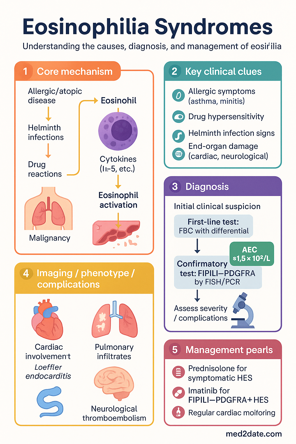
Causes & Classification
The 2012 Revised International Classification of eosinophilic disorders uses the terms hypereosinophilia (HE — blood or tissue criterion) and hypereosinophilic syndrome (HE + end-organ damage). Eosinophilia is stratified by aetiology into three major categories:
| Category | Subtype | Key Examples |
|---|---|---|
| Secondary (Reactive) | Allergic/Atopic | Asthma, allergic rhinitis, atopic dermatitis, drug hypersensitivity (penicillins, NSAIDs, carbamazepine) |
| Infectious | Helminths (Strongyloides, hookworm, Toxocara, Schistosoma), Strongyloides hyperinfection, visceral larva migrans | |
| Neoplastic | T-cell lymphoma (Sézary syndrome), Hodgkin lymphoma, solid tumours, mast cell neoplasia with eosinophilia | |
| Primary (Clonal) | Myeloproliferative | FIP1L1-PDGFRA+ myeloid neoplasm, PDGFRB rearrangement, FGFR1 rearrangement (8p11 syndrome) |
| Lymphoproliferative | Lymphocytic variant HES (clonal T-helper cells producing IL-5), chronic eosinophilic leukaemia NOS | |
| Idiopathic | — | Diagnosis of exclusion; all reactive and clonal causes excluded |
| Organ-restricted | — | Eosinophilic oesophagitis, eosinophilic granulomatosis with polyangiitis (EGPA/Churg-Strauss), eosinophilic fasciitis |
Stepwise Diagnostic Approach
A systematic approach to unexplained eosinophilia reduces the time to diagnosis and prevents inappropriate immunosuppression:
Hypereosinophilic Syndrome
Hypereosinophilic syndrome (HES) is diagnosed when all three criteria are met: (1) AEC ≥1.5 × 10⁹/L on ≥2 occasions or tissue HE with marked eosinophilic infiltration; (2) exclusion of secondary/reactive causes; and (3) signs and symptoms of end-organ damage attributable to eosinophilia. HES is further sub-classified by the presence or absence of identifiable clonal markers.
Myeloproliferative HES (M-HES)
The FIP1L1-PDGFRA fusion gene results from an interstitial deletion on chromosome 4q12 (detected by FISH or RT-PCR). It encodes a constitutively active tyrosine kinase driving eosinophil proliferation. Features include male predominance, splenomegaly, elevated serum B12 (>1500 pmol/L), elevated tryptase (>20 µg/L), anaemia, and thrombocytopenia. Cardiac involvement occurs in 50–60% and is the leading cause of morbidity and mortality.
Lymphocytic Variant HES (L-HES)
Clonal T-helper lymphocytes (often CD3⁻CD4⁺) produce IL-5 and other eosinophilopoietic cytokines. Distinguishing features: normal or mildly elevated B12/tryptase, absence of PDGFRA mutations, and elevated IL-5 or TARC/CCL17. There is a 10–15% risk of transformation to T-cell lymphoma over 10–15 years of follow-up. Management: corticosteroids first-line; mepolizumab (anti-IL-5 monoclonal antibody) or interferon-α as steroid-sparing agents.
Idiopathic HES
Diagnosed when all reactive, clonal, and lymphoproliferative causes have been excluded. Requires ongoing surveillance (6–12 monthly) for clonal evolution. Corticosteroids are first-line treatment. Hydroxyurea, interferon-α, and mepolizumab are second-line steroid-sparing options.
End-Organ Manifestations
End-organ damage in HES results from direct eosinophilic infiltration and release of cytotoxic granule proteins (major basic protein, eosinophil cationic protein, eosinophil peroxidase). Any organ can be involved, but cardiac, pulmonary, neurological, dermatological, and gastrointestinal manifestations are most clinically significant.
Cardiac Involvement — Loeffler Endocarditis
Cardiac disease is the most important prognostic determinant in HES. The pathophysiology progresses through three stages:
Pulmonary Involvement
Pulmonary eosinophilia may present as simple peripheral infiltrates (Löffler syndrome — often parasitic), chronic eosinophilic pneumonia (bilateral peripheral opacities, "photographic negative of pulmonary oedema"), or as part of EGPA. PFTs should include spirometry and DLCO. Bronchoalveolar lavage eosinophilia (>25% eosinophils) supports diagnosis when tissue sampling is not feasible.
Neurological Involvement
Peripheral neuropathy (mononeuritis multiplex or symmetric polyneuropathy) affects 5–15% of HES patients. Central nervous system involvement is usually thromboembolic (secondary to cardiac mural thrombi) rather than direct eosinophilic infiltration. Cerebral embolisation warrants cardiac MRI and consideration of long-term anticoagulation.
Dermatological Involvement
Cutaneous manifestations are often the presenting feature of HES. Eosinophilic cellulitis (Wells syndrome) presents with tender, oedematous plaques that may evolve through violaceous to granulomatous phases. Skin biopsy shows flame figures (collagen coated with degranulated eosinophil major basic protein). Treatment is with topical or systemic corticosteroids.
Investigations & Treatment
Investigations
Investigations should be tiered according to the severity and suspected aetiology. All patients with unexplained eosinophilia (AEC ≥1.0 × 10⁹/L) require a minimum baseline panel. Patients with AEC ≥1.5 × 10⁹/L or end-organ damage require the extended panel.
Treatment Algorithm
Treatment is determined by the underlying aetiology and presence of end-organ damage:
Pharmacological Agents
Monitoring Plan
| Parameter | Frequency | Action Threshold |
|---|---|---|
| FBC with differential | Weekly during active treatment; monthly when stable | AEC >1.5 × 10⁹/L → escalate therapy |
| Troponin | Every 3–6 months if prior cardiac involvement | Any rise → urgent echo + cardiology referral |
| Echocardiography | Baseline, 3 months, then annually | New wall motion abnormality, EF decline, or valvular disease |
| FIP1L1-PDGFRA transcript | Every 3–6 months on imatinib | Rising transcript → assess compliance, consider dose escalation |
| T-cell markers + IL-5 | Every 6–12 months for L-HES | Clonal expansion → re-evaluate for lymphoma transformation |
Special Populations
Aboriginal and Torres Strait Islander Health
Eosinophilia syndromes in Aboriginal and Torres Strait Islander populations are disproportionately driven by infectious aetiologies, particularly soil-transmitted helminths. Strongyloides stercoralis seroprevalence ranges from 15–60% in some remote NT and Queensland communities, compared to <1% in non-Indigenous metropolitan populations. Hookworm, Toxocara, and Schistosoma species (in communities with travel to endemic regions) should be considered.
📚 References
- 1. Valent P, Klion AD, Horny HP, et al. Contemporary consensus proposal on criteria and classification of eosinophilic disorders and related syndromes. Journal of Allergy and Clinical Immunology. 2012;130(3):607-612.e9.
- 2. Gotlib J. World Health Organization-defined eosinophilic disorders: 2022 update on diagnosis, risk stratification, and management. American Journal of Hematology. 2022;97(8):1089-1108.
- 3. Cools J, DeAngelo DJ, Gotlib J, et al. A tyrosine kinase created by fusion of the PDGFRA and FIP1L1 genes as a target of imatinib in idiopathic hypereosinophilic syndrome. New England Journal of Medicine. 2003;348(13):1201-1214.
- 4. Klion AD, Bochner BS, Gleich GJ, et al. Approaches to the treatment of hypereosinophilic syndromes: a workshop summary report. Journal of Allergy and Clinical Immunology. 2006;117(6):1292-1302.
- 5. Rothenberg ME, Klion AD, Roufosse FE, et al. Treatment of patients with the hypereosinophilic syndrome with mepolizumab. New England Journal of Medicine. 2008;358(12):1215-1228.
- 6. Helbig G, Hus M, Francuz T, et al. Characteristics and clinical outcome of patients with hypereosinophilic syndrome: a single-centre experience. Advances in Medical Sciences. 2013;58(2):285-291.
- 7. Currie BJ, McCarthy JS. Parasitic infections in tropical Australia. Medical Journal of Australia. 2010;192(8):432-437.
- 8. Shield JM, Vatui G, Betuela I. Strongyloides stercoralis infection in Australia: a neglected disease of Indigenous communities. Australian and New Zealand Journal of Public Health. 2005;29(1):71-76.
- 9. O'Connell EM, Nutman TB. Eosinophilia in infectious diseases. Immunology and Allergy Clinics of North America. 2015;35(3):493-522.
- 10. Bain BJ. Eosinophilia — idiopathic or secondary? Blood. 2021;137(22):3033-3034.
- 11. Roufosse F, Weller PF. Practical approach to the patient with hypereosinophilia. Journal of Allergy and Clinical Immunology. 2010;126(1):39-44.
- 12. RHDAustralia. Strongyloides stercoralis: Guidelines for the Diagnosis, Treatment and Prevention of Strongyloidiasis. Darwin: RHDAustralia; 2022.
- 13. Australian Institute of Health and Welfare (AIHW). Aboriginal and Torres Strait Islander Health Performance Framework 2020 Summary Report. Canberra: AIHW; 2020.
- 14. Schwartz LB, Metcalfe DD, Miller JS, et al. Tryptase levels as an indicator of mast-cell activation in systemic anaphylaxis and mastocytosis. New England Journal of Medicine. 1987;316(26):1622-1626.