Home Immunology Cellular Immunity

Cellular Immunity

📋 Key Information Summary

📋
  • Cellular immunity — T cell-mediated adaptive responses — is essential for defence against intracellular pathogens (mycobacteria, fungi, viruses), tumour surveillance, and transplant rejection.
  • CD4⁺ helper T cells coordinate immunity through cytokine release: Th1 (cellular), Th2 (humoral/allergic), Th17 (mucosal), Tfh (germinal centre), and Treg (suppression).
  • CD8⁺ cytotoxic T lymphocytes (CTLs) kill via perforin/granzyme exocytosis and Fas/FasL signalling — critical for viral clearance and anti-tumour immunity.
  • Memory T cells (Tcm, Tem, Trm) provide rapid recall responses; Trm cells reside in mucosal tissue and skin for frontline defence.
  • Regulatory T cells (Tregs, CD4⁺CD25⁺FOXP3⁺) maintain self-tolerance; deficiency causes IPEX syndrome and multi-organ autoimmunity.
  • Lymphocyte subset enumeration (CD3, CD4, CD8, CD19, CD16/56) by flow cytometry is MBS-listed (Item 69332) and essential for immunodeficiency workup.
  • Absolute CD4 count remains the principal marker for HIV staging and opportunistic infection risk; <200 cells/µL defines AIDS.
  • T cell functional assays (PHA/anti-CD3 lymphocyte proliferation, intracellular cytokine staining) are available at tertiary centres and reference labs.
  • Primary immunodeficiencies affecting T cells (DiGeorge, SCID, STAT3/STAT1 GOF, IPEX) require urgent immunology referral for haematopoietic stem cell transplant assessment.
  • Secondary T cell dysfunction is most commonly caused by HIV, immunosuppressive therapy (corticosteroids, calcineurin inhibitors, biologics), malnutrition, and haematological malignancy.
  • T cell-directed therapies include calcineurin inhibitors (cyclosporin, tacrolimus), anti-thymocyte globulin, anti-CD52 (alemtuzumab), and emerging checkpoint inhibitors (anti-PD-1/PD-L1, anti-CTLA-4).
  • Aboriginal and Torres Strait Islander peoples have higher rates of infections requiring robust cellular immunity (TB, invasive pneumococcal disease, rheumatic fever); culturally safe immunology referral pathways are essential.
  • Live vaccines (MMR, varicella, BCG, yellow fever) are contraindicated when CD4 <200 cells/µL or in severe T cell immunodeficiency — assess prior to vaccination.
  • Flow cytometry turnaround in most Australian public hospital labs is 3–5 business days; specialist lymphocyte function testing may take 2–4 weeks.

Introduction & Australian Epidemiology

Cellular immunity — the arm of adaptive immunity mediated by T lymphocytes — is central to host defence against intracellular pathogens, tumour surveillance, graft rejection, and regulation of the broader immune response. Unlike humoral (antibody-mediated) immunity, cellular immunity requires direct cell-to-cell contact and cytokine-mediated signalling to eliminate infected or malignant cells and to coordinate downstream effector functions.

T cells originate from haematopoietic precursors in the bone marrow, undergo positive and negative selection in the thymus, and emerge as naïve CD4⁺ or CD8⁺ single-positive cells bearing unique T cell receptors (TCRs). Upon antigen encounter in secondary lymphoid organs, naïve T cells differentiate into effector and memory subsets that mediate both immediate and long-term protection.

⚠️
Australian clinical relevance: T cell-mediated diseases account for a significant burden of morbidity in Australia. HIV prevalence is approximately 29,000 people (Kirby Institute, 2023), with ~900 new diagnoses per year. Tuberculosis notifications average 1,400 per year (NNDSS), disproportionately affecting migrants from high-burden countries and Aboriginal and Torres Strait Islander communities. Organ transplantation — approximately 1,500 procedures annually — depends entirely on understanding and managing T cell alloreactivity.

The Australian landscape for cellular immunology is shaped by several factors:

  • HIV programme: Australia's universal access to antiretroviral therapy (ART) through the PBS Pharmaceutical Benefits Scheme has achieved viral suppression in >95% of those on treatment, preserving CD4 counts and reducing opportunistic infections.
  • Transplant immunology: National transplant programmes rely on calcineurin inhibitor-based regimens; tertiary centres perform HLA typing and donor-specific antibody (DSA) monitoring with virtual crossmatching.
  • Primary immunodeficiency: The Australian National Immunodeficiency Registry estimates a prevalence of 1 in 1,200 for combined variable immunodeficiency (CVID) and related disorders; severe T cell deficiencies (SCID, DiGeorge) are identified through newborn screening programmes now operational in NSW, ACT, and expanding nationally.
  • Checkpoint immunotherapy: Immune checkpoint inhibitors (nivolumab, pembrolizumab, ipilimumab) are PBS-listed for melanoma, NSCLC, renal cell carcinoma, and other malignancies; immune-related adverse events (irAEs) from T cell over-activation are managed by multidisciplinary teams.
  • ATSI health: Aboriginal and Torres Strait Islander peoples experience higher rates of conditions requiring intact cellular immunity, including TB, rheumatic heart disease, and invasive Group A Streptococcus disease.

Core Concepts in T Cell Biology

The T cell compartment is defined by surface markers and functional capacity:

T Cell Subset Surface Markers Primary Function Key Cytokines / Molecules
CD4⁺ Naïve CD3⁺CD4⁺CD45RA⁺CCR7⁺ Precursor to helper subsets IL-7 (survival), TCR engagement required for activation
Th1 CD3⁺CD4⁺IFN-γ⁺ Intracellular pathogen defence, macrophage activation IFN-γ, TNF-α, IL-2; T-bet transcription factor
Th2 CD3⁺CD4⁺IL-4⁺ Helminth defence, B cell class switching to IgE IL-4, IL-5, IL-13; GATA3 transcription factor
Th17 CD3⁺CD4⁺IL-17A⁺ Mucosal defence, neutrophil recruitment IL-17A, IL-17F, IL-22; RORγt transcription factor
Tfh CD3⁺CD4⁺CXCR5⁺PD-1⁺ Germinal centre B cell help, antibody affinity maturation IL-21, ICOS, CD40L; Bcl-6 transcription factor
Treg CD3⁺CD4⁺CD25⁺FOXP3⁺ Immune suppression, tolerance IL-10, TGF-β, CTLA-4, IL-35
CD8⁺ CTL CD3⁺CD8⁺ Killing of virus-infected and tumour cells Perforin, granzymes, IFN-γ, FasL
MAIT CD3⁺CD161⁺Vα7.2⁺ Mucosal innate-like defence against bacteria IFN-γ, IL-17; MR1-restricted
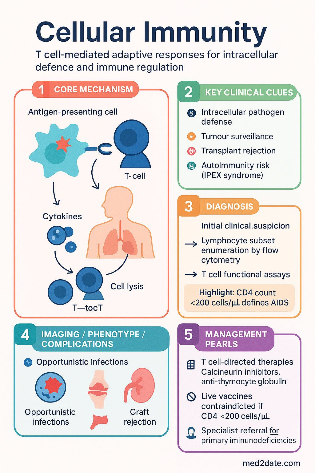
Cellular Immunity clinical infographic — pathophysiology, clinical clues, diagnosis, imaging, and management
Tap or click image to enlarge — Cellular Immunity: pathophysiology, clinical clues, diagnosis, imaging, and management.
Cellular Immunity infographic, full size

T Cell Subsets

Thymic Development & Selection

T cell precursors migrate from the bone marrow to the thymus, where they undergo a tightly regulated maturation programme:

1
DN (Double Negative) Stage
Thymocytes are CD4⁻CD8⁻. TCR β-chain rearrangement occurs (V(DJ recombination). Successful β-chain expression with pre-Tα forms the pre-TCR, triggering β-selection and proliferation.
2
DP (Double Positive) Stage
Thymocytes become CD4⁺CD8⁺. TCR α-chain rearrangement generates diverse TCRαβ combinations. Cells now express both co-receptors and low-level TCR.
3
Positive Selection (Cortex)
DP thymocytes must recognise self-MHC on cortical thymic epithelial cells (cTECs) with low-to-moderate affinity to survive. Cells that fail die by neglect (~90% of DP cells).
4
Negative Selection (Medulla)
Thymocytes with high-affinity TCR for self-peptide/MHC complexes presented by medullary TECs (mTECs) and dendritic cells undergo apoptosis (clonal deletion). AIRE gene expression in mTECs drives tissue-restricted antigen presentation, central to self-tolerance.
5
SP (Single Positive) Maturation
Surviving cells commit to either CD4⁺ (MHC-II restricted) or CD8⁺ (MHC-I restricted) lineage via ThPOK or Runx3 transcription factors, respectively. Mature naïve T cells exit the thymus.

Effector T Cell Subsets in Peripheral Blood

Once in the periphery, naïve T cells encounter antigen on antigen-presenting cells (APCs) in secondary lymphoid organs and differentiate into effector subsets. The prevailing cytokine milieu during activation determines subset commitment:

Subset Activating Cytokines Key Transcription Factor Effector Cytokines Target Pathogens / Roles Dysregulation
Th1 IL-12, IFN-γ T-bet IFN-γ, TNF-α, IL-2 Mycobacteria, viruses, intracellular bacteria Autoimmunity (MS, T1DM, RA)
Th2 IL-4 GATA3 IL-4, IL-5, IL-13 Helminths Asthma, atopic dermatitis, allergic rhinitis
Th17 IL-6 + TGF-β (mice); IL-1β, IL-6, IL-23 (humans) RORγt IL-17A, IL-17F, IL-22 Extracellular bacteria (Klebsiella, Staph), fungi (Candida) Psoriasis, IBD, ankylosing spondylitis
Tfh IL-6, IL-21, ICOS Bcl-6 IL-21, IL-4 Germinal centre reactions, high-affinity antibody SLE, angioimmunoblastic T cell lymphoma
Treg TGF-β, IL-2 FOXP3 IL-10, TGF-β, IL-35 Immune suppression, self-tolerance IPEX syndrome, autoimmunity
🔬
Australian laboratory note: T helper subset profiling by intracellular cytokine staining (ICS) after PMA/ionomycin stimulation is available at reference immunology laboratories including the Royal Melbourne Hospital, Westmead Hospital, and SA Pathology. Results should be interpreted with knowledge that ICS is a functional readout and may not reflect in vivo subset proportions. Cost: approximately 0–450 (not MBS-listed; request via specialist referral).

Unconventional T Cells

Beyond classical αβ T cells, several unconventional T cell populations contribute to immune defence:

  • γδ T cells: Constitute 1–5% of peripheral blood T cells; enriched in mucosal tissues. Recognise phosphoantigens and stress-induced ligands (MICA/MICB) without classical MHC restriction. Important in defence against mycobacteria and epithelial tumours.
  • MAIT cells: Mucosal-associated invariant T cells recognise microbial vitamin B metabolites presented by MR1. Abundant in human blood (1–10% of T cells) and liver. Rapidly produce IFN-γ and IL-17 upon bacterial encounter. Depleted in HIV infection despite ART.
  • NKT cells: Recognise glycolipid antigens presented by CD1d. Invariant NKT (iNKT) cells produce large cytokine bursts early in infection. Deficiency associated with increased susceptibility to certain infections.
  • Double-negative (DN) T cells: CD3⁺CD4⁻CD8⁻ T cells that can suppress autoreactive T cells and are expanded in autoimmune lymphoproliferative syndrome (ALPS).

CD4⁺ Helper T Cell Functions

CD4⁺ T helper cells are the conductors of adaptive immunity. Their functions extend far beyond simple cytokine production — they direct the type, magnitude, and location of immune responses. Understanding CD4⁺ T cell biology is essential for managing HIV infection, autoimmune diseases, transplant rejection, and vaccine design.

Mechanisms of Help

Cognate Help to B Cells

Tfh cells in germinal centres provide signals to B cells via CD40L–CD40 interaction and IL-21 secretion, driving class-switch recombination, somatic hypermutation, and affinity maturation. Without Tfh help, germinal centre reactions fail, and long-lived plasma cells and memory B cells do not form.

Clinical correlate: Patients with Hyper-IgM syndrome (CD40L deficiency) have absent class switching and are susceptible to Pneumocystis jirovecii and Cryptosporidium.

Macrophage Activation (Th1)

IFN-γ produced by Th1 cells activates macrophages, enhancing phagolysosome fusion, reactive oxygen/nitrogen species production, and MHC-II expression. This classical activation is critical for killing intracellular pathogens such as Mycobacterium tuberculosis and Leishmania.

Clinical correlate: Patients on anti-TNF therapy (infliximab, adalimumab) have impaired granuloma maintenance and risk of TB reactivation.

CD4⁺ T Cell Roles in Specific Diseases

Protective
Anti-pathogen Defence
Th1 cells activate macrophages against TB and Leishmania. Th17 cells recruit neutrophils against Candida and Staphylococcus. Tfh cells drive protective antibody responses in vaccination.
Setting: Infectious disease, vaccination programmes
Pathological
Autoimmune Disease
Th1/Th17 skewing drives organ-specific autoimmunity (T1DM, MS, RA). Loss of Treg function or Treg/Th17 plasticity exacerbates disease. Molecular mimicry can activate autoreactive clones.
Setting: Rheumatology, neurology, endocrinology
Critical
HIV/AIDS — CD4 Depletion
HIV preferentially infects activated CD4⁺ T cells via CCR5/CXCR4 co-receptors. Progressive CD4 depletion (<200 cells/µL) leads to AIDS-defining illnesses: PCP, toxoplasmosis, CMV retinitis, disseminated MAC.
Setting: HIV medicine, infectious disease

CD4 Count Interpretation in Australian Practice

CD4 Count (cells/µL) Risk Level Clinical Implications Prophylaxis Required
500–1,500 (normal) Normal Adequate immune function Standard vaccination schedule
350–500 Mild reduction ART initiation recommended (all PLHIV); increased infection risk minimal Nil additional
200–350 Moderate reduction Increased risk of TB, oral candidiasis, herpes zoster, bacterial pneumonia TB screening (IGRA/TST); consider Bactrim if CD4 approaching 200
100–200 Severe (AIDS if <200) PCP, oesophageal candidiasis, disseminated HSV Co-trimoxazole 480 mg daily (PCP prophylaxis)
50–100 Very severe Toxoplasma encephalitis, Cryptococcal meningitis, disseminated MAC Co-trimoxazole + azithromycin 1,200 mg weekly (MAC prophylaxis if CD4 <50)
<50 Critical CMV retinitis, disseminated MAC, CNS lymphoma (EBV-driven) Full opportunistic infection prophylaxis; ophthalmology review
🚨
Safety alert — live vaccines: Live vaccines (MMR, varicella/zoster, BCG, yellow fever, oral polio, rotavirus) are contraindicated in patients with CD4 counts <200 cells/µL or known severe T cell immunodeficiency. In Australia, zoster vaccination (Shingrix®, recombinant, non-live) is preferred over Zostavax® (live) in immunocompromised patients aged ≥50 years and is PBS-listed for this indication.

Therapeutic Modulation of CD4⁺ T Cells

Multiple drug classes target CD4⁺ T cell function in transplantation, autoimmunity, and lymphoma:

💊
Cyclosporin
Sandimmun®, Neoral® · Calcineurin inhibitor
Mechanism Inhibits calcineurin → blocks NFAT nuclear translocation → suppresses IL-2 transcription in CD4⁺ T cells
Adult dose 2–5 mg/kg/day PO in 2 divided doses; titrate to trough 150–300 ng/mL (transplant)
Renal adjustment Nephrotoxic — monitor serum creatinine; reduce dose if eGFR <30
PBS status ✔ PBS General Benefit
💊
Tacrolimus
Prograf®, Advagraf® · Calcineurin inhibitor
Mechanism Binds FKBP12 → inhibits calcineurin → suppresses IL-2; 50–100× more potent than cyclosporin in vitro
Adult dose 0.05–0.1 mg/kg/day PO in 2 divided doses; trough 5–15 ng/mL (transplant)
Renal adjustment Nephrotoxic; monitor renal function closely; no specific dose table for CKD
PBS status ✔ PBS General Benefit
💊
Anti-thymocyte globulin (rabbit)
Thymoglobulin® · Polyclonal antibody
Mechanism Polyclonal antibodies against multiple T cell surface molecules → complement-dependent lysis and opsonisation of T cells
Dose 1.5 mg/kg/day IV for 3–7 days (induction); 1.5 mg/kg on days 0, 3, 7 (treatment of rejection)
PBS status ⚠ PBS Authority Required

CD8⁺ Cytotoxic Mechanisms

CD8⁺ cytotoxic T lymphocytes (CTLs) are the primary effectors for eliminating virus-infected cells, intracellular bacteria-harbouring cells, and tumour cells. CTL killing is exquisitely specific — each CTL recognises its cognate peptide presented on MHC class I molecules, which are expressed on virtually all nucleated cells.

Killing Mechanisms

CTLs employ two major cytotoxic pathways:

1. Perforin–Granzyme Pathway (Primary)
  • Upon TCR engagement, the immunological synapse forms and lytic granules polarise toward the target cell.
  • Perforin polymerises in the target cell membrane, forming pores (~16 nm diameter).
  • Granzyme B enters through perforin pores and directly cleaves caspase-3, -7, and -10, triggering apoptosis.
  • Granzyme B also cleaves Bid → tBid → mitochondrial outer membrane permeabilisation → cytochrome c release → caspase-9 activation (intrinsic apoptotic pathway).
  • Families with biallelic PRF1 mutations (perforin deficiency) develop familial haemophagocytic lymphohistiocytosis (FHL type 2) — a life-threatening hyperinflammatory syndrome.
2. Fas/FasL (CD95/CD95L) Pathway
  • Fas ligand (CD95L, TNFSF6) on CTLs engages Fas (CD95, TNFRSF6) on target cells.
  • Fas trimerisation recruits FADD and procaspase-8 to form the death-inducing signalling complex (DISC).
  • Caspase-8 activation → cleavage of caspase-3 → apoptosis.
  • Important for immune contraction (activation-induced cell death, AICD) and liver immunopathology.
  • Mutations in FAS or FASL cause autoimmune lymphoproliferative syndrome (ALPS) — characterised by lymphadenopathy, splenomegaly, and accumulation of double-negative T cells.

Non-Cytolytic Functions of CD8⁺ T Cells

Beyond direct killing, CTLs contribute to immunity through:

  • IFN-γ production: Enhances MHC-I expression on neighbouring cells, upregulates antigen processing machinery, and activates macrophages.
  • TNF-α secretion: Promotes inflammation, endothelial activation, and has direct anti-viral and anti-tumour effects.
  • Cytotoxic degranulation without killing: Partial degranulation can deliver granzymes that induce non-apoptotic effects (pyroptosis via gasdermin E cleavage in some contexts).
  • Immunosurveillance: Tissue-resident memory CD8⁺ T cells (Trm) positioned in epithelia provide rapid responses to re-infection at barrier sites.

CD8⁺ T Cells in Key Clinical Scenarios

Clinical Scenario CD8⁺ Role Australian Relevance
HIV infection HIV-specific CTLs suppress viraemia (elite controllers); escape mutations in immunodominant epitopes drive viral evolution ~29,000 PLHIV; CD8 responses correlate with set-point viral load
SARS-CoV-2 Robust CD8⁺ responses correlate with mild disease; impaired responses in severe COVID-19 T cell immunity persists after antibody waning; relevant to booster strategy
Solid organ transplant Donor-specific CD8⁺ T cells mediate acute cellular rejection (ACR); controlled by calcineurin inhibitors and anti-metabolites ~1,500 transplants/year; protocol biopsies monitor for ACR
Melanoma Tumour-infiltrating lymphocytes (TILs) with CD8⁺ predominance predict response to checkpoint inhibitors Australia has highest melanoma incidence globally; checkpoint immunotherapy PBS-listed
CMV reactivation CMV-specific CD8⁺ T cells control latent CMV; depletion in transplant/HIV leads to reactivation CMV PCR monitoring post-transplant standard in Australian centres
Hepatitis B HBV-specific CTL responses determine clearance vs. chronicity; dysfunctional/exhausted in chronic HBV ~220,000 people living with chronic HBV in Australia; higher prevalence in ATSI and CALD communities
⚠️
Haemophagocytic lymphohistiocytosis (HLH): When CD8⁺ T cell activation is uncontrolled (e.g., EBV-driven, perforin deficiency, or during checkpoint inhibitor therapy), massive cytokine release and impaired CTL killing lead to HLH — a medical emergency. Diagnostic criteria (HLH-2004) include fever, splenomegaly, cytopenias, hypertriglyceridaemia, hyperferritinaemia (often >10,000 µg/L), haemophagocytosis on biopsy, low/absent NK cell activity, and elevated soluble CD25 (sIL-2R). First-line treatment: dexamethasone + etoposide (per HLH-94 protocol). Seek haematology/immunology consultation urgently.

Checkpoint Inhibitors & CD8⁺ T Cell Reinvigoration

Immune checkpoint inhibitors (ICIs) reinvigorate exhausted CD8⁺ T cells in the tumour microenvironment:

💊
Nivolumab
Opdivo® · Anti-PD-1 monoclonal antibody
Mechanism Blocks PD-1/PD-L1 interaction → prevents CD8⁺ T cell exhaustion → reinvigorates anti-tumour cytotoxicity
Dose 240 mg IV q2 weeks or 480 mg IV q4 weeks
PBS status ⚠ PBS Authority Required (melanoma, NSCLC, RCC)
💊
Pembrolizumab
Keytruda® · Anti-PD-1 monoclonal antibody
Mechanism Blocks PD-1; similar to nivolumab with distinct binding epitope
Dose 200 mg IV q3 weeks or 400 mg IV q6 weeks
PBS status ⚠ PBS Authority Required (melanoma, NSCLC, head & neck, urothelial)
💊
Ipilimumab
Yervoy® · Anti-CTLA-4 monoclonal antibody
Mechanism Blocks CTLA-4 → enhances CD28 co-stimulation → expands T cell repertoire and effector function
Dose 3 mg/kg IV q3 weeks × 4 cycles (monotherapy); 1 mg/kg q6 weeks (combination with nivolumab)
PBS status ⚠ PBS Authority Required (melanoma)

Immune-related adverse events (irAEs) from checkpoint inhibitors reflect unrestrained T cell activation against self-antigens. Common irAEs include colitis, hepatitis, pneumonitis, thyroiditis, hypophysitis, and dermatitis. Severe irAEs (Grade 3–4) require high-dose corticosteroids (methylprednisolone 1–2 mg/kg IV) and may necessitate infliximab (colitis) or mycophenolate (hepatitis).

Memory & Regulation

Memory T Cell Subsets

Immunological memory is the foundation of vaccination and long-term pathogen protection. Memory T cells persist for decades and provide faster, stronger, and qualitatively superior responses upon antigen re-encounter.

Memory Subset Surface Markers Location Function Lifespan
Tcm (Central memory) CD45RO⁺CCR7⁺CD62L⁺ Secondary lymphoid organs (lymph nodes, spleen) High proliferative capacity upon re-stimulation; self-renewing via IL-7/IL-15 Decades
Tem (Effector memory) CD45RO⁺CCR7⁻CD62L⁻ Peripheral tissues, blood Rapid effector function (cytokine production, cytotoxicity) upon re-encounter; less proliferative than Tcm Years to decades
Trm (Tissue-resident memory) CD69⁺CD103⁺ (variable) Non-lymphoid tissues (skin, lung, gut, reproductive tract) First-line barrier defence; do not recirculate; produce IFN-γ and TNF-α rapidly; recruit circulating immune cells Years (self-maintained locally)
Tscm (Stem cell memory) CD45RA⁺CCR7⁺CD62L⁺CD95⁺CD122⁺ Blood, lymphoid organs Stem-like self-renewal; can differentiate into Tcm, Tem, and effector cells; rare population (~2–3% of CD4⁺) Lifetime
💉
Vaccination implications: Trm cells generated by mucosal or intradermal vaccination may provide superior protection at the site of pathogen entry compared to intramuscular vaccination (which primarily generates circulating Tem). This principle is being explored in next-generation COVID-19 and influenza vaccine platforms. In Australia, the National Immunisation Programme prioritises vaccines that generate robust T cell memory, particularly for pertussis (acellular vaccines generate less Trm than whole-cell, contributing to waning protection).

Maintenance of Memory T Cells

Memory T cell persistence depends on:

  • Homeostatic cytokines: IL-7 (produced by stromal cells) and IL-15 (produced by DCs and macrophages) drive slow homeostatic proliferation of memory T cells, maintaining the pool without antigen stimulation.
  • Metabolic reprogramming: Memory T cells shift from glycolysis (effector cells) to fatty acid oxidation and oxidative phosphorylation, enabling long-term survival with minimal nutrient requirements.
  • Epigenetic imprinting: Memory T cells carry permissive chromatin marks at effector gene loci, allowing rapid transcription upon restimulation (poised chromatin state).
  • Antigen-independent signals: MHC-II interactions (for CD4⁺ memory) and tonic TCR signalling contribute to memory maintenance.

Regulatory T Cells (Tregs)

Tregs are indispensable for maintaining immune homeostasis and preventing autoimmunity. They constitute 5–10% of circulating CD4⁺ T cells in healthy adults.

Thymic Tregs (tTregs)

Develop in the thymus during negative selection. High-affinity TCR engagement with self-antigen/MHC on mTECs drives FOXP3 expression and commitment to the Treg lineage. These cells are critical for dominant tolerance — actively suppressing autoreactive T cells that escape deletion.

Marker profile: CD4⁺CD25^(hi)CD127^(lo)FOXP3⁺Helios⁺Neuropilin-1⁺

Peripheral Tregs (pTregs)

Induced in the periphery from naïve CD4⁺ T cells by TGF-β and retinoic acid, particularly in mucosal tissues (gut). Essential for tolerance to commensal flora, food antigens, and fetal antigens during pregnancy.

Marker profile: CD4⁺CD25^(hi)CD127^(lo)FOXP3⁺Helios⁻Neuropilin-1⁻

Mechanisms of Treg Suppression

  • IL-2 consumption: Tregs express high-affinity IL-2Rα (CD25) but do not produce IL-2, acting as an "IL-2 sink" that deprives effector T cells of this essential growth factor.
  • CTLA-4-mediated suppression: Constitutive CTLA-4 expression on Tregs strips CD80/CD86 from APCs (trans-endocytosis), reducing co-stimulation for effector T cells.
  • Immunosuppressive cytokines: Secretion of IL-10, TGF-β, and IL-35 inhibits APC function and effector T cell proliferation.
  • Granzyme/perforin: Some Tregs can directly kill effector T cells and APCs via cytolytic mechanisms.
  • Metabolic disruption: CD39/CD73 ectoenzymes on Tregs convert ATP to adenosine, which suppresses effector T cell function via A2A receptor signalling.
  • Dendritic cell modulation: Tregs induce tolerogenic DCs via LAG-3–MHC-II interaction, promoting anergy in subsequently activated T cells.

Treg Dysfunction in Disease

🧬
IPEX Syndrome
Immune dysregulation, Polyendocrinopathy, Enteropathy, X-linked
Genetics X-linked; FOXP3 loss-of-function mutations
Presentation Early-onset (neonatal/infantile): severe diarrhoea (enteropathy), type 1 diabetes, eczema, cytopenias, thyroiditis
Treatment Haematopoietic stem cell transplant (HSCT) — curative; immunosuppression as bridge
Prognosis Without HSCT, most die in infancy/childhood from infections or organ failure
🩸
Treg Deficiency in Cancer
Tumour-associated Tregs
Mechanism Tumours recruit and expand Tregs (via CCL22, TGF-β, adenosine) to suppress anti-tumour CD8⁺ CTL responses
Therapeutic target Anti-CTLA-4 (ipilimumab) depletes intratumoural Tregs via ADCC; low-dose cyclophosphamide preferentially depletes Tregs

Clinical Assessment of Memory & Regulatory T Cells

In Australian clinical practice, assessment of T cell memory and regulatory compartments is performed by:

  • Flow cytometry (MBS Item 69332): Lymphocyte subset panel includes CD45RA/RO and CCR7 to delineate naïve, Tcm, and Tem populations. CD25/CD127 ratio approximates Treg frequency (FOXP3 intracellular staining confirmatory).
  • TREC (T cell receptor excision circle) assay: Measures thymic output; low TRECs indicate reduced naïve T cell production (DiGeorge, post-chemotherapy, ageing). Available at select reference labs.
  • TCR repertoire analysis: Next-generation sequencing of TCRβ CDR3 regions assesses clonality and diversity. Expanded clones may indicate chronic infection, malignancy, or alloreactivity. Available through specialised genomics services.
  • Vaccine response testing: T cell responses to recall antigens (tetanus, CMV, EBV) by ELISpot (IFN-γ) confirm functional immunological memory. Available at Westmead, RMH, and SA Pathology.

Pathophysiology

Disorders of cellular immunity arise from defects at multiple levels of T cell biology — from thymic development through effector function and regulation. Understanding the pathophysiology is essential for targeted diagnosis and management.

Classification of T Cell Immunodeficiencies

Category Examples Pathophysiological Basis Inheritance
Combined immunodeficiencies Severe combined immunodeficiency (SCID): X-linked (IL2RG), ADA deficiency, Artemis, RAG1/2 Absent or profoundly reduced T cells ± B/NK cells; failed V(D)J recombination or cytokine signalling X-linked, AR
Thymic defects DiGeorge syndrome (22q11.2 deletion) Thymic hypoplasia/aplasia → reduced T cell production; severity proportional to thymic tissue present Sporadic (de novo) or AD
Predominantly T cell defects CD3 deficiency, ZAP-70 deficiency, MHC-II deficiency (Bare lymphocyte syndrome) Defective TCR signalling or antigen presentation AR
Immune dysregulation IPEX (FOXP3), ALPS (FAS/FASL/CASP10), CTLA-4 haploinsufficiency Loss of peripheral tolerance; uncontrolled T cell proliferation or impaired apoptosis X-linked (IPEX), AD/AR (ALPS)
Syndromic immunodeficiencies Ataxia-telangiectasia (ATM), Wiskott-Aldrich (WASP), CHARGE syndrome DNA repair defects, cytoskeletal dysfunction, or developmental genes affecting multiple lineages AR (AT, WAS), AD (CHARGE)
Secondary (acquired) HIV/AIDS, iatrogenic (corticosteroids, calcineurin inhibitors, anti-CD20, biologics), malnutrition, malignancy Direct T cell destruction, impaired production, or functional suppression N/A

T Cell Exhaustion

Chronic antigen stimulation (persistent viral infection, tumour) drives T cell exhaustion — a state of progressive loss of effector function characterised by:

  • Upregulation of inhibitory receptors: PD-1, LAG-3, TIM-3, TIGIT, CTLA-4
  • Hierarchical loss of cytokine production: IL-2 first, then TNF-α, then IFN-γ (functional avidity decreases)
  • Reduced proliferative capacity and cytotoxicity
  • Distinct epigenetic landscape (TOX transcription factor drives exhaustion programme)
  • Presence of progenitor exhausted cells (TCF1⁺PD-1⁺) that can be reinvigorated — the target of checkpoint immunotherapy

Investigations

Assessment of cellular immunity involves quantification of T cell populations, assessment of T cell function, and evaluation of underlying aetiology. Investigation selection depends on clinical context.

Tier 1 — Initial Assessment (Primary Care / General Pathology)

Available
Full blood count with differential
Lymphocyte count — absolute lymphopenia (<1.0 × 10⁹/L in adults) triggers further immunological workup. MBS Item 65070.
Available
Serum immunoglobulins (IgG, IgA, IgM, IgE)
Low IgG/IgA may suggest T cell defect (impaired T cell help for class switching). MBS Item 65090.
Available
HIV serology (4th-generation Ag/Ab combo)
First-line for unexplained T cell lymphopenia in adults. MBS Item 69345.
Available
Liver and renal function
Baseline for secondary causes (hepatic/renal disease, drug toxicity).

Tier 2 — Immunology Specialist Assessment

Available
Lymphocyte subset enumeration by flow cytometry
CD3⁺ (total T cells), CD4⁺, CD8⁺, CD19⁺ (B cells), CD16/56⁺ (NK cells), CD4:CD8 ratio. MBS Item 69332. Turnaround 3–5 days at most public hospital labs.
Available
Naïve/memory T cell subset analysis
CD45RA/CCR7 for Tnaïve, Tcm, Tem, Temra. CD31 for recent thymic emigrants (RTE). Available at tertiary centres.
Available
Treg enumeration
CD4⁺CD25^(hi)CD127^(lo)FOXP3⁺ by flow cytometry. Important for IPEX screening, ALPS workup, post-transplant monitoring.
Referral
T cell proliferation assay (lyocyte transformation test)
PHA, anti-CD3, tetanus, candida stimulation with ³H-thymidine incorporation or CFSE dilution. Assesses T cell functional capacity. Tertiary centres only. Turnaround 2–4 weeks.
Referral
Intracellular cytokine staining (ICS)
PMA/ionomycin stimulation → IFN-γ, IL-4, IL-17, IL-21 detection in CD4⁺ and CD8⁺ subsets. Research/reference labs (RMH, Westmead, SA Pathology). Not MBS-listed.
Specialist
TCR excision circles (TRECs)
Quantitative PCR for sjTRECs — measures thymic output. Used in newborn SCID screening (now active in NSW, ACT) and post-HSCT engraftment monitoring.
Specialist
Genetic testing (primary immunodeficiency panels)
Targeted gene panels or whole-exome sequencing for SCID (IL2RG, JAK3, RAG1/2, ADA, Artemis), IPEX (FOXP3), ALPS (FAS, FASL, CASP10), STAT3 GOF/LOF. Available through clinical genetics services; turnaround 4–12 weeks.
Essential
IGRA (QuantiFERON-TB Gold Plus) or TST
Tuberculosis screening when CD4 <350 cells/µL or commencing anti-TNF therapy. MBS Item 69378 (IGRA).

Tier 3 — Advanced / Research-Level

Specialist
TCR repertoire sequencing (NGS)
TCRβ CDR3 deep sequencing for clonality assessment, post-transplant chimerism, and tumour-infiltrating lymphocyte analysis. Research and specialised clinical labs.
Specialist
ELISpot (IFN-γ)
Antigen-specific T cell responses (CMV, EBV, adenovirus, SARS-CoV-2). Used in transplant virology and vaccine immunogenicity studies. Available at select centres.
Specialist
Mass cytometry (CyTOF) / Spectral flow cytometry
40+ parameter phenotyping of T cell subsets simultaneously. Research setting at major university centres.

Risk Stratification & Severity Scoring

Severity and risk stratification of T cell disorders guides urgency of referral, treatment intensity, and monitoring frequency.

Mild
Isolated Lymphopenia
Absolute lymphocyte count 0.5–1.5 × 10⁹/L with normal subset distribution. Asymptomatic. May be drug-related or incidental finding.
Setting: GP follow-up; repeat in 4–6 weeks; consider drug review
Moderate
T Cell Dysfunction / Partial Deficiency
Recurrent sinopulmonary infections, oral candidiasis, low-normal CD4 (200–500 cells/µL). May include DiGeorge (partial), CVID with T cell involvement, drug-related immunosuppression.
Setting: Immunology outpatient referral; prophylactic antimicrobials; vaccination review
Severe
Severe T Cell Immunodeficiency
SCID, IPEX, severe DiGeorge (complete athymia), AIDS with CD4 <50. Life-threatening infections (PCP, CMV, disseminated BCG). Failure to thrive in infants.
Setting: Tertiary/quaternary centre; HSCT assessment; protective isolation; urgent ART (HIV)

HIV-Specific Risk Stratification

CDC Stage CD4 Count Clinical Features Management Priority
Stage 1 ≥500 cells/µL Asymptomatic or persistent generalised lymphadenopathy Commence ART regardless of CD4 (Treat All); STI screening; hepatitis B/C co-testing
Stage 2 200–499 cells/µL Weight loss <10%, oral ulcers, herpes zoster, seborrhoeic dermatitis ART urgent; TB screening (IGRA); vaccination catch-up (inactivated); PCP prophylaxis if CD4 <200
Stage 3 (AIDS) <200 cells/µL PCP, toxoplasmosis, CMV, cryptococcosis, MAC, Kaposi sarcoma, NHL ART (watch for IRIS); OI prophylaxis; ART initiation timing: start within 2 weeks (except cryptococcal meningitis: delay 4–6 weeks)
🚨
SCID — medical emergency in neonates: Severe combined immunodeficiency presents in the first 3–6 months of life with failure to thrive, chronic diarrhoea, recurrent severe infections (PCP, CMV, persistent candidiasis), and absent thymic shadow on chest X-ray. Without HSCT, death usually occurs by age 1–2 years. Australian newborn screening (heel-prick TREC assay) is operational in NSW and ACT, with national expansion underway. Any infant with lymphocyte count <3.0 × 10⁹/L at birth should be urgently referred to a paediatric immunologist.

Directed / Pathogen- or Mechanism-Specific Therapy

T Cell-Directed Therapies by Indication

Indication Agent T Cell Target Australian Access
Transplant rejection prophylaxis Mycophenolate mofetil + tacrolimus ± prednisolone Calcineurin inhibition (IL-2 suppression) + purine synthesis inhibition (T & B cell proliferation) PBS General Benefit
Acute cellular rejection Methylprednisolone pulse 500 mg–1 g IV × 3 days; anti-thymocyte globulin for steroid-resistant Broad T cell depletion (ATG); cytokine suppression (steroids) Hospital PBS; ATG Authority Required
Autoimmune disease (RA, SLE) Abatacept (CTLA-4-Ig) Blocks CD80/86–CD28 co-stimulation → T cell anergy PBS Authority Required (RA)
Melanoma, NSCLC, RCC Nivolumab, pembrolizumab, ipilimumab PD-1/PD-L1 or CTLA-4 blockade → T cell reinvigoration PBS Authority Required
CLL, T cell lymphoma Alemtuzumab (anti-CD52) Depletes T cells, B cells, NK cells via ADCC/CDC Hospital supply; limited PBS
GvHD prophylaxis Ciclosporin + methotrexate; post-transplant cyclophosphamide Donor T cell suppression and depletion PBS General Benefit
SCID (definitive) Haematopoietic stem cell transplant (HSCT) Reconstitutes T (and B/NK) cell compartment from donor stem cells Funded through state transplant programmes (Sydney Children's, RCH Melbourne)
IPEX syndrome HSCT (curative); sirolimus (bridge) Replaces FOXP3-deficient Tregs with donor-derived FOXP3⁺ cells HSCT: state-funded; sirolimus: PBS Authority Required

Emerging T Cell Therapies

  • Chimeric antigen receptor T cells (CAR-T): CD19-directed CAR-T (tisagenlecleucel, axicabtagene ciloleucel) is TGA-approved and available through the Life Saving Drugs Programme (LSDP) for relapsed/refractory B-ALL and DLBCL. T cells are genetically engineered to express a synthetic receptor targeting CD19. Australian manufacturing at specialised cell therapy facilities (Peter MacCallum, Westmead).
  • Bispecific T cell engagers (BiTEs): Blinatumomab (CD3×CD19) redirects T cells to kill CD19⁺ tumour cells. Available via LSDP.
  • Treg cell therapy: Clinical trials of ex vivo expanded polyclonal or antigen-specific Tregs for transplant tolerance and autoimmunity are underway internationally; early-phase trials in Australian centres.
  • Next-generation checkpoint inhibitors: Anti-LAG-3 (relatlimab — FDA-approved with nivolumab for melanoma), anti-TIGIT, anti-TIM-3 agents in Phase II/III trials with Australian recruitment sites.

Monitoring

Monitoring Strategies by Condition

HIV — Ongoing

CD4 count every 3–6 months until stable >500 for >2 years on ART, then annually. Viral load every 6 months (or 3-monthly if non-adherence concern). Resistance genotyping at treatment failure.

Post-transplant — Weekly to Monthly

Trough drug levels (tacrolimus 5–15 ng/mL, ciclosporin 150–300 ng/mL) twice weekly initially, then weekly, then monthly. Lymphocyte subsets monthly for first 6 months. Donor-specific antibodies (DSA) at 1, 3, 6, 12 months. Protocol biopsies per unit protocol.

Checkpoint inhibitor therapy — Each cycle

TFTs (TSH, fT4) every cycle for first 6 months. LFTs each cycle. Cortisol if symptomatic (fatigue, hyponatraemia). CT chest if new cough/dyspnoea (pneumonitis). Faecal calprotectin if diarrhoea (colitis).

Primary immunodeficiency — 3–6 monthly

Lymphocyte subsets (CD3, CD4, CD8, CD19, CD16/56) every 6 months. Immunoglobulin levels if on IgRT. Lung function (FEV₁/FVC) annually. Growth and development in children. Infection log review.

When to Refer to Specialist Immunology

  • Absolute lymphocyte count <1.0 × 10⁹/L on two occasions in adults
  • Absolute lymphocyte count <3.0 × 10⁹/L in children under 2 years
  • Recurrent or severe infections (≥2 systemic infections, ≥3 respiratory infections/year) with suspected immunodeficiency
  • Opportunistic infections (PCP, disseminated candidiasis, atypical mycobacteria) without HIV or known immunosuppression
  • Family history of primary immunodeficiency or unexplained infant deaths
  • Autoimmune cytopenias with lymphoproliferation (ALPS screen)
  • Newborn screening TREC abnormality

Special Populations

🤰 Pregnancy
Immunological changes
Pregnancy involves Th2 bias (protects fetus from Th1-mediated rejection) with relative suppression of Th1 and CD8⁺ CTL responses. Tregs expand at the maternal-fetal interface. This increases susceptibility to intracellular pathogens (Listeria, TB, malaria) in pregnancy.
Tacrolimus / ciclosporin
Calcineurin inhibitors are continued in pregnant transplant recipients (Category C). No clear teratogenic signal. Monitor levels closely as volume of distribution changes. Breastfeeding: limited data; generally considered compatible.
Checkpoint inhibitors
Contraindicated in pregnancy (risk of fetal immune activation, miscarriage). Effective contraception required during and for 5 months after last dose.
👶 Paediatrics
Age-specific reference ranges
Infants have higher absolute lymphocyte counts (age-specific reference ranges must be used). CD4 count at birth reflects maternal cells initially; true infant CD4 by 3–6 months. Neonatal SCID screening (TREC) now in NSW/ACT.
DiGeorge syndrome
22q11.2 deletion syndrome prevalence ~1:4,000 births. T cell counts vary from near-normal to severe T cell lymphopenia. Cardiac anomaly, hypocalcaemia, developmental delay. Flow cytometry at diagnosis and annually. Live vaccines contraindicated if CD4 <200.
SCID — HSCT
Early HSCT (before 3.5 months of age, before first infection) gives best outcomes (>90% survival). Referred to Sydney Children's Hospital or RCH Melbourne. Gene therapy available for ADA-SCID in clinical trials.
👴 Elderly (≥65 years)
Immunosenescence
Ageing involves thymic involution (reduced naïve T cell output), accumulation of terminally differentiated memory/effector cells, reduced TCR diversity, and chronic low-grade inflammation ("inflammageing"). Contributes to increased infection susceptibility, reduced vaccine efficacy, and cancer risk.
Practical implications
Higher doses or adjuvanted vaccines needed (Fluad Quad® adjuvanted influenza). Zoster vaccination (Shingrix®) recommended ≥50 years. Watch for drug-induced T cell suppression (polypharmacy with corticosteroids, DMARDs).
🫘 Renal Impairment
Uraemic immunodeficiency
CKD/ESKD causes T cell lymphopenia, impaired proliferative responses, and reduced cytokine production. Dialysis does not fully correct. Contributes to poor vaccine responses and increased infection risk.
Calcineurin inhibitors
Both ciclosporin and tacrolimus are nephrotoxic. Dose adjustment in renal impairment is primarily guided by trough monitoring rather than eGFR-based tables. Target troughs may be lower in CKD.
🫁 Hepatic Impairment
Cirrhosis-associated immune dysfunction (CAID)
Liver cirrhosis causes systemic immunodeficiency including T cell lymphopenia, monocyte dysfunction, and impaired phagocytosis. Contributes to spontaneous bacterial peritonitis and sepsis risk.
Drug metabolism
Tacrolimus is extensively hepatically metabolised (CYP3A4). Reduce dose in hepatic impairment; monitor levels closely. Mycophenolate: no hepatic dose adjustment needed.
🛡️ Immunocompromised
Biologic-induced T cell suppression
Anti-TNF (infliximab, adalimumab): impairs granuloma maintenance — TB reactivation risk. Screen with IGRA before initiation. JAK inhibitors (tofacitinib, baricitinib): block cytokine signalling including IL-2/IL-7 (T cell homeostatic cytokines) — increased herpes zoster risk.
Pre-vaccination assessment
Before starting immunosuppression, check CD4 count, vaccinate with live vaccines (if safe), check varicella/zoster/CMV serostatus, and screen for hepatitis B/C and TB.

Aboriginal and Torres Strait Islander Health Considerations

Aboriginal and Torres Strait Islander Health
Infection burden
Aboriginal and Torres Strait Islander peoples experience disproportionately higher rates of infections requiring robust cellular immunity. Tuberculosis incidence is 6–8× higher than the non-Indigenous population in some jurisdictions. Invasive Group A Streptococcus (iGAS) and rheumatic fever remain significant, particularly in remote communities in the Northern Territory, Queensland, and Western Australia.
Rheumatic heart disease
Australia has among the highest rates of acute rheumatic fever (ARF) and rheumatic heart disease (RHD) globally, almost exclusively affecting Aboriginal and Torres Strait Islander peoples. ARF is a Th1/Th17-mediated autoimmune response to Group A Streptococcus. The RHDAustralia registry and ARF/RHD clinical guidelines (2020) guide management, including secondary prophylaxis with benzathine penicillin G (BPG) every 21–28 days.
HIV
HIV prevalence is lower in Aboriginal and Torres Strait Islander peoples than non-Indigenous Australians, but diagnoses have increased in recent years, particularly among men who have sex with men and people who inject drugs. Late diagnosis (lower CD4 at presentation) is more common, reflecting barriers to testing. Culturally safe, community-controlled health service engagement is essential.
Remote access to immunology
Access to specialist immunology services is severely limited in remote and very remote areas. Telehealth immunology consultations are increasingly available through partnerships between Aboriginal Community Controlled Health Organisations (ACCHOs) and tertiary centres. Specimen transport for flow cytometry from remote communities requires coordination with state pathology services.
Vaccination
National Immunisation Programme coverage for Aboriginal and Torres Strait Islander children is comparable to or exceeds non-Indigenous rates in many jurisdictions, but catch-up vaccination for adolescents and adults remains a priority. Special recommendations include pneumococcal vaccination (13vPCV + 23vPPV) for Aboriginal and Torres Strait Islander adults aged 50+ and those aged 15–49 with risk factors.
Cultural safety in immunology care
Immunological investigations and chronic disease management must be delivered in a culturally safe framework. This includes engagement with local Elders and community, use of Aboriginal Health Workers and Aboriginal Liaison Officers, respect for sorry business and kinship obligations, health literacy-appropriate education materials (including in language where relevant), and integration with ACCHO-led care models.

📚 References

  1. 1. Kirby Institute. HIV, viral hepatitis and sexually transmissible infections in Australia: Annual surveillance report 2023. Sydney: UNSW; 2023.
  2. 2. Australian Technical Advisory Group on Immunisation (ATAGI). Australian Immunisation Handbook. Australian Government Department of Health; 2024. Available from: https://immunisationhandbook.health.gov.au
  3. 3. Tangye SG, Al-Herz W, Bousfiha A, et al. Human inborn errors of immunity: 2022 update on the classification from the International Union of Immunological Societies Expert Committee. J Clin Immunol. 2022;42(7):1473–1507.
  4. 4. RHDAustralia (ARF/RHD writing group). The 2020 Australian guideline for prevention, diagnosis and management of acute rheumatic fever and rheumatic heart disease. 3rd ed. Darwin: Menzies School of Health Research; 2020.
  5. 5. Australian Institute of Health and Welfare (AIHW). Aboriginal and Torres Strait Islander health performance framework 2023 summary report. Canberra: AIHW; 2023.
  6. 6. Farber DL, Netea MG, Radbruch A, Rajewsky K, Zinkernagel RM. Immunological memory: lessons from the past and a look to the future. Nat Rev Immunol. 2016;16(2):124–128.
  7. 7. Wherry EJ, Kurachi M. Molecular and cellular insights into T cell exhaustion. Nat Rev Immunol. 2015;15(8):486–499.
  8. 8. Sakaguchi S, Mikami N, Wing JB, Tanaka A, Ichiyama K, Ohkura N. Regulatory T cells and human disease. Annu Rev Immunol. 2020;38:541–566.
  9. 9. Society of Transplantation of Australia. The CARI guidelines: KDIGO clinical practice guideline for the care of kidney transplant recipients. Nephrology. 2010;15(Suppl 1):S1–155. (Updated guidance in collaboration with Transplantation Society of Australia and New Zealand.)
  10. 10. National Health and Medical Research Council (NHMRC). Australian guidelines for the clinical care of people with COVID-19. 9th ed. Canberra: NHMRC; 2024.
  11. 11. Vantourout P, Hayday A. Six-of-the-best: unique contributions of γδ T cells to immunology. Nat Rev Immunol. 2013;13(2):88–100.
  12. 12. Wong EB, Ndung'u T, Kasprowicz VO. The role of mucosal-associated invariant T cells in infectious diseases. PLoS Pathog. 2017;13(1):e1006063.
  13. 13. Kinnunen T, Chamberlain N, Morbach H, et al. Accumulation of peripheral autoreactive B cells in the absence of functional human regulatory T cells. Blood. 2013;121(9):1608–1616. (Related to FOXP3 and IPEX pathophysiology.)
  14. 14. Currow DC, You H, Aranda S, et al. What is the impact of comorbidities on cancer care? A population-based study. Med J Aust. 2023;219(6):268–274. (Context for immunological comorbidities in cancer care.)
  15. 15. Australian Commission on Safety and Quality in Health Care (ACSQHC). National Safety and Quality Health Service Standards. 2nd ed. Sydney: ACSQHC; 2021.
for PBS-listed medicines at participating pharmacies.
Cultural safety
Engagement with Aboriginal Community Controlled Health Organisations (ACCHOs) is essential. Cultural safety training for non-Indigenous clinicians, use of Aboriginal Health Workers and Liaison Officers, and incorporation of traditional healing practices alongside Western medicine improve treatment adherence and outcomes. Avoidance of eye contact, respect for gender-sensitive examination practices, and understanding of sorry business protocols are critical elements of culturally safe care.
Medication adherence
Complex DMARD regimens with frequent monitoring requirements present adherence challenges. Long-acting depot injections (e.g., methotrexate SC) may improve adherence compared to oral regimens. Community pharmacy partnerships through the Indigenous Pharmacy Programmes improve medication management.
Specific conditions
Rheumatic heart disease (RHD) requires secondary prophylaxis with benzathine penicillin G (BPG) 1.2 MU IM every 3–4 weeks for a minimum of 10 years or until age 21 (whichever is longer). RHD registers (e.g., NT RHD Register) facilitate recall and follow-up. The Australian RHD Endgame Strategy targets elimination by 2031.
Referral pathways
Referral through ACCHOs and Aboriginal Hospital Liaison Officers (AHLOs) improves engagement. The Specialist Outreach Assistance Programme provides funded specialist visits to remote communities. NT, WA, and QLD have specific rheumatology outreach programmes targeting Indigenous communities.

📚 References

  1. 1. Australian Institute of Health and Welfare (AIHW). Autoimmune disease in Australia. Cat. no. PHE 312. Canberra: AIHW; 2023.
  2. 2. Fraenkel L, Bathon JM, England BR, et al. 2021 American College of Rheumatology guideline for the treatment of rheumatoid arthritis. Arthritis Care Res. 2021;73(7):924–939.
  3. 3. Fanouriakis A, Kostopoulou M, Alber K, et al. 2019 update of the EULAR recommendations for the management of systemic lupus erythematosus. Ann Rheum Dis. 2019;78(6):736–745.
  4. 4. Chung SA, Langford CA, Maz M, et al. 2021 American College of Rheumatology/Vasculitis Foundation guideline for the management of antineutrophil cytoplasmic antibody-associated vasculitis. Arthritis Care Res. 2021;73(11):1583–1599.
  5. 5. Smolen JS, Landewé RBM, Bijlsma JWJ, et al. EULAR recommendations for the management of rheumatoid arthritis with synthetic and biological disease-modifying antirheumatic drugs: 2022 update. Ann Rheum Dis. 2023;82(1):3–18.
  6. 6. Australian Technical Advisory Group on Immunisation (ATAGI). Australian Immunisation Handbook. Australian Government Department of Health; 2024. Available from: immunisationhandbook.health.gov.au.
  7. 7. Rheumatic Heart Disease Australia (RHDAustralia). The 2020 Australian guideline for prevention, diagnosis, and management of acute rheumatic fever and rheumatic heart disease. 3rd ed. Darwin: Menzies School of Health Research; 2020.
  8. 8. Pharmaceutical Benefits Scheme (PBS). PBS Schedule. Australian Government Department of Health. Available from: pbs.gov.au. Accessed 2024.
  9. 9. Agarwal S, Cunnington J, Nossent J. Autoimmune disease in Indigenous Australians: a systematic review. Int J Rheum Dis. 2021;24(12):1487–1498.
  10. 10. Pisetsky DS. Antinuclear antibody testing — misunderstood or misused? Clin Immunol. 2023;255:109717.
  11. 11. Bertsias GK, Tektonidou M, Amoura Z, et al. Joint European League Against Rheumatism and European Renal Association–European Dialysis and Transplant Association (EULAR/ERA-EDTA) recommendations for the management of adult and paediatric lupus nephritis. Ann Rheum Dis. 2012;71(11):1771–1782.
  12. 12. Ledingham J, Deighton C; British Society for Rheumatology Standards, Audit and Guidelines Working Group. Update on the British Society for Rheumatology guidelines for prescribing TNFα blockers in adults with rheumatoid arthritis. Rheumatology. 2005;44(2):155–158.
  13. 13. National Health and Medical Research Council (NHMRC). National statement on ethical conduct in human research. Canberra: NHMRC; 2023 (updated).
for PBS-listed medicines at participating pharmacies.
Cultural safety
Engagement with Aboriginal Community Controlled Health Organisations (ACCHOs) is essential. Cultural safety training for non-Indigenous clinicians, use of Aboriginal Health Workers and Liaison Officers, and incorporation of traditional healing practices alongside Western medicine improve treatment adherence and outcomes. Avoidance of eye contact, respect for gender-sensitive examination practices, and understanding of sorry business protocols are critical elements of culturally safe care.
Medication adherence
Complex DMARD regimens with frequent monitoring requirements present adherence challenges. Long-acting depot injections (e.g., methotrexate SC) may improve adherence compared to oral regimens. Community pharmacy partnerships through the Indigenous Pharmacy Programmes improve medication management.
Specific conditions
Rheumatic heart disease (RHD) requires secondary prophylaxis with benzathine penicillin G (BPG) 1.2 MU IM every 3–4 weeks for a minimum of 10 years or until age 21 (whichever is longer). RHD registers (e.g., NT RHD Register) facilitate recall and follow-up. The Australian RHD Endgame Strategy targets elimination by 2031.
Referral pathways
Referral through ACCHOs and Aboriginal Hospital Liaison Officers (AHLOs) improves engagement. The Specialist Outreach Assistance Programme provides funded specialist visits to remote communities. NT, WA, and QLD have specific rheumatology outreach programmes targeting Indigenous communities.

📚 References

  1. 1. Australian Institute of Health and Welfare (AIHW). Autoimmune disease in Australia. Cat. no. PHE 312. Canberra: AIHW; 2023.
  2. 2. Fraenkel L, Bathon JM, England BR, et al. 2021 American College of Rheumatology guideline for the treatment of rheumatoid arthritis. Arthritis Care Res. 2021;73(7):924–939.
  3. 3. Fanouriakis A, Kostopoulou M, Alber K, et al. 2019 update of the EULAR recommendations for the management of systemic lupus erythematosus. Ann Rheum Dis. 2019;78(6):736–745.
  4. 4. Chung SA, Langford CA, Maz M, et al. 2021 American College of Rheumatology/Vasculitis Foundation guideline for the management of antineutrophil cytoplasmic antibody-associated vasculitis. Arthritis Care Res. 2021;73(11):1583–1599.
  5. 5. Smolen JS, Landewé RBM, Bijlsma JWJ, et al. EULAR recommendations for the management of rheumatoid arthritis with synthetic and biological disease-modifying antirheumatic drugs: 2022 update. Ann Rheum Dis. 2023;82(1):3–18.
  6. 6. Australian Technical Advisory Group on Immunisation (ATAGI). Australian Immunisation Handbook. Australian Government Department of Health; 2024. Available from: immunisationhandbook.health.gov.au.
  7. 7. Rheumatic Heart Disease Australia (RHDAustralia). The 2020 Australian guideline for prevention, diagnosis, and management of acute rheumatic fever and rheumatic heart disease. 3rd ed. Darwin: Menzies School of Health Research; 2020.
  8. 8. Pharmaceutical Benefits Scheme (PBS). PBS Schedule. Australian Government Department of Health. Available from: pbs.gov.au. Accessed 2024.
  9. 9. Agarwal S, Cunnington J, Nossent J. Autoimmune disease in Indigenous Australians: a systematic review. Int J Rheum Dis. 2021;24(12):1487–1498.
  10. 10. Pisetsky DS. Antinuclear antibody testing — misunderstood or misused? Clin Immunol. 2023;255:109717.
  11. 11. Bertsias GK, Tektonidou M, Amoura Z, et al. Joint European League Against Rheumatism and European Renal Association–European Dialysis and Transplant Association (EULAR/ERA-EDTA) recommendations for the management of adult and paediatric lupus nephritis. Ann Rheum Dis. 2012;71(11):1771–1782.
  12. 12. Ledingham J, Deighton C; British Society for Rheumatology Standards, Audit and Guidelines Working Group. Update on the British Society for Rheumatology guidelines for prescribing TNFα blockers in adults with rheumatoid arthritis. Rheumatology. 2005;44(2):155–158.
  13. 13. National Health and Medical Research Council (NHMRC). National statement on ethical conduct in human research. Canberra: NHMRC; 2023 (updated).