📋 Key Information Summary
- The Major Histocompatibility Complex (MHC) is a genetic region on chromosome 6p21.3 encoding cell-surface glycoproteins critical for adaptive immune recognition.
- MHC Class I molecules (HLA-A, -B, -C) are expressed on virtually all nucleated cells and present endogenous peptide fragments (8–10 amino acids) to CD8⁺ cytotoxic T lymphocytes.
- MHC Class II molecules (HLA-DR, -DQ, -DP) are constitutively expressed on antigen-presenting cells (dendritic cells, macrophages, B cells) and present exogenous peptide fragments (13–25 amino acids) to CD4⁺ helper T lymphocytes.
- Antigen presentation via MHC is the fundamental mechanism by which the immune system discriminates self from non-self, driving both protective immunity and tolerance.
- The HLA system is the human MHC — the most polymorphic gene family known, with >35 000 alleles described, making tissue matching between unrelated individuals exceptionally challenging.
- HLA compatibility is the single most important determinant of solid organ and haematopoietic stem cell transplant rejection risk in Australia.
- HLA typing by next-generation sequencing (NGS) is the current gold standard for donor–recipient matching, replacing older serological and low-resolution molecular methods.
- Strong HLA-disease associations exist: HLA-B*27 with ankylosing spondylitis, HLA-DQ2/DQ8 with coeliac disease, HLA-B*57:01 with abacavir hypersensitivity — all clinically actionable in Australian practice.
- Aboriginal and Torres Strait Islander populations exhibit distinct HLA allele frequencies that may influence transplant matching, disease susceptibility, and pharmacogenomic screening strategies.
- Anti-HLA antibodies (donor-specific antibodies) are the primary mediators of antibody-mediated rejection and are detected by single-antigen bead (SAB) assays in Australian transplant centres.
- PBS-funded pharmacogenomic HLA testing (e.g. HLA-B*57:01 before abacavir, HLA-B*15:02 before carbamazepine) is now standard of care and reduces serious adverse drug reactions.
- Cross-reactive group (CREG) epitope matching and virtual crossmatch algorithms are increasingly used in Australian allocation systems to optimise organ utilisation.
- Understanding MHC biology is essential for interpreting flow cytometry crossmatch results, managing desensitisation protocols, and guiding immunosuppression in transplant recipients.
Introduction & Australian Context
The Major Histocompatibility Complex (MHC) is a tightly linked cluster of genes located on the short arm of chromosome 6 (6p21.3) that encodes cell-surface glycoproteins essential to the adaptive immune response. First described in the context of murine transplantation biology — hence the name — the MHC's primary physiological role is not transplantation but the presentation of peptide antigens to T lymphocytes, thereby enabling the immune system to survey cellular integrity and mount appropriate responses against pathogens, tumours, and other threats.
In humans, the MHC is termed the Human Leukocyte Antigen (HLA) system. It spans approximately 3.6 megabases and contains over 200 genes, of which the classical HLA class I and class II loci are the most clinically significant. The extreme polymorphism of HLA genes — exceeding 35 000 named alleles across the major loci — is the product of balancing selection driven by pathogen diversity over evolutionary time, and it underlies the formidable challenge of finding compatible donors for organ and tissue transplantation.
In the Australian healthcare context, knowledge of HLA/MHC biology is directly relevant to:
- Solid organ transplantation: Kidney, liver, heart, and lung transplantation programmes across all Australian states rely on HLA typing and donor-specific antibody (DSA) monitoring. OrganMatch and DonateLife coordinate allocation with HLA data.
- Haematopoietic stem cell transplantation (HSCT): Australia's bone marrow donor registries (Australian Bone Marrow Donor Registry — ABMDR) catalogue HLA types to facilitate unrelated donor searches, critical for patients with haematological malignancies.
- Pharmacogenomic screening: PBS-funded pre-prescription HLA testing (HLA-B*57:01 for abacavir, HLA-B*15:02 for carbamazepine in at-risk populations) has been standard Australian practice since the early 2010s.
- Autoimmune disease risk stratification: HLA typing supports diagnosis and prognostication in coeliac disease, ankylosing spondylitis, type 1 diabetes, and rheumatoid arthritis.
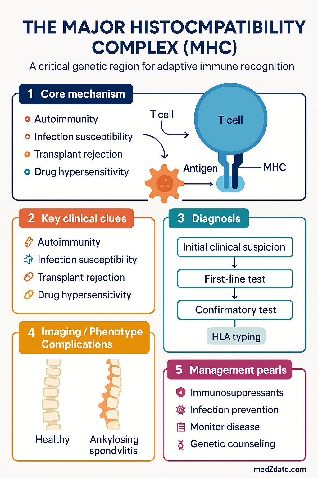
MHC Class I & Class II Molecules
MHC Class I
MHC class I molecules consist of a polymorphic α heavy chain (encoded by HLA-A, -B, or -C loci) non-covalently associated with the invariant β₂-microglobulin (β₂M) light chain (encoded on chromosome 15). The peptide-binding groove is formed by the α₁ and α₂ domains, which accommodate short peptides of 8–10 amino acids.
- Expression: Constitutively expressed on virtually all nucleated cells; highest density on lymphocytes, lowest on specialised tissues such as placental trophoblast (which uses HLA-G for immune evasion).
- Function: Presents endogenous (intracellular) peptides — typically derived from cytoplasmic proteins degraded by the proteasome — to CD8⁺ cytotoxic T lymphocytes (CTLs).
- Pathway: Proteasomal degradation → TAP transport into ER → peptide loading onto MHC-I via the peptide-loading complex (TAP, tapasin, ERp57, calreticulin) → surface transport via Golgi.
- Immunological role: Enables CTLs to detect virus-infected cells, intracellular bacteria, and tumour cells displaying aberrant peptides. Also serves as the ligand for killer-cell immunoglobulin-like receptors (KIRs) on natural killer (NK) cells.
MHC Class II
MHC class II molecules are heterodimers of an α chain and a β chain, both polymorphic, encoded by the HLA-DR, -DQ, and -DP loci. The peptide-binding groove, formed by the α₁ and β₁ domains, accommodates longer peptides of 13–25 amino acids (and occasionally longer, with overhanging ends).
- Expression: Constitutively expressed on professional antigen-presenting cells (APCs) — dendritic cells, macrophages, and B lymphocytes. Can be induced on other cell types (e.g. endothelial cells, epithelial cells) by interferon-γ (IFN-γ) during inflammation.
- Function: Presents exogenous (extracellular) peptides — derived from endocytosed and lysosomal-processed antigens — to CD4⁺ helper T lymphocytes.
- Pathway: Endocytosis → lysosomal/endosomal proteolysis (cathepsins) → CLIP removal from MHC-II by HLA-DM → peptide loading in MIIC compartment → surface transport.
- Immunological role: Activates CD4⁺ T cells, which orchestrate humoral immunity (via B cell help), macrophage activation (Th1), allergic/eosinophilic responses (Th2), and regulatory functions (Tregs via MHC-II on thymic epithelium).
| Feature | MHC Class I | MHC Class II |
|---|---|---|
| Loci | HLA-A, -B, -C | HLA-DR, -DQ, -DP |
| Chain structure | α chain + β₂-microglobulin | α chain + β chain |
| Peptide length | 8–10 amino acids | 13–25 amino acids |
| Peptide source | Endogenous (cytoplasmic) | Exogenous (endocytic) |
| T cell receptor | CD8⁺ cytotoxic T cells | CD4⁺ helper T cells |
| Expression | All nucleated cells | Professional APCs (constitutive); others (inducible by IFN-γ) |
| Processing pathway | Proteasome → TAP → ER peptide loading | Endosome/lysosome → MIIC peptide loading |
| Clinical transplantation role | HLA-A, -B matched (solid organ); HLA-C (HSCT, KIR ligand) | HLA-DR matched (solid organ); HLA-DQ (coeliac); HLA-DP (HSCT) |
Non-Classical MHC Molecules
Several MHC-encoded molecules have specialised immune functions:
- HLA-E: Presents leader peptides from classical HLA class I molecules to NK cell inhibitory receptor NKG2A/CD94; important in maternal–foetal tolerance.
- HLA-F: Expressed on activated lymphocytes; role in NK cell regulation under investigation.
- HLA-G: Restricted primarily to trophoblast; provides immune privilege at the maternal–foetal interface by inhibiting NK cells, T cells, and dendritic cells. Soluble HLA-G (sHLA-G) is measurable in serum and may serve as a biomarker in transplantation tolerance research.
- MICA/MICB: MHC class I chain-related genes encoding stress-induced ligands for the activating NK cell receptor NKG2D; upregulated on tumour and virus-infected cells. MICA antibodies are clinically relevant in solid organ transplant rejection.
Antigen Presentation Pathways
The central function of MHC molecules is to display peptide fragments on the cell surface for surveillance by T lymphocytes. The route by which a protein antigen reaches the MHC molecule determines which class of MHC presents it and, consequently, which T cell subset is activated.
Classical MHC Class I Pathway (Endogenous/Cytosolic)
Classical MHC Class II Pathway (Exogenous/Endocytic)
Cross-Presentation
Cross-presentation is a specialised pathway in which dendritic cells load exogenous antigens onto MHC class I molecules — a critical mechanism for initiating CD8⁺ T cell responses against viruses and tumours that do not directly infect APCs. Two principal models exist:
- Cytosolic pathway: Exogenous antigens escape from endosomes into the cytosol, are degraded by the proteasome, and enter the classical MHC-I loading pathway via TAP.
- Vacuolar pathway: Antigens are processed by cathepsins within endosomes and loaded onto MHC-I molecules recycling through the endosomal compartment.
Cross-presentation is the mechanistic basis for most cancer vaccine strategies and is exploited by adjuvants (e.g. STING agonists, poly I:C) in clinical trials at Australian cancer centres including the Peter MacCallum Cancer Centre and the Chris O'Brien Lifehouse.
Non-Classical Antigen Presentation
- CD1-restricted presentation: CD1 molecules (CD1a–d) present lipid and glycolipid antigens (e.g. mycobacterial mycolic acids) to specialised T cell subsets including NKT cells and γδ T cells. Relevant to tuberculosis and leprosy in ATSI communities.
- MR1-restricted presentation: MHC-related protein 1 (MR1) presents microbial vitamin B metabolites (e.g. 5-OP-RU from riboflavin synthesis) to mucosal-associated invariant T (MAIT) cells, an innate-like population enriched at mucosal surfaces.
The HLA System
Genomic Organisation
The HLA region spans approximately 3.6 Mb on chromosome 6p21.3 and is conventionally divided into three regions:
- Class I region (telomeric): Contains the classical loci HLA-A, -B, -C and the non-classical loci HLA-E, -F, -G, as well as MICA and MICB.
- Class III region (central): Encodes complement components (C2, C4A/B, factor B), TNF-α, TNF-β, heat shock proteins (HSP70), and other immune-related genes. Does not encode classical HLA molecules.
- Class II region (centromeric): Contains the classical loci HLA-DR, -DQ, -DP and the non-classical HLA-DM and HLA-DO loci, plus TAP1, TAP2, LMP2, and LMP7 (antigen processing genes).
Polymorphism
HLA is the most polymorphic genetic system in the human genome. As of the IPD-IMGT/HLA Database (Release 3.54, October 2024), there are >35 000 named alleles. Key features of HLA polymorphism include:
- Codominant expression: An individual expresses both maternal and paternal alleles at each locus, yielding up to six distinct HLA class I molecules and six (or more) class II molecules.
- Polygeny: Multiple class I and class II loci are expressed simultaneously, increasing the diversity of peptides presented.
- Linkage disequilibrium: HLA alleles at adjacent loci are frequently inherited together as haplotypes (e.g. A1-B8-DR3 in Northern Europeans). This has implications for transplant matching and disease association studies.
- Ethnic and population-specific variation: Allele frequencies differ markedly between populations. Certain alleles are enriched in specific ethnic groups — an important consideration in Australia's multicultural transplant programmes.
HLA Nomenclature & Typing Resolution
Modern HLA nomenclature follows the IPD-IMGT/HLA system:
- Two-digit (allele group): e.g. HLA-A*02 — encodes the A2 serological specificity.
- Four-digit (HLA protein): e.g. HLA-A*02:01 — specifies a unique amino acid sequence.
- Six-digit (synonymous substitution): e.g. HLA-A*02:01:01 — specifies a unique nucleotide sequence.
- Eight-digit (non-coding variation): e.g. HLA-A*02:01:01:01 — includes intron and UTR variation.
Typing resolution requirements vary by clinical application:
| Clinical Application | Required Resolution | Method |
|---|---|---|
| Solid organ transplant (deceased donor) | Two-digit (low/intermediate) | SSO, SSP, or NGS |
| HSCT (unrelated donor) | Four-digit or higher (high resolution) | NGS (gold standard) |
| Pharmacogenomic screening (HLA-B*57:01) | Four-digit (allele-specific) | PCR-SSP or NGS |
| HLA disease association testing | Four-digit | NGS, SSO, or SSP |
| Donor-specific antibody (DSA) identification | Four-digit (epitope-level preferred) | Single-antigen bead (SAB) assay |
HLA Typing Methods Available in Australia
HLA Inheritance & Family Typing
HLA genes are inherited as haplotypes (sets of linked alleles across the HLA region). Each individual inherits one haplotype from each parent, yielding a 25% chance that siblings are HLA-identical, a 50% chance of a haploidentical match, and a 25% chance of no shared haplotypes. Family HLA typing is the first step in identifying related donors for HSCT and living-donor solid organ transplantation.
In Australia, family typing for living-related kidney donation is coordinated through transplant centres and typically involves HLA-A, -B, -C, -DR, -DQ typing at high resolution.
Clinical Relevance of MHC/HLA
Transplantation Immunology
HLA matching is the cornerstone of transplantation medicine. The degree of HLA compatibility between donor and recipient directly influences graft survival, rejection risk, and immunosuppression requirements.
Solid Organ Transplantation
- Kidney: HLA-A, -B, and -DR matching are standard. Zero-mismatch (0MM) kidney transplants have significantly better long-term graft survival. Australia's national allocation algorithm (OrganMatch) incorporates HLA match grade, waiting time, sensitisation status, and geography.
- Heart/Lung: HLA matching is less prioritised due to organ scarcity and ischaemia time constraints, but DSA screening is mandatory pre-transplant.
- Liver: Historically considered relatively "HLA-permissive," but emerging data show that pre-formed DSAs are associated with acute antibody-mediated rejection and worse outcomes.
Haematopoietic Stem Cell Transplantation
HLA matching at high resolution is critical for HSCT outcomes. Mismatching at HLA-A, -B, -C, -DRB1, or -DQB1 increases the risk of graft-versus-host disease (GVHD), graft failure, and transplant-related mortality. Current practice in Australia:
- Matched unrelated donor (MUD): 10/10 or 12/12 match at HLA-A, -B, -C, -DRB1, -DQB1 (± DPB1) is preferred. Searched via ABMDR and international registries.
- Haploidentical transplant: Increasingly used with post-transplant cyclophosphamide (PTCy) regimens when no matched donor is available. One Australian centre performing haplo-HSCT is the Royal Adelaide Hospital.
- HLA-DPB1 permissive matching: DPB1 is in weaker linkage disequilibrium with other class II loci. Permissive mismatches (based on T-cell epitope classification) are acceptable and expand the donor pool.
Pharmacogenomic HLA Testing
Several HLA alleles are strongly associated with severe adverse drug reactions. Pre-prescription screening is now standard Australian practice and in some cases PBS-funded:
HLA-Disease Associations
Specific HLA alleles confer significant risk for autoimmune and inflammatory diseases. The strength of association is expressed as an odds ratio (OR) or relative risk (RR). Selected clinically important associations in Australian practice:
| Disease | HLA Association | Odds Ratio / Relative Risk | Clinical Utility |
|---|---|---|---|
| Ankylosing spondylitis | HLA-B*27 | OR ~100–200 | Supportive in diagnosis (BASDAI/modified New York criteria); ~90% of AS patients are B27-positive |
| Coeliac disease | HLA-DQ2 (DQA1*05/DQB1*02) and DQ8 (DQA1*03/DQB1*03:02) | NPV >99% | High negative predictive value; HLA-DQ2/8 negativity essentially excludes coeliac disease; useful for ruling out in equivocal cases |
| Type 1 diabetes | HLA-DR3, DR4, DQ2, DQ8 | OR 3–30 (haplotype dependent) | Risk stratification in first-degree relatives; research screening (TrialNet) |
| Rheumatoid arthritis | HLA-DRB1 shared epitope (SE) | OR 3–6 | Associated with anti-CCP antibody-positive, erosive disease; prognostic marker |
| Narcolepsy type 1 | HLA-DQB1*06:02 | OR >250 | Near-universal association; supports diagnosis in conjunction with hypocretin-1 levels |
| Psoriatic arthritis | HLA-C*06:02, HLA-B*27 | OR 2–10 | C*06:02 associated with skin psoriasis; B27 with axial disease |
| Behçet's disease | HLA-B*51 | OR 5–10 | Supportive; more prevalent in Mediterranean, Middle Eastern, and East Asian populations |
| Drug hypersensitivity (abacavir) | HLA-B*57:01 | PPV ~55%; NPV ~100% | Mandatory pre-prescription screening (PBS-funded) |
Anti-HLA Antibodies & Sensitisation
Pre-formed anti-HLA antibodies develop through prior pregnancy, blood transfusion, or previous transplant. These antibodies are detected by panel-reactive antibody (PRA) testing and characterised by single-antigen bead (SAB) assays. Key clinical concepts:
- Panel-Reactive Antibody (PRA): The percentage of the donor population against which a recipient has pre-formed antibodies. PRA >80% defines a "highly sensitised" patient, who faces prolonged wait-list times and limited compatible donor options.
- Donor-Specific Antibodies (DSAs): Anti-HLA antibodies directed against a specific donor's HLA antigens. Pre-formed DSAs cause hyperacute rejection (solid organ) or graft failure (HSCT). De novo DSAs developing post-transplant mediate chronic antibody-mediated rejection.
- Virtual crossmatch: Prediction of crossmatch result by comparing the recipient's antibody profile (from SAB) against the donor's HLA type. Reduces need for physical crossmatches and accelerates deceased donor organ allocation.
- Mean Fluorescence Intensity (MFI): SAB assays report MFI as a semi-quantitative measure of antibody strength. MFI >1 000–2 000 is generally considered clinically significant, though thresholds vary by centre.
Desensitisation Protocols
Highly sensitised transplant candidates may require desensitisation to reduce DSA levels before transplantation. Australian transplant centres employ regimens including:
- Plasmapheresis / Plasma exchange: Physically removes circulating antibodies. Typically performed 3–5 sessions pre-transplant.
- Intravenous immunoglobulin (IVIg): 2 g/kg over 2–4 days; immunomodulatory and anti-idiotypic effects. PBS Authority for transplant desensitisation.
- Rituximab: Anti-CD20 monoclonal antibody; depletes B cells. Off-label but widely used in desensitisation protocols. PBS-listed for other indications.
- Bortezomib: Proteasome inhibitor targeting plasma cells. Used for refractory antibody-mediated rejection. PBS authority may be required.
- Eculizumab / Ravulizumab: Complement C5 inhibitors; used in selected cases of antibody-mediated rejection at specialised centres.
MHC in Immunodeficiency & Immune Dysregulation
Genetic defects in MHC or antigen-processing genes cause primary immunodeficiencies:
- Bare lymphocyte syndrome type I (BLS-I): Mutations in TAP1, TAP2, or TAPBP (tapasin) result in deficient MHC class I expression. Presents with sinopulmonary infections and granulomatous skin lesions. CD8⁺ T cell counts reduced.
- Bare lymphocyte syndrome type II (BLS-II): Mutations in transcription factors CIITA (MHC-II transactivator), RFXANK, RFX5, or RFXAP result in absent MHC class II expression. Presents in infancy with severe recurrent infections, chronic diarrhoea, and failure to thrive. CD4⁺ T cell counts profoundly reduced. Requires HSCT for survival.
Special Populations
📚 References
- 1. Robinson J, Barker DJ, Georgiou X, Cooper MA, Flicek P, Marsh SGE. IPD-IMGT/HLA Database. Nucleic Acids Res. 2020;48(D1):D948–D955. doi:10.1093/nar/gkz950.
- 2. Wiebe C, Kosmoliaptsis V, Pochinco D, et al. HLA-DR/DQ molecular mismatch: a prognostic biomarker for primary alloimmunity. Am J Transplant. 2019;19(6):1708–1719. doi:10.1111/ajt.15257.
- 3. Mallal S, Phillips E, Carosi G, et al. HLA-B*5701 screening for hypersensitivity to abacavir. N Engl J Med. 2008;358(6):568–579. doi:10.1097/QCO.0b013e32832f6a60.
- 4. McCormack M, Alfirevic A, Bourgeois S, et al. HLA-A*3101 and carbamazepine-induced hypersensitivity reactions in Europeans. N Engl J Med. 2011;364(12):1134–1143. doi:10.1056/NEJMoa1013297.
- 5. Australian Institute of Health and Welfare (AIHW). Organ and tissue donation and transplantation activities in Australia. Canberra: AIHW; 2023.
- 6. Australian Red Cross Lifeblood. HLA Laboratory Services: Technical Manual. Melbourne: Lifeblood; 2024.
- 7. Brown MA, Li Z, Cao KL, et al. A genome-wide association study of ankylosing spondylitis identifies non-MHC susceptibility loci. Nat Genet. 2010;42(2):123–127. doi:10.1038/ng.513.
- 8. Kidney Health Australia. Guidelines for the management of chronic kidney disease in adults. Melbourne: Kidney Health Australia; 2024.
- 9. Marsh SGE, Albert ED, Bodmer WF, et al. Nomenclature for factors of the HLA system, 2010. Tissue Antigens. 2010;75(4):291–455. doi:10.1111/j.1399-0039.2010.01466.x.
- 10. Neefjes J, Jongsma MLM, Paul P, Bakke O. Towards a systems understanding of MHC class I and MHC class II antigen presentation. Nat Rev Immunol. 2011;11(12):823–836. doi:10.1038/nri3084.
- 11. Hughes DA, Vilar FJ, Ward CC, Alfirevic A, Park BK, Pirmohamed M. Cost-effectiveness analysis of HLA-B*5701 genotyping in preventing abacavir hypersensitivity. Pharmacogenetics. 2004;14(5):335–342. doi:10.1097/00008571-200405000-00008.
- 12. Australian Bone Marrow Donor Registry (ABMDR). Annual Report 2023–2024. Sydney: ABMDR; 2024.
- 13. RHDAustralia (Australian Government Department of Health). The 2020 Australian guideline for prevention, diagnosis, and management of acute rheumatic fever and rheumatic heart disease. 2nd edn. Darwin: Menzies School of Health Research; 2020.
- 14. Wiebe C, Gibson IW, Blydt-Hansen TD, et al. Evolution and clinical pathologic correlations of de novo donor-specific HLA antibody post kidney transplant. Am J Transplant. 2012;12(5):1157–1167. doi:10.1111/j.1600-6143.2012.04013.x.
- 15. Tait BD, Süsal C, Gebel HM, et al. Consensus guidelines on the testing and clinical management issues associated with HLA and non-HLA antibodies in transplantation. Transplantation. 2013;95(1):19–47. doi:10.1097/TP.0b013e31827a19cc.