Home Immunology B Cells and T Cells

B Cells and T Cells

📋 Key Information Summary

📋
  • B cells and T cells are the adaptive immune system's primary effectors; their cooperation generates high-affinity antibodies and durable immunological memory
  • CD4⁺ T helper cells provide critical cognate help to B cells via CD40L–CD40 interaction, ICOS–ICOSL costimulation, and cytokine signals
  • T follicular helper (Tfh) cells are the specialised CD4 subset essential for germinal centre formation and affinity maturation
  • Germinal centres are organised lymphoid structures where B cells undergo somatic hypermutation and class-switch recombination to produce high-affinity, class-switched antibodies
  • The germinal centre reaction separates into a dark zone (proliferation/mutation) and light zone (selection via follicular dendritic cell antigen display)
  • B cell class switching is directed by cytokines: IL-4 → IgG1/IgE, IFN-γ → IgG2/IgG3, TGF-β → IgA, BAFF/APRIL → IgA at mucosal sites
  • Long-lived plasma cells migrate to bone marrow survival niches and secrete antibody for years to decades after immunisation or infection
  • Memory B cells and memory T cells are formed both within and outside germinal centres, providing rapid secondary responses
  • Defects in B–T cell collaboration underlie primary immunodeficiencies (e.g., Hyper-IgM syndrome, CVID) and are exploited in autoimmune disease
  • Australian vaccination programmes (NIP schedule) rely on intact germinal centre reactions to generate protective antibody titres
  • ATSI populations experience higher rates of invasive pneumococcal disease and influenza-related hospitalisation, making understanding of humoral immunity clinically important

Introduction & Australian Context

Cooperation between B lymphocytes and T lymphocytes within germinal centres drives the generation of high-affinity antibody responses and long-lived immunological memory — the foundation of protective immunity conferred by vaccination and natural infection.

Understanding B–T cell collaboration is essential for Australian clinicians managing immunodeficiency, autoimmune disease, transplant immunology, and vaccine programmes. Australia's National Immunisation Program (NIP) schedules vaccines across the lifespan, relying on robust germinal centre responses for protection against pneumococcal disease, influenza, measles, pertussis, and SARS-CoV-2.

Primary immunodeficiency affecting B–T cell cooperation — including X-linked agammaglobulinaemia, common variable immunodeficiency (CVID), and combined immunodeficiencies — affects approximately 1 in 1,200 Australians, though many remain undiagnosed. The Australasian Society of Clinical Immunology and Allergy (ASCIA) maintains diagnostic and management guidelines for these conditions.

This guideline provides a clinical overview of B–T cell biology with emphasis on the germinal centre reaction, CD4 helper functions, and memory formation, framed for primary care physicians, immunologists, haematologists, and infectious diseases specialists practising in Australia.
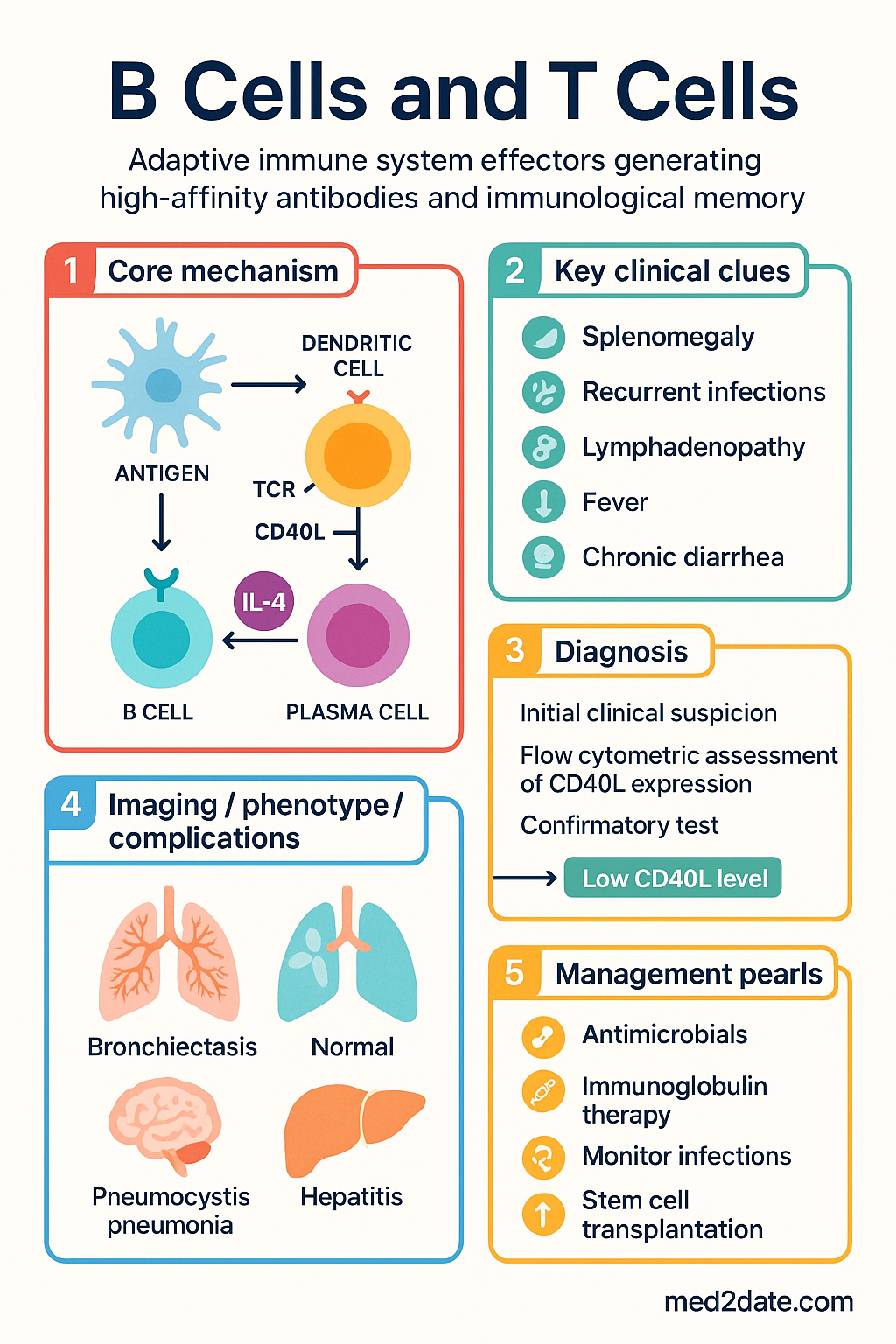
B Cells and T Cells clinical infographic — pathophysiology, clinical clues, diagnosis, imaging, and management
Tap or click image to enlarge — B Cells and T Cells: pathophysiology, clinical clues, diagnosis, imaging, and management.
B Cells and T Cells infographic, full size

B–T Cell Collaboration

The adaptive immune response requires intimate cooperation between antigen-specific B cells and CD4⁺ T helper cells. This collaboration occurs primarily in secondary lymphoid organs (lymph nodes, spleen, mucosal-associated lymphoid tissue) and is essential for the production of class-switched, high-affinity antibodies.

Antigen Presentation and T Cell Priming

The collaboration begins when dendritic cells capture antigen in peripheral tissues and migrate to draining lymph nodes. There, they present processed peptide–MHC class II complexes to naïve CD4⁺ T cells. Successful T cell activation requires three signals:

  • Signal 1: TCR engagement with peptide–MHC II complex on the dendritic cell
  • Signal 2: Costimulation via CD28 (T cell) binding B7-1/B7-2 (CD80/CD86) on the dendritic cell
  • Signal 3: Cytokine milieu that directs T helper cell differentiation

The Immunological Synapse

Activated CD4⁺ T cells migrate to the T–B cell border of lymphoid follicles. B cells that have captured and processed antigen via their B cell receptor (BCR) present peptide–MHC II complexes to cognate T helper cells. This interaction forms an immunological synapse characterised by:

  • TCR–MHC II recognition: Antigen-specific, MHC-restricted contact
  • CD40L (T cell) – CD40 (B cell): Critical costimulatory signal that rescues B cells from apoptosis and promotes proliferation, germinal centre entry, and class switching
  • ICOS (T cell) – ICOSL (B cell): Essential for Tfh cell function and germinal centre maintenance
  • SAP/SLAM interactions: Regulate stable T–B conjugate formation; deficiency causes X-linked lymphoproliferative disease
⚠️
Clinical Pearl: Defects in the CD40–CD40L axis cause Hyper-IgM syndrome type 1 (X-linked, CD40L deficiency) or type 3 (autosomal recessive, CD40 deficiency). Patients present with recurrent sinopulmonary infections, opportunistic infections (Pneumocystis jirovecii, Cryptosporidium), and absent class-switched immunoglobulins (low IgG, IgA, IgE with normal or elevated IgM). Diagnosis requires flow cytometric assessment of CD40L expression on activated T cells. PBS-listed immunoglobulin replacement therapy (Intragam P® or Privigen®) is available as Authority Required for these patients.

Thymus-Dependent vs Thymus-Independent Responses

Antigens are classified based on their T cell dependence:

Feature Thymus-Dependent (TD) Thymus-Independent (TI)
Antigen type Proteins (most vaccines, toxins) Polysaccharides, repetitive epitopes (LPS, flagellin)
T cell help required Yes — CD4⁺ Tfh cells No
Germinal centre formation Yes No (TI-1) or minimal (TI-2)
Class switching Yes — all isotypes Limited
Affinity maturation Yes No
Memory formation Robust, long-lived Limited
Clinical example Tetanus toxoid, hepatitis B vaccine Pneumococcal polysaccharide (23vPPV)
💡
Vaccination Implication: Children under 2 years have an immature marginal zone and respond poorly to polysaccharide antigens (TI-2 responses). Conjugate vaccines (e.g., 13vPCV pneumococcal, Hib, meningococcal) overcome this limitation by covalently linking polysaccharide to a carrier protein, converting the response to T cell–dependent with germinal centre formation, class switching, and memory. This is why the NIP uses 13vPCV (Prevenar 13®) rather than 23vPPV for infants.

CD4⁺ T Helper Cell Functions

CD4⁺ T helper cells are the orchestrators of adaptive immunity. Upon activation, naïve CD4⁺ T cells differentiate into specialised subsets based on the cytokine environment, each with distinct effector functions relevant to B cell help and broader immune regulation.

Major CD4⁺ T Helper Subsets

Subset Key Transcription Factor Signature Cytokines Primary Function B Cell Relevance
Th1 T-bet IFN-γ, TNF-α, IL-2 Intracellular pathogens, cell-mediated immunity Promotes IgG2 (opsonisation, complement fixation)
Th2 GATA-3 IL-4, IL-5, IL-13 Helminth defence, allergy IL-4 drives IgG1 and IgE class switching
Th17 RORγt IL-17A, IL-17F, IL-22 Extracellular bacteria, fungi; mucosal defence Indirect role in mucosal IgA
Tfh Bcl-6 IL-21, IL-4 Germinal centre support, B cell help Primary B cell helper — essential for GC reaction
Treg FoxP3 IL-10, TGF-β, IL-35 Immune tolerance, suppression Regulates GC output, prevents autoantibody production

T Follicular Helper (Tfh) Cells — The Master B Cell Helpers

Tfh cells are the dominant CD4⁺ subset providing direct help to B cells within germinal centres. They are identified by surface expression of CXCR5 (chemokine receptor directing migration to follicles), PD-1, ICOS, and CD40L, with Bcl-6 as the lineage-defining transcription factor.

Tfh cell functions in germinal centres:

  • IL-21 secretion: Major B cell growth and differentiation factor; promotes plasma cell generation and class-switch recombination
  • IL-4 secretion: Drives IgG1 and IgE switching
  • CD40L expression: Provides survival, proliferation, and differentiation signals to B cells; upregulates AID (activation-induced cytidine deaminase) for somatic hypermutation
  • ICOS–ICOSL interaction: Sustains Tfh function within the germinal centre; ICOS deficiency causes a combined immunodeficiency with absent germinal centres
  • CXCL13 production: Chemokine that attracts CXCR5⁺ B cells and maintains follicular architecture
⚠️
SARS-CoV-2 and Tfh Cells: Circulating Tfh (cTfh) cells are measurable in peripheral blood and correlate with neutralising antibody titres after SARS-CoV-2 infection and vaccination. In Australia, cTfh13⁺ cells (CXCR3⁻CCR6⁻) are a biomarker of effective germinal centre responses following COVID-19 vaccination. Patients on B cell–depleting therapies (e.g., rituximab, ocrelizumab) have impaired GC responses and reduced vaccine efficacy — ATAGI recommends vaccination timing relative to B cell reconstitution.

Cytokine-Directed Class Switch Recombination

The isotype of antibody produced by B cells is dictated by cytokines from T helper cells during the germinal centre reaction:

🧬
IL-4 / IL-13
Th2 / Tfh-derived · Switch to IgG1, IgE
Clinical relevance Allergic disease, helminth defence, IgE-mediated hypersensitivity
Pathology Excessive IL-4 → atopic disease, anaphylaxis
🧬
IFN-γ
Th1 / NK-derived · Switch to IgG2, IgG3
Clinical relevance Opsonisation, complement activation, antimicrobial defence
Deficiency Impaired IgG2 → susceptibility to encapsulated bacteria
🧬
TGF-β / BAFF / APRIL
Mucosal · Switch to IgA
Clinical relevance Mucosal immunity, gut and respiratory tract defence
Deficiency Selective IgA deficiency (1/300–700 Australians); most common primary immunodeficiency

Germinal Centre Reaction

The germinal centre (GC) is a specialised microanatomical structure within secondary lymphoid follicles where antigen-activated B cells undergo rapid proliferation, somatic hypermutation (SHM), affinity-based selection, and class-switch recombination (CSR) to generate high-affinity, class-switched antibodies. The GC reaction is the cornerstone of effective humoral immunity and the immunological basis for vaccination.

Germinal Centre Architecture

GCs form in B cell follicles 7–10 days after antigen exposure and consist of two functionally distinct zones:

Zone 1
Dark Zone
Dense packing of rapidly proliferating B cells (centroblasts) expressing CXCR4. Site of somatic hypermutation catalysed by activation-induced cytidine deaminase (AID). High mutation rate (~10⁻³ per bp per division) introduces random point mutations in immunoglobulin variable region genes.
Key cells: Centroblasts, CXCL12-producing reticular cells
Zone 2
Light Zone
Less dense region containing centrocytes (B cells that have ceased dividing) and a network of follicular dendritic cells (FDCs). FDCs display intact antigen as immune complexes on their surface. B cells compete for antigen binding; those with higher BCR affinity capture more antigen, present more peptide–MHC II to Tfh cells, and receive stronger survival signals.
Key cells: Centrocytes, FDCs, Tfh cells

The Cyclic Re-entry Model

GC B cells cycle between dark and light zones in a process of iterative mutation and selection:

1
Activation & Dark Zone Entry
Antigen-activated B cells upregulate Bcl-6 and AID, enter the dark zone, and undergo rapid proliferation (cell cycle ~6–8 hours). AID introduces somatic mutations in Ig V-region genes.
2
Migration to Light Zone
Centroblasts cease dividing, downregulate CXCR4, upregulate CXCR5, and migrate to the light zone guided by CXCL13 chemokine gradient.
3
Affinity-Based Selection
Centrocytes test mutated BCRs against antigen displayed on FDCs. Higher-affinity BCRs capture more antigen, process it, and present more peptide–MHC II to Tfh cells. Cells with low-affinity or autoreactive BCRs undergo apoptosis (death by neglect).
4
Tfh Rescue & Re-entry
Surviving centrocytes receive Tfh help (CD40L, IL-21) and can either re-enter the dark zone for further mutation cycles or differentiate into memory B cells or plasma cells.
💡
Clinical Significance: The GC reaction requires 2–4 weeks to generate peak affinity-matured antibody responses. This is why vaccine booster doses are typically spaced ≥4 weeks apart (e.g., primary COVID-19 vaccination schedule) to allow full GC maturation. Rapid booster schedules may truncate the GC reaction and compromise long-lived memory formation.

Somatic Hypermutation and Class-Switch Recombination

Both SHM and CSR are catalysed by AID (encoded by AICDA gene):

Process Mechanism Outcome Clinical Deficiency
Somatic Hypermutation AID deaminates cytosine → uracil in Ig V-region DNA; error-prone repair introduces point mutations Altered BCR affinity (increase or decrease); selection retains high-affinity variants AID deficiency (Hyper-IgM syndrome type 2): absent SHM, impaired CSR, elevated IgM, susceptibility to infections
Class-Switch Recombination AID targets switch (S) regions upstream of constant-region genes; double-strand breaks allow deletion of intervening DNA and recombination to downstream isotype (IgG, IgA, IgE) Change from IgM/IgD to IgG, IgA, or IgE while retaining the same antigen specificity UNG deficiency (Hyper-IgM syndrome type 5), NEMO deficiency (X-linked ectodermal dysplasia with immunodeficiency)

Memory Formation

Immunological memory — the ability to mount faster, stronger, and more specific responses upon re-exposure to a previously encountered antigen — is the fundamental goal of vaccination. Memory is mediated by two major cellular compartments arising from the germinal centre and extrafollicular responses: memory B cells and long-lived plasma cells. Memory T cells provide cellular recall immunity.

Memory B Cell Formation

Memory B cells emerge from germinal centres (GC-derived memory) or from early extrafollicular responses. Key characteristics:

  • Surface phenotype: CD27⁺ (in humans), class-switched (IgG⁺, IgA⁺, IgE⁺) or unswitched (IgM⁺IgD⁺CD27⁺ — "IgM memory" from T-independent or marginal zone responses)
  • Affinity status: Carry somatically mutated, affinity-matured BCR from GC selection
  • Longevity: Can persist for decades; maintenance does not require continuous antigen exposure (debate ongoing; bone marrow niches may play a role)
  • Secondary response: Upon re-encounter with antigen, memory B cells rapidly differentiate into antibody-secreting plasma cells or re-enter GCs for further affinity maturation
  • Class switching flexibility: Can undergo further class switching upon restimulation (e.g., switch from IgG1 to IgG4 with repeated antigen exposure — relevant in allergen immunotherapy)

Long-Lived Plasma Cells (LLPCs)

LLPCs are terminally differentiated antibody-secreting cells that migrate to survival niches in the bone marrow and constitutively secrete antibody for years to decades without requiring further antigen stimulation.

  • Surface phenotype: CD38⁺⁺ CD138⁺ (syndecan-1), CD19⁻/low, CD20⁻, CD27⁺⁺
  • Survival signals: BAFF, APRIL (from stromal cells), CXCL12 (from bone marrow niches), IL-6, contact with bone marrow stromal cells
  • Antibody secretion rate: Each LLPC secretes ~10,000 antibody molecules per second
  • Lifespan: Estimated years to decades; recent evidence from smallpox-vaccinated individuals shows detectable antibodies >50 years post-vaccination
  • Niche competition: LLPCs compete for limited bone marrow survival niches; new GC reactions may displace older LLPCs (relevant to repeated vaccination)
💡
Booster Vaccination Rationale: The primary vaccine series establishes a pool of LLPCs and memory B cells. Booster doses serve to (1) expand pre-existing memory B cells, (2) drive further affinity maturation in secondary GC reactions, and (3) replenish LLPC numbers that may have declined due to niche competition or natural attrition. Australia's NIP booster schedule (e.g., 12-month and 4-year diphtheria–tetanus–pertussis boosters) is designed around these principles.

Memory T Cell Subsets

Memory T cells provide rapid effector function upon re-encounter with antigen and are categorised by location and function:

Subset Location Surface Markers Function
Central Memory (Tcm) Secondary lymphoid organs, blood CCR7⁺ CD62L⁺ CD45RO⁺ High proliferative capacity; provide sustained effector supply upon restimulation
Effector Memory (Tem) Peripheral tissues, blood CCR7⁻ CD62L⁻ CD45RO⁺ Rapid effector function (cytokine production, cytotoxicity); patrol peripheral tissues
Tissue-Resident Memory (Trm) Non-lymphoid tissues (lung, gut, skin) CD69⁺ CD103⁺ First-line defence at barrier sites; do not recirculate; provide immediate local immunity
Stem Cell Memory (Tscm) Blood, lymphoid organs CD45RA⁺ CD95⁺ CD122⁺ Self-renewing; longest-lived memory subset; can regenerate all other memory subsets

Clinical Applications of Memory Biology

Understanding memory formation informs clinical practice in several domains:

  • Vaccine design: Adjuvants (e.g., AS01 in Shingrix®, MF59 in Fluad®) enhance GC reactions and memory formation; aluminium salts promote Th2-biased responses with strong antibody but limited T cell memory
  • Immunosuppressive therapy: Rituximab (anti-CD20) depletes memory B cells but spares LLPCs (CD20⁻); patients maintain pre-existing antibodies but cannot mount new humoral responses
  • Transplant immunology: Donor-specific memory T and B cells mediate accelerated rejection in sensitised recipients; donor-specific antibody (DSA) from LLPCs causes antibody-mediated rejection
  • Primary immunodeficiency: Patients with CVID or XLA have impaired memory formation; rely on regular immunoglobulin replacement (Intragam P® 300–600 mg/kg IV every 3–4 weeks, PBS Authority Required)

Pathophysiology of B–T Cell Collaboration Defects

Defects at any step in B–T cell collaboration can result in immunodeficiency, autoimmunity, or lymphoproliferative disorders. Understanding the specific molecular defect informs diagnosis and management.

Defect Gene/Protein Consequence Inheritance
X-linked agammaglobulinaemia (XLA) BTK (Bruton tyrosine kinase) Block in B cell maturation at pre-B stage; absent mature B cells and immunoglobulins X-linked recessive
Hyper-IgM type 1 CD40LG (CD40 ligand) Absent T–B cooperation; no class switching; elevated IgM, absent IgG/IgA/IgE X-linked recessive
Hyper-IgM type 2 AICDA (AID) Absent SHM and CSR; elevated IgM Autosomal recessive
Common variable immunodeficiency (CVID) Multiple (ICOS, TACI, BAFF-R, CD19) Hypogammaglobulinaemia, impaired GC formation, reduced class-switched antibodies Variable (often polygenic)
ICOS deficiency ICOS Absent GC formation, CVID phenotype Autosomal recessive
SAP/SH2D1A deficiency (XLP) SH2D1A (SAP) Unstable T–B conjugates; absent GC formation; EBV-triggered haemophagocytic lymphohistiocytosis X-linked recessive
Selective IgA deficiency Unknown (polygenic) Absent serum IgA (<0.07 g/L); IgG and IgM normal; mucosal infections Variable
🚨
Red Flags for Primary Immunodeficiency: Consider B–T cell collaboration defects in patients with: ≥4 ear infections in 12 months, ≥2 serious sinus infections, ≥2 months on antibiotics with little effect, recurrent deep-seated infections (pneumonia, osteomyelitis), family history of PID, failure to thrive in infants. Refer to clinical immunology for investigation. Australian diagnostic pathway: serum immunoglobulins (IgG, IgA, IgM, IgE), lymphocyte subsets (CD3, CD4, CD8, CD19, CD16/56), specific antibody responses (anti-tetanus, anti-pneumococcal), and referral to an ASCIA-accredited clinical immunologist.

Investigations

Assessment of B–T cell collaboration and humoral immunity involves a tiered investigation approach available through Australian pathology services.

Available Serum Immunoglobulin Quantification (IgG, IgA, IgM, IgE) MBS Item 66622. Widely available. First-line screen for antibody deficiency. Age-specific paediatric reference ranges essential.
Available Lymphocyte Subset Analysis (CD3, CD4, CD8, CD19, CD16/56) MBS Item 66619. Flow cytometry. Essential for quantifying T and B cell compartments. CD19⁺ B cell count assesses B cell compartment.
Available Specific Antibody Responses (Anti-tetanus, Anti-pneumococcal serotypes) MBS Item 66625 (functional antibody assessment). Assess T cell–dependent antibody responses; pre- and post-vaccination titres determine protective immunity.
Available IgG Subclass Quantification (IgG1–4) MBS Item 66622. Useful when total IgG is low-normal but clinical suspicion of class-switching defect persists.
Referral CD40L Expression on Activated T Cells (Flow Cytometry) Specialist immunology laboratory. Diagnoses X-linked Hyper-IgM syndrome (CD40L deficiency). Requires stimulation with PMA/ionomycin.
Referral Tfh Cell Phenotyping (CXCR5⁺ PD-1⁺ ICOS⁺ CD4⁺) Research/specialist laboratory. Circulating Tfh (cTfh) cells correlate with GC activity and vaccine responses. Not routine clinical.
Specialist AID Gene Sequencing (AICDA) Molecular genetics laboratory. Diagnoses Hyper-IgM syndrome type 2 (AID deficiency). Medicare-eligible under genomic testing criteria.
Specialist Lymph Node Biopsy with Immunohistochemistry Histopathology. Assess GC architecture, Bcl-6 staining, follicular dendritic cell networks. Indicated for suspected immunodeficiency with lymphoproliferation or lymphoma workup.

Special Populations

👶 Paediatric
Neonatal and infant immune system
Neonates have immature GC reactions and limited somatic hypermutation. Maternal IgG transferred transplacentally provides passive protection for the first 6 months of life. B cell numbers reach adult levels by 6 months but functional maturity (including marginal zone B cells) is not complete until 2–5 years of age.
NIP vaccine implications
Conjugate vaccines are essential in infancy to convert polysaccharide antigens to T cell–dependent responses. Primary series at 2, 4, 6 months generates protective IgG responses. Boosters at 12 months consolidate memory. Delayed vaccine schedules leave infants vulnerable to encapsulated organisms.
XLA presentation
X-linked agammaglobulinaemia typically presents at 6–9 months when maternal antibodies wane. Male infants with recurrent sinopulmonary infections, absent B cells (CD19 <1%), and very low immunoglobulins should be investigated urgently.
🤰 Pregnancy
Immunological changes in pregnancy
Pregnancy involves a Th2 shift (increased IL-4, IL-10) to promote fetal tolerance. This enhances antibody production but may exacerbate Th2-mediated diseases (e.g., SLE flares). Treg cells expand to maintain fetal tolerance.
Transplacental antibody transfer
IgG1 crosses the placenta most efficiently via FcRn-mediated transport. Maternal vaccination (influenza, pertussis, COVID-19) during pregnancy generates protective IgG1 that transfers to the fetus, providing neonatal protection. The NIP recommends pertussis vaccination at 20–32 weeks gestation.
🛡️ Immunocompromised
B cell–depleting therapies (Rituximab, Ocrelizumab)
Anti-CD20 antibodies deplete CD20⁺ B cells including memory B cells. GC reactions are impaired; new antibody responses to vaccines are markedly reduced. Existing LLPCs (CD20⁻) and pre-formed antibodies may persist. ATAGI recommends vaccination ≥4 weeks before planned rituximab or ≥6 months after the last dose.
T cell–depleting therapies (Corticosteroids, calcineurin inhibitors, anti-thymocyte globulin)
Broad T cell suppression impairs Tfh function and GC reactions. Live vaccines are contraindicated. Inactivated vaccine responses may be suboptimal. Monitor immunoglobulin levels and consider immunoglobulin replacement if recurrent infections develop.
👴 Elderly
Immunosenescence
Ageing reduces naïve T cell output from the involuting thymus, shrinks the naïve B cell repertoire, and impairs GC reactions. Aged GCs produce lower-affinity antibodies with reduced somatic hypermutation. This contributes to poorer vaccine responses in the elderly and underpins the NIP recommendation for higher-dose influenza vaccines (Fluzone High-Dose®) in adults ≥65 years.
🫘 Renal Impairment
Uraemic immunodeficiency
Chronic kidney disease causes a combined B and T cell immunodeficiency with impaired GC function, reduced vaccine responses, and increased infection risk. Haemodialysis patients show particularly poor responses to hepatitis B vaccination; double-dose or additional booster schedules are recommended. Peritoneal dialysis patients generally respond better.

ATSI Health Considerations

Aboriginal and Torres Strait Islander Health
Infection burden
ATSI Australians experience significantly higher rates of invasive pneumococcal disease (IPD), Haemophilus influenzae type b (Hib), and influenza-related hospitalisation compared to non-Indigenous Australians. These infections are directly relevant to understanding humoral immunity and vaccine responses. IPD incidence in ATSI children <2 years remains 3–5 times the non-Indigenous rate despite vaccination (AIHW, 2023).
Vaccine programme access
The NIP provides free vaccination for all Australians; however, ATSI populations in remote and very remote areas face barriers including limited healthcare access, cold-chain management challenges, and workforce shortages. Aboriginal Health Workers and Practitioners play a critical role in vaccine delivery. Enhanced schedule vaccines (additional meningococcal B and pneumococcal boosters) are funded for ATSI children in some jurisdictions.
Immunodeficiency screening
Primary immunodeficiency may be underdiagnosed in ATSI communities due to limited access to specialist immunology services. The closest ASCIA-accredited clinical immunologist may be hundreds of kilometres away. Telehealth services (Medicare Benefits Schedule items 91822, 91823) provide essential specialist access. Serum immunoglobulins and lymphocyte subsets can be performed through regional pathology services.
Environmental factors
High rates of environmental enteropathy in remote communities may alter mucosal IgA responses and gut-associated lymphoid tissue (GALT) function. Chronic helminth exposure promotes Th2/Tfh2 bias with elevated IgE, potentially modulating responses to vaccines and infections. Overcrowded housing facilitates respiratory pathogen transmission, increasing the burden on humoral immunity.
Cultural safety
Investigations involving blood collection should be conducted with cultural sensitivity, with appropriate consent and involvement of Aboriginal Health Workers. Yarning-based education about vaccination and immune function should be delivered by culturally safe healthcare providers. Acknowledge the strengths of ATSI community-controlled health services (ACCHS) in delivering comprehensive primary care including immunisation programmes.

📚 References

  1. 1. Victora GD, Nussenzweig MC. Germinal centers. Annual Review of Immunology. 2022;40:413–442. doi:10.1146/annurev-immunol-120419-022408
  2. 2. Crotty S. T follicular helper cell biology: a decade of discovery and diseases. Immunity. 2019;50(5):1132–1148. doi:10.1016/j.immuni.2019.04.011
  3. 3. Tangye SG, Ma CS, Brink R, Deenick EK. The good, the bad and the ugly — TFH cells in human health and disease. Nature Reviews Immunology. 2013;13(6):412–426. doi:10.1038/nri3447
  4. 4. Australia Government Department of Health and Aged Care. National Immunisation Program Schedule. Canberra: Australian Government; 2024.
  5. 5. Australasian Society of Clinical Immunology and Allergy (ASCIA). Primary Immunodeficiency Guidelines. Sydney: ASCIA; 2024. Available from: https://www.allergy.org.au
  6. 6. Australian Technical Advisory Group on Immunisation (ATAGI). Australian Immunisation Handbook. Canberra: Australian Government Department of Health and Aged Care; 2024. Available from: https://immunisationhandbook.health.gov.au
  7. 7. Australian Institute of Health and Welfare (AIHW). Aboriginal and Torres Strait Islander Health Performance Framework 2023. Canberra: AIHW; 2023.
  8. 8. Slifka MK, Antia R, Whitmire JK, Ahmed R. Humoral immunity due to long-lived plasma cells. Immunity. 1998;8(3):363–372. doi:10.1016/S1074-7613(00)80541-5
  9. 9. Hammarlund E, Lewis MW, Hansen SG, et al. Duration of antiviral immunity after smallpox vaccination. Nature Medicine. 2003;9(9):1131–1137. doi:10.1038/nm917
  10. 10. Tangye SG, Al-Herz W, Bousfiha A, et al. Human inborn errors of immunity: 2022 update on the classification from the International Union of Immunological Societies Expert Committee. Journal of Clinical Immunology. 2022;42(7):1473–1507. doi:10.1007/s10875-022-01289-3
  11. 11. Nutt SL, Hodgkin PD, Tarlinton DM, Corcoran LM. The generation of antibody-secreting plasma cells. Nature Reviews Immunology. 2015;15(3):160–171. doi:10.1038/nri3795
  12. 12. RHDAustralia (Northern Territory Department of Health and Charles Darwin University). Australian Guidelines for the Prevention, Diagnosis and Management of Acute Rheumatic Fever and Rheumatic Heart Disease. 3rd edition. Darwin: RHDAustralia; 2020. [Referenced for ATSI health context and vaccination strategies]
for PBS-listed medicines at participating pharmacies.
Cultural safety
Engagement with Aboriginal Community Controlled Health Organisations (ACCHOs) is essential. Cultural safety training for non-Indigenous clinicians, use of Aboriginal Health Workers and Liaison Officers, and incorporation of traditional healing practices alongside Western medicine improve treatment adherence and outcomes. Avoidance of eye contact, respect for gender-sensitive examination practices, and understanding of sorry business protocols are critical elements of culturally safe care.
Medication adherence
Complex DMARD regimens with frequent monitoring requirements present adherence challenges. Long-acting depot injections (e.g., methotrexate SC) may improve adherence compared to oral regimens. Community pharmacy partnerships through the Indigenous Pharmacy Programmes improve medication management.
Specific conditions
Rheumatic heart disease (RHD) requires secondary prophylaxis with benzathine penicillin G (BPG) 1.2 MU IM every 3–4 weeks for a minimum of 10 years or until age 21 (whichever is longer). RHD registers (e.g., NT RHD Register) facilitate recall and follow-up. The Australian RHD Endgame Strategy targets elimination by 2031.
Referral pathways
Referral through ACCHOs and Aboriginal Hospital Liaison Officers (AHLOs) improves engagement. The Specialist Outreach Assistance Programme provides funded specialist visits to remote communities. NT, WA, and QLD have specific rheumatology outreach programmes targeting Indigenous communities.

📚 References

  1. 1. Australian Institute of Health and Welfare (AIHW). Autoimmune disease in Australia. Cat. no. PHE 312. Canberra: AIHW; 2023.
  2. 2. Fraenkel L, Bathon JM, England BR, et al. 2021 American College of Rheumatology guideline for the treatment of rheumatoid arthritis. Arthritis Care Res. 2021;73(7):924–939.
  3. 3. Fanouriakis A, Kostopoulou M, Alber K, et al. 2019 update of the EULAR recommendations for the management of systemic lupus erythematosus. Ann Rheum Dis. 2019;78(6):736–745.
  4. 4. Chung SA, Langford CA, Maz M, et al. 2021 American College of Rheumatology/Vasculitis Foundation guideline for the management of antineutrophil cytoplasmic antibody-associated vasculitis. Arthritis Care Res. 2021;73(11):1583–1599.
  5. 5. Smolen JS, Landewé RBM, Bijlsma JWJ, et al. EULAR recommendations for the management of rheumatoid arthritis with synthetic and biological disease-modifying antirheumatic drugs: 2022 update. Ann Rheum Dis. 2023;82(1):3–18.
  6. 6. Australian Technical Advisory Group on Immunisation (ATAGI). Australian Immunisation Handbook. Australian Government Department of Health; 2024. Available from: immunisationhandbook.health.gov.au.
  7. 7. Rheumatic Heart Disease Australia (RHDAustralia). The 2020 Australian guideline for prevention, diagnosis, and management of acute rheumatic fever and rheumatic heart disease. 3rd ed. Darwin: Menzies School of Health Research; 2020.
  8. 8. Pharmaceutical Benefits Scheme (PBS). PBS Schedule. Australian Government Department of Health. Available from: pbs.gov.au. Accessed 2024.
  9. 9. Agarwal S, Cunnington J, Nossent J. Autoimmune disease in Indigenous Australians: a systematic review. Int J Rheum Dis. 2021;24(12):1487–1498.
  10. 10. Pisetsky DS. Antinuclear antibody testing — misunderstood or misused? Clin Immunol. 2023;255:109717.
  11. 11. Bertsias GK, Tektonidou M, Amoura Z, et al. Joint European League Against Rheumatism and European Renal Association–European Dialysis and Transplant Association (EULAR/ERA-EDTA) recommendations for the management of adult and paediatric lupus nephritis. Ann Rheum Dis. 2012;71(11):1771–1782.
  12. 12. Ledingham J, Deighton C; British Society for Rheumatology Standards, Audit and Guidelines Working Group. Update on the British Society for Rheumatology guidelines for prescribing TNFα blockers in adults with rheumatoid arthritis. Rheumatology. 2005;44(2):155–158.
  13. 13. National Health and Medical Research Council (NHMRC). National statement on ethical conduct in human research. Canberra: NHMRC; 2023 (updated).
for PBS-listed medicines at participating pharmacies.
Cultural safety
Engagement with Aboriginal Community Controlled Health Organisations (ACCHOs) is essential. Cultural safety training for non-Indigenous clinicians, use of Aboriginal Health Workers and Liaison Officers, and incorporation of traditional healing practices alongside Western medicine improve treatment adherence and outcomes. Avoidance of eye contact, respect for gender-sensitive examination practices, and understanding of sorry business protocols are critical elements of culturally safe care.
Medication adherence
Complex DMARD regimens with frequent monitoring requirements present adherence challenges. Long-acting depot injections (e.g., methotrexate SC) may improve adherence compared to oral regimens. Community pharmacy partnerships through the Indigenous Pharmacy Programmes improve medication management.
Specific conditions
Rheumatic heart disease (RHD) requires secondary prophylaxis with benzathine penicillin G (BPG) 1.2 MU IM every 3–4 weeks for a minimum of 10 years or until age 21 (whichever is longer). RHD registers (e.g., NT RHD Register) facilitate recall and follow-up. The Australian RHD Endgame Strategy targets elimination by 2031.
Referral pathways
Referral through ACCHOs and Aboriginal Hospital Liaison Officers (AHLOs) improves engagement. The Specialist Outreach Assistance Programme provides funded specialist visits to remote communities. NT, WA, and QLD have specific rheumatology outreach programmes targeting Indigenous communities.

📚 References

  1. 1. Australian Institute of Health and Welfare (AIHW). Autoimmune disease in Australia. Cat. no. PHE 312. Canberra: AIHW; 2023.
  2. 2. Fraenkel L, Bathon JM, England BR, et al. 2021 American College of Rheumatology guideline for the treatment of rheumatoid arthritis. Arthritis Care Res. 2021;73(7):924–939.
  3. 3. Fanouriakis A, Kostopoulou M, Alber K, et al. 2019 update of the EULAR recommendations for the management of systemic lupus erythematosus. Ann Rheum Dis. 2019;78(6):736–745.
  4. 4. Chung SA, Langford CA, Maz M, et al. 2021 American College of Rheumatology/Vasculitis Foundation guideline for the management of antineutrophil cytoplasmic antibody-associated vasculitis. Arthritis Care Res. 2021;73(11):1583–1599.
  5. 5. Smolen JS, Landewé RBM, Bijlsma JWJ, et al. EULAR recommendations for the management of rheumatoid arthritis with synthetic and biological disease-modifying antirheumatic drugs: 2022 update. Ann Rheum Dis. 2023;82(1):3–18.
  6. 6. Australian Technical Advisory Group on Immunisation (ATAGI). Australian Immunisation Handbook. Australian Government Department of Health; 2024. Available from: immunisationhandbook.health.gov.au.
  7. 7. Rheumatic Heart Disease Australia (RHDAustralia). The 2020 Australian guideline for prevention, diagnosis, and management of acute rheumatic fever and rheumatic heart disease. 3rd ed. Darwin: Menzies School of Health Research; 2020.
  8. 8. Pharmaceutical Benefits Scheme (PBS). PBS Schedule. Australian Government Department of Health. Available from: pbs.gov.au. Accessed 2024.
  9. 9. Agarwal S, Cunnington J, Nossent J. Autoimmune disease in Indigenous Australians: a systematic review. Int J Rheum Dis. 2021;24(12):1487–1498.
  10. 10. Pisetsky DS. Antinuclear antibody testing — misunderstood or misused? Clin Immunol. 2023;255:109717.
  11. 11. Bertsias GK, Tektonidou M, Amoura Z, et al. Joint European League Against Rheumatism and European Renal Association–European Dialysis and Transplant Association (EULAR/ERA-EDTA) recommendations for the management of adult and paediatric lupus nephritis. Ann Rheum Dis. 2012;71(11):1771–1782.
  12. 12. Ledingham J, Deighton C; British Society for Rheumatology Standards, Audit and Guidelines Working Group. Update on the British Society for Rheumatology guidelines for prescribing TNFα blockers in adults with rheumatoid arthritis. Rheumatology. 2005;44(2):155–158.
  13. 13. National Health and Medical Research Council (NHMRC). National statement on ethical conduct in human research. Canberra: NHMRC; 2023 (updated).