📋 Key Information Summary
- Tumour markers are biological substances — proteins, hormones, enzymes, or genetic material — measurable in blood, urine, or tissue that may indicate malignancy, disease burden, or treatment response.
- Major categories include oncofetal antigens (AFP, CEA), carbohydrate antigens (CA-125, CA 19-9, CA 15-3), glycoproteins (PSA), hormones (β-hCG), enzymes (LDH, ALP), and molecular/genomic markers (ctDNA, MSI, PD-L1).
- PSA remains the cornerstone of prostate cancer screening and monitoring but carries significant limitations due to false positives from benign prostatic hyperplasia and prostatitis.
- CEA is used primarily for monitoring colorectal cancer post-surgery rather than as a diagnostic screen; rising CEA post-resection warrants investigation for recurrence.
- AFP is elevated in hepatocellular carcinoma, non-seminomatous germ cell tumours, and normal pregnancy; serial monitoring is more informative than single values.
- CA-125 is a key marker for ovarian cancer monitoring and response assessment, though it is elevated in many benign gynaecological and non-gynaecological conditions.
- No tumour marker is sufficiently sensitive and specific for population-level cancer screening in asymptomatic individuals (except PSA in selected shared-decision contexts).
- Serial measurements trending in one direction are far more clinically meaningful than single values; always compare against the patient's own baseline.
- Tumour marker results must be interpreted in the full clinical context — including imaging, histopathology, and patient demographics — never in isolation.
- Liquid biopsy and circulating tumour DNA (ctDNA) are emerging Australian-available tools for minimal residual disease detection and targeted therapy selection.
- MBS item numbers exist for PSA, CEA, AFP, CA-125, β-hCG, and LDH; some molecular marker assays require specialist referral or are performed at tertiary centres only.
- Aboriginal and Torres Strait Islander peoples have higher cancer mortality and later-stage diagnosis; equitable access to tumour marker testing and follow-up must be prioritised.
Introduction & Australian Epidemiology
Tumour markers are biological substances — proteins, hormones, enzymes, antigens, or nucleic acids — that can be measured in blood, urine, cerebrospinal fluid, or tissue. Their presence, concentration, or change over time may reflect the presence of a malignant neoplasm, disease burden, or response to anticancer therapy. While no single tumour marker is pathognomonic for any cancer, they serve as valuable adjuncts to imaging, clinical assessment, and histopathological diagnosis in oncology practice.
Cancer remains a leading cause of morbidity and mortality in Australia. According to the Australian Institute of Health and Welfare (AIHW), an estimated 162,000 new cancer cases were diagnosed in 2023, with over 50,000 cancer-related deaths annually. Prostate cancer, colorectal cancer, breast cancer, lung cancer, and melanoma collectively account for the majority of diagnoses. Hepatocellular carcinoma incidence is rising, driven in part by metabolic dysfunction-associated steatotic liver disease (MASLD). Ovarian cancer, while less common, carries a high case-fatality rate due to late-stage presentation.
In this context, tumour markers play defined roles across the cancer care continuum — from risk stratification and early detection in selected settings, through treatment response monitoring and recurrence surveillance, to prognostication and targeted therapy selection. Their use is governed by established guidelines from Cancer Council Australia, the Royal Australasian College of Physicians (RACP), and international bodies such as the American Society of Clinical Oncology (ASCO) and the European Society for Medical Oncology (ESMO).
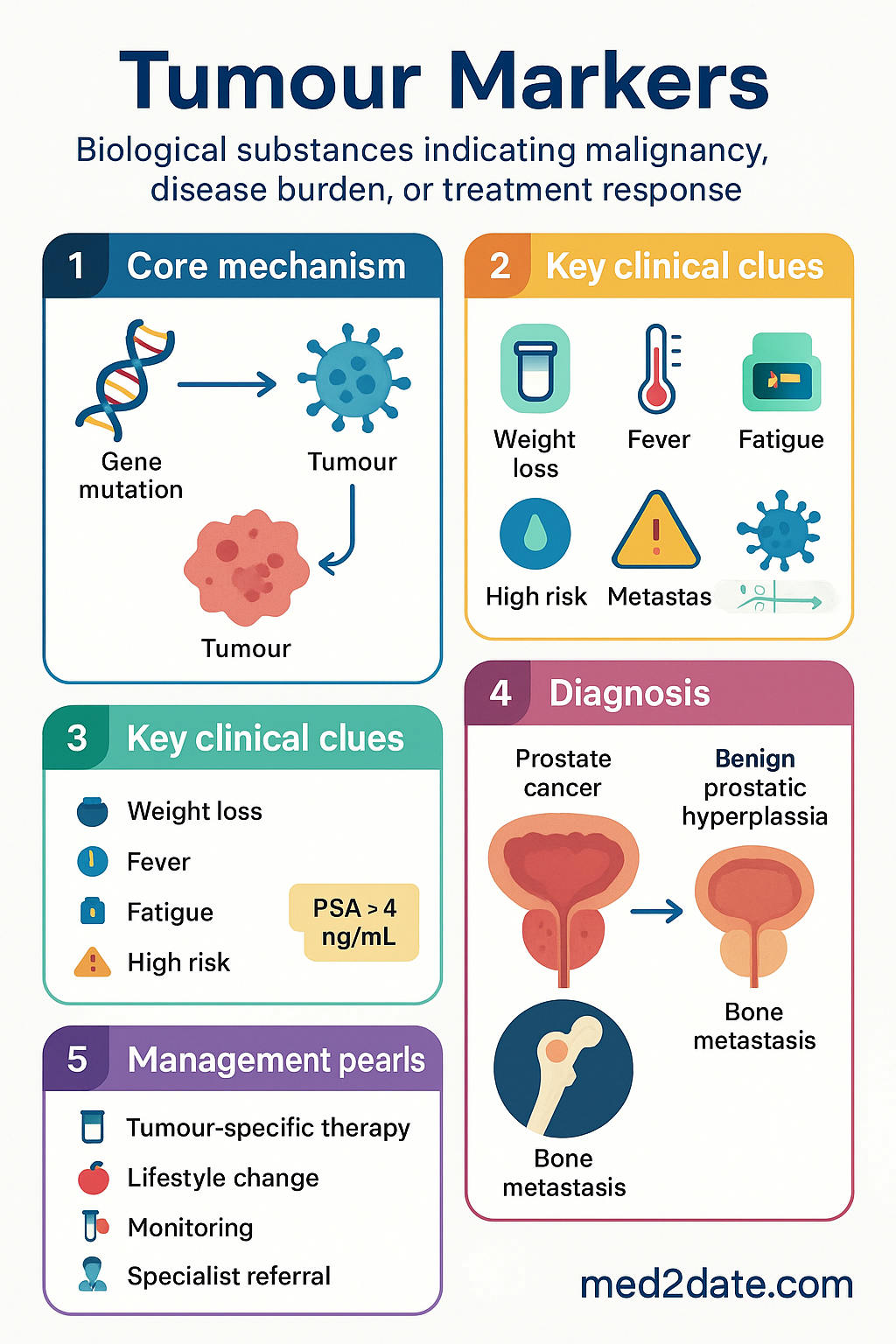
Types of Tumour Markers
Tumour markers are broadly classified by their biochemical nature and clinical utility. Understanding the category of a marker aids in interpreting results and selecting appropriate clinical actions.
Oncofetal Antigens
Proteins normally expressed during foetal development that are re-expressed or overexpressed in certain malignancies. Examples include alpha-fetoprotein (AFP) and carcinoembryonic antigen (CEA). These tend to be organ-associated but not organ-specific.
Carbohydrate (Mucin) Antigens
High-molecular-weight glycoproteins shed by tumour cells into the circulation. This group includes CA-125 (ovarian), CA 19-9 (pancreatic/biliary), CA 15-3 (breast), and CA 27-29 (breast). They are frequently elevated in benign conditions, limiting their specificity.
Hormones
Ectopic or excessive production of hormones by tumours. Beta-human chorionic gonadotropin (β-hCG) is the classic example, elevated in gestational trophoblastic disease and germ cell tumours. Calcitonin (medullary thyroid carcinoma) and catecholamine metabolites (phaeochromocytoma) are other examples.
Enzymes
Lactate dehydrogenase (LDH), alkaline phosphatase (ALP), prostate-specific acid phosphatase, and neuron-specific enolase (NSE) fall into this category. LDH is a non-specific marker of tissue turnover and is prognostically significant in lymphoma, germ cell tumours, and melanoma.
Receptors & Surface Proteins
Tissue-based markers including HER2/neu (breast/gastric cancer), oestrogen receptor (ER) and progesterone receptor (PR) in breast cancer, and PD-L1 expression across multiple tumour types. These guide targeted therapy selection and are assessed immunohistochemically on biopsy specimens.
Molecular & Genomic Markers
Circulating tumour DNA (ctDNA), microsatellite instability (MSI) status, BRAF V600E, KRAS/NRAS mutations, EGFR mutations, and tumour mutational burden (TMB). These markers are increasingly used for treatment selection, minimal residual disease (MRD) monitoring, and prognosis. Liquid biopsy (ctDNA) is available through several Australian pathology providers.
Other Markers
S-100 protein (melanoma, neural tumours), immunoglobulins (multiple myeloma), beta-2 microglobulin (myeloma, lymphoma), and prostate-specific membrane antigen (PSMA) used in both serum assays and PET imaging (⁶⁸Ga-PSMA PET, now widely available in Australia).
| Category | Examples | Primary Cancer Association |
|---|---|---|
| Oncofetal antigens | AFP, CEA | HCC, germ cell, colorectal |
| Carbohydrate antigens | CA-125, CA 19-9, CA 15-3 | Ovarian, pancreatic, breast |
| Glycoproteins | PSA | Prostate |
| Hormones | β-hCG, calcitonin | Germ cell, gestational, medullary thyroid |
| Enzymes | LDH, ALP, NSE | Lymphoma, bone metastases, neuroendocrine |
| Molecular/genomic | ctDNA, MSI, BRAF, EGFR, HER2 | Multiple — therapy selection |
Common Markers — PSA, CEA, AFP, CA-125
Prostate-Specific Antigen (PSA)
PSA is a kallikrein-related serine protease produced by prostatic epithelial cells. It is the most widely used tumour marker in Australia, predominantly for prostate cancer detection, risk stratification, and post-treatment surveillance.
Age-specific reference ranges improve specificity: 0–2.5 ng/mL for men <50 years; 0–3.5 ng/mL (50–59); 0–4.5 ng/mL (60–69); 0–6.5 ng/mL (≥70). PSA density (PSA ÷ prostate volume on TRUS), PSA velocity, and free-to-total PSA ratio can improve diagnostic accuracy. A free:total PSA ratio <10% suggests a higher likelihood of malignancy; >25% favours benign disease.
Post-radical prostatectomy, PSA should fall to undetectable levels (<0.1 ng/mL). Any detectable or rising PSA indicates biochemical recurrence and warrants further investigation. Prostate Health Index (PHI) and 4Kscore are additional risk stratification tools available through specialist referral in Australia.
Carcinoembryonic Antigen (CEA)
CEA is a glycoprotein involved in cell adhesion, normally produced during foetal development. In adults, elevated CEA levels are most commonly associated with colorectal cancer (CRC), though elevations occur in pancreatic, gastric, lung, breast, and medullary thyroid cancers.
CEA is not recommended for CRC screening due to poor sensitivity for early-stage disease (only 5–10% of Dukes A/B tumours have elevated CEA). Its principal role is in post-operative surveillance: CEA should be measured every 3–6 months for at least 5 years following curative CRC resection. Rising CEA warrants CT imaging to evaluate recurrence before committing to second-look surgery. Smoking cessation should be confirmed, as chronic smoking elevates CEA independently.
Alpha-Fetoprotein (AFP)
AFP is a major plasma protein produced by the yolk sac and foetal liver. In adults, elevated AFP is associated with hepatocellular carcinoma (HCC), non-seminomatous germ cell tumours (NSGCT), and, rarely, gastric and pancreatic cancers. AFP is also physiologically elevated in pregnancy.
For HCC surveillance in high-risk patients (chronic hepatitis B, hepatitis C with advanced fibrosis, cirrhosis of any aetiology), Cancer Council Australia and the Gastroenterological Society of Australia (GESA) recommend 6-monthly AFP combined with liver ultrasound. AFP alone has a sensitivity of approximately 60–70% for HCC; combined with ultrasound, sensitivity rises to approximately 90%. In germ cell tumours, AFP (alongside β-hCG and LDH) is essential for staging, risk stratification (IGCCCG classification), and treatment monitoring. AFP half-life of approximately 5–7 days is used to assess chemotherapy response.
Cancer Antigen 125 (CA-125)
CA-125 is a high-molecular-weight glycoprotein encoded by the MUC16 gene. It is the most widely used serum marker for epithelial ovarian cancer (EOC), particularly high-grade serous histology. CA-125 is also elevated in endometriosis, pelvic inflammatory disease, menstruation, pregnancy, liver cirrhosis, and peritoneal disease.
CA-125 is elevated in approximately 80% of advanced-stage EOC but only 50% of stage I disease, limiting its use as a standalone screening tool. The UK Collaborative Trial of Ovarian Cancer Screening (UKCTOCS) demonstrated that CA-125-based screening algorithms did not reduce ovarian cancer mortality. In Australia, CA-125 is not recommended for population screening in asymptomatic women.
Its established roles include: (1) assessment of adnexal masses using risk prediction models such as the Risk of Malignancy Index (RMI), which combines CA-125, menopausal status, and ultrasound score; (2) monitoring treatment response during chemotherapy — normalisation after 3 cycles predicts favourable outcome; (3) post-treatment surveillance with serial measurements every 2–4 months; and (4) distinguishing benign from malignant pleural/peritoneal effusions.
Clinical Applications
Tumour markers serve distinct clinical functions across the cancer care continuum. Their utility varies significantly depending on the clinical scenario.
Screening & Early Detection
No tumour marker currently meets the criteria for population-level cancer screening in asymptomatic individuals (high sensitivity, high specificity, acceptable positive predictive value, demonstrated mortality reduction). PSA screening for prostate cancer is the most debated example; Australian guidelines endorse shared decision-making rather than population-based screening. AFP + ultrasound for HCC in high-risk groups (chronic hepatitis B/C, cirrhosis) is a recommended surveillance strategy. CA-125 is not recommended for ovarian cancer screening.
Diagnosis & Differential Diagnosis
Markers may support — but never confirm — a cancer diagnosis. Elevated AFP in a liver mass is highly suggestive of HCC. Elevated β-hCG in a testicular mass indicates a germ cell tumour component. Elevated PSA in a man with an abnormal DRE prompts prostate biopsy. However, no marker alone can establish a diagnosis; tissue confirmation remains the gold standard.
Prognosis & Risk Stratification
Several markers have independent prognostic value. LDH is incorporated into the International Germ Cell Cancer Collaborative Group (IGCCCG) prognostic classification for germ cell tumours. Pre-operative CEA level independently predicts CRC recurrence risk. PSA at diagnosis, Gleason score, and T-stage form the basis of prostate cancer risk stratification (D'Amico classification, NCCN risk groups). Beta-2 microglobulin and serum free light chain ratio are prognostic in multiple myeloma (ISS/R-ISS staging).
Treatment Response Monitoring
Serial marker measurement is one of the most clinically valuable applications. Key examples include:
- Germ cell tumours: AFP and β-hCG half-life calculations during chemotherapy — failure to decline by expected half-life indicates chemoresistance and may prompt treatment intensification.
- Ovarian cancer: CA-125 normalisation during first-line chemotherapy correlates with surgical complete response and improved PFS.
- Colorectal cancer: CEA reduction following surgery or chemotherapy indicates treatment efficacy.
- Prostate cancer: PSA decline ≥50% (PCWG3 criteria) is a standard response endpoint in metastatic castration-resistant prostate cancer (mCRPC) trials.
Post-Treatment Surveillance & Recurrence Detection
Regular marker testing in the post-treatment setting enables early detection of recurrence, potentially before radiological or symptomatic evidence. CEA monitoring post-CRC resection (every 3–6 months for 5 years) is standard of care. PSA post-radical prostatectomy or radiotherapy detects biochemical recurrence. Rising CA-125 post-treatment for ovarian cancer precedes clinical recurrence by 2–6 months in most patients.
Guiding Targeted Therapy Selection
Molecular markers increasingly direct therapy choice:
- HER2 amplification: Trastuzumab, pertuzumab, T-DXd (breast and gastric cancer).
- EGFR mutations: Osimertinib and other EGFR-TKIs in NSCLC.
- BRAF V600E: Dabrafenib + trametinib in melanoma and other solid tumours.
- MSI-H/dMMR: Pembrolizumab across tumour types (TGA-approved tissue-agnostic indication).
- PD-L1 expression: Guides immunotherapy use in NSCLC, urothelial, and other cancers.
- ALK/ROS1 rearrangements: Crizotinib, alectinib, lorlatinib in NSCLC.
Minimal Residual Disease (MRD) & Liquid Biopsy
Circulating tumour DNA (ctDNA) analysis is an emerging application in Australian oncology. Tumour-informed ctDNA assays (e.g., for CRC and lung cancer) can detect molecular residual disease after curative surgery with high sensitivity. Rising ctDNA post-resection predicts radiological relapse by weeks to months. Several Australian pathology providers offer ctDNA testing; MBS item availability is limited, and out-of-pocket costs apply in most settings. The DYNAMIC-II trial (Australian-led, published 2024) demonstrated ctDNA-guided adjuvant chemotherapy in stage II CRC reduced treatment without compromising recurrence-free survival.
| Clinical Scenario | Primary Marker(s) | Recommended Action |
|---|---|---|
| CRC post-surgical surveillance | CEA | Every 3–6 months × 5 years; rising → CT |
| Prostate cancer — active surveillance | PSA | Every 3–6 months; rise triggers MRI/biopsy |
| HCC surveillance (high-risk) | AFP + liver USS | Every 6 months |
| Ovarian cancer — chemo response | CA-125 | Every cycle; normalisation = good response |
| Germ cell tumour — chemo response | AFP, β-hCG, LDH | Half-life monitoring; non-decline = poor response |
| NSCLC — EGFR therapy selection | ctDNA / tissue EGFR | Mutation-positive → osimertinib |
Limitations & Interpretation
Tumour marker interpretation is fraught with potential pitfalls. Clinicians must understand the inherent limitations to avoid inappropriate clinical decisions.
Lack of Cancer Specificity
Most tumour markers are not unique to malignant cells. PSA is elevated in benign prostatic hyperplasia (BPH) and prostatitis. CA-125 rises with endometriosis, menstruation, and cirrhosis. CEA is elevated by smoking and inflammatory bowel disease. An elevated marker alone should never prompt a cancer diagnosis or invasive investigation without supporting clinical context.
Insufficient Sensitivity for Early-Stage Disease
The majority of circulating tumour markers are produced in greater quantities by larger, more advanced tumours. CA-125 is elevated in only ~50% of stage I ovarian cancers. CEA is elevated in <10% of Dukes A colorectal cancers. This limits their utility in early detection and underscores the need for complementary diagnostic modalities (imaging, endoscopy, biopsy).
Variability & Assay Differences
Tumour marker values can vary between laboratories due to differences in assay platforms, antibodies, and calibrators. When monitoring serial values, it is essential to use the same laboratory and assay method. Changes of <20–25% may reflect assay variability rather than true biological change. Results should always be compared against the laboratory's own reference range.
Lead-Time Bias
Detecting a rising tumour marker before clinical recurrence does not automatically translate to improved survival. Lead-time bias — the apparent prolongation of survival from earlier detection without true benefit — must be considered when interpreting surveillance marker trends. Only marker-guided interventions that demonstrably improve outcomes (e.g., surgical resection of oligometastatic recurrence detected by rising CEA) confer genuine benefit.
Hook Effect
In sandwich immunoassays, extremely high analyte concentrations can saturate both capture and detection antibodies, paradoxically yielding falsely low results. This is clinically relevant for β-hCG in gestational trophoblastic disease and large-volume germ cell tumours. If a result is unexpectedly low despite strong clinical suspicion, the laboratory should be asked to repeat the test with sample dilution.
Interfering Substances
Heterophilic antibodies (e.g., human anti-mouse antibodies from prior monoclonal antibody therapy) can cause false-positive results in immunoassays. Biotin supplements (>5 mg/day) can interfere with streptavidin-based assays, affecting results for several markers. Haemolysis, lipaemia, and icterus may also impact certain assays. Clinical correlation and repeat testing with a different platform can resolve discrepant results.
Common Pitfalls — Quick Reference
Optimal Monitoring Intervals
Investigations & Australian Laboratory Availability
All commonly used serum tumour markers are available through major Australian pathology networks (Sullivan Nicolaides Pathology, Douglass Hanly Moir Pathology, Laverty Pathology, Western Diagnostic Pathology, and public hospital laboratories). Molecular and genomic testing availability varies by state and tumour type.
Special Populations
Aboriginal and Torres Strait Islander Health Considerations
Aboriginal and Torres Strait Islander peoples experience a disproportionate cancer burden compared to non-Indigenous Australians. According to AIHW data, cancer is the third leading cause of death among Indigenous Australians, with mortality rates 1.4 times higher than the non-Indigenous population. Cancers are frequently diagnosed at a later stage, contributing to poorer outcomes. Liver cancer (HCC), lung cancer, cervical cancer, and head and neck cancers are among those with the greatest disparity.
Key Considerations for Tumour Marker Use
📚 References
- 1. Australian Institute of Health and Welfare. Cancer data in Australia 2023. Canberra: AIHW; 2023. Available from: https://www.aihw.gov.au/reports/cancer/cancer-data-in-australia
- 2. Cancer Council Australia. Clinical practice guidelines for the management of colorectal cancer. 4th ed. Sydney: Cancer Council Australia; 2023.
- 3. Duff J, Engel A, Valentine D, et al. Prostate Cancer Foundation of Australia / Royal Australian College of General Practitioners. Prostate cancer screening policy. Sydney; 2022.
- 4. Gastroenterological Society of Australia (GESA). Clinical guidelines for hepatocellular carcinoma surveillance. Melbourne: GESA; 2023.
- 5. Duffy MJ, Lamerz R, Haglund C, et al. Tumor markers in colorectal cancer, gastric cancer and gastrointestinal stromal cancers: European Group on Tumor Markers 2014 guidelines update. Int J Cancer. 2014;134(11):2513–2522.
- 6. Sölétormos G, Duffy MJ, Hayes DF, et al. Design of tumor biomarker monitoring trials: a proposal by the European Group on Tumor Markers. Clin Chem. 2013;59(1):52–59.
- 7. Cancer Council Australia. National framework for gynaemic cancer services. Sydney: Cancer Council Australia; 2021.
- 8. Tie J, Cohen JD, Lahouel K, et al. Circulating tumor DNA analysis guiding adjuvant treatment in stage II colon cancer. N Engl J Med. 2022;386(24):2261–2272. (DYNAMIC Trial)
- 9. Tie J, Cohen JD, Lo SN, et al. Recurrence surveillance in stage II–III colorectal cancer using circulating tumour DNA (DYNAMIC-II). Nat Med. 2024. doi:10.1038/s41591-024-03009-2.
- 10. Australian Institute of Health and Welfare. Aboriginal and Torres Strait Islander health performance framework 2023 summary report. Canberra: AIHW; 2023.
- 11. Conroy T, Desseigne F, Ychou M, et al. FOLFIRINOX versus gemcitabine for metastatic pancreatic cancer. N Engl J Med. 2011;364(19):1817–1825. (CA 19-9 response data)
- 12. National Health and Medical Research Council. National statement on ethical conduct in human research. Canberra: NHMRC; 2023 (Updated).
- 13. Cancer Australia. A national cancer control framework and strategy for Australia. Surry Hills: Cancer Australia; 2022.
- 14. NHS Cancer Screening Programmes. United Kingdom Collaborative Trial of Ovarian Cancer Screening (UKCTOCS). Lancet. 2021;397(10270):218–228.
- 15. Australian Government Department of Health. Medical Benefits Schedule — Pathology items. Canberra: DoH; 2024. Available from: http://www.mbsonline.gov.au